

The Journal of Practical Medicine ›› 2026, Vol. 42 ›› Issue (4): 668-676.doi: 10.3969/j.issn.1006-5725.2026.04.018
• Original Articles • Previous Articles
Shouquan CHENG1,Naifeng LIU2( ),Ruoshui LI1
Received:2025-11-20
Online:2026-02-25
Published:2026-02-25
Contact:
Naifeng LIU
E-mail:tigetige@163.com
CLC Number:
Shouquan CHENG,Naifeng LIU,Ruoshui LI. Analysis of risk factors for aortic valve calcification in patients with coronary artery calcification and construction of a predictive model[J]. The Journal of Practical Medicine, 2026, 42(4): 668-676.
Tab.1
comparison of general data between the training set and the validation set"
| 变量 | 训练集(n = 1 020) | 验证集(n = 438) | χ2 /Z值 | P值 |
|---|---|---|---|---|
| 主动脉瓣钙化/[例(%)] | 312(30.6) | 122(27.9) | 1.10 | 0.295 |
| 年龄/岁 | 69.00(61.00,77.00) | 68.00(60.25,75.00) | 0.70 | 0.487 |
| 性别/[例(%)] | 1.62 | 0.203 | ||
| 男 | 553(54.22) | 254(57.99) | ||
| 女 | 467(45.78) | 184(42.01) | ||
| 身高/cm | 165.00(159.75,171.00) | 165.00(158.25,171.00) | 0.09 | 0.924 |
| 体质量/kg | 66.00(60.00,75.00) | 67.25(59.00,75.00) | 0.66 | 0.509 |
| BMI/(kg/m2) | 24.44(22.49,26.73) | 24.61(22.57,27.05) | 0.77 | 0.438 |
| LM钙化积分 | 0.00(0.00,1.00) | 0.00(0.00,0.00) | 0.65 | 0.52 |
| LAD钙化积分 | 45.50(6.00,181.25) | 52.23(4.94,211.00) | 0.14 | 0.888 |
| LCX钙化积分 | 0.00(0.00,35.00) | 0.00(0.00,46.75) | 0.57 | 0.565 |
| RCA钙化积分 | 7.47(0.00,100.00) | 7.00(0.00,99.00) | 0.27 | 0.789 |
| 总钙化积分 | 104.50(23.00,388.00) | 102.00(21.00,440.14) | 0.11 | 0.915 |
| AO/cm | 2.20(2.00,2.30) | 2.20(2.00,2.35) | 0.05 | 0.959 |
| AAO/cm | 3.42(3.20,3.70) | 3.40(3.13,3.68) | 1.74 | 0.082 |
| LAD/cm | 3.90(3.60,4.20) | 3.85(3.50,4.24) | 0.88 | 0.377 |
| RVEDd/cm | 2.40(2.20,2.59) | 2.34(2.20,2.50) | 1.03 | 0.301 |
| IVST/cm | 1.05(0.94,1.20) | 1.01(0.92,1.18) | 2.41 | 0.016 |
| LVEDd/cm | 4.60(4.30,5.00) | 4.60(4.30,5.00) | 0.19 | 0.851 |
| LVPW/cm | 1.00(0.90,1.10) | 0.98(0.89,1.06) | 2.29 | 0.022 |
| RWT | 0.42(0.38,0.48) | 0.42(0.38,0.47) | 1.64 | 0.101 |
| IVS/LVPW | 1.05(1.00,1.16) | 1.04(0.99,1.15) | 1.11 | 0.269 |
| LVEF/% | 69.00 (63.00,74.00) | 68.00(63.00,73.00) | 1.26 | 0.205 |
| 高血压病史/[例(%)] | 761(74.61) | 305(69.63) | 3.66 | 0.058 |
| 2型糖尿病/[例(%)] | 302(29.61) | 116(26.48) | 1.32 | 0.252 |
| 他汀类药物/[例(%)] | 546(53.53) | 208(47.49) | 4.26 | 0.039 |
| 吸烟/[例(%)] | 217(21.27) | 106(24.20) | 1.35 | 0.244 |
| UA/(umol/L) | 338.00(277.00,409.00) | 339.28(280.00,420.00) | 0.42 | 0.670 |
| TG/(mmol/L) | 1.29(0.93,1.90) | 1.26(0.90,1.93) | 0.03 | 0.976 |
| HDL-C/(mmol/L) | 1.24(1.06,1.44) | 1.21(1.04,1.45) | 0.83 | 0.405 |
| LDL-C/(mmol/L) | 2.56(1.98,3.17) | 2.46(1.95,3.04) | 1.58 | 0.112 |
| Lp(a)/(mg/L) | 127.50(62.00,262.25) | 128.00(5.25,274.75) | 0.21 | 0.836 |
Tab.2
Basic information of patients with/without aortic valve calcification in the training group"
| 变量 | 主动脉瓣无钙化(n = 708) | 主动脉瓣钙化(n = 312) | χ2 /Z值 | P值 |
|---|---|---|---|---|
| 年龄/岁 | 66.00(59.00,74.00) | 75.00(66.75,83.00) | 8.67 | < 0.001 |
| 性别/[例(%)] | 7.24 | 0.007 | ||
| 男 | 404(57.06) | 149(47.8) | ||
| 女 | 304(42.94) | 163(52.2) | ||
| 身高/cm | 165.00(160.00,172.00) | 163.00(157.00,170.00) | 3.32 | 0.001 |
| 体质量/kg | 67.50(60.00,75.00) | 65.00(58.00,73.00) | 3.04 | 0.003 |
| BMI/(kg/m2) | 24.44(22.60,26.73) | 24.39(22.49,26.73) | 0.26 | 0.794 |
| LM钙化积分 | 0.00(0.00,0.00) | 0.00(0.00,29.25) | 7.89 | < 0.001 |
| LAD钙化积分 | 36.00(5.00,141.00) | 86.50(11.96,325.00) | 6.98 | < 0.001 |
| LCX钙化积分 | 0.00(0.00,19.25) | 4.50(0.00,71.77) | 6.71 | <0.001 |
| RCA钙化积分 | 2.00(0.00,64.04) | 45.00(0.00,245.00) | 7.53 | <0.001 |
| 总钙化积分 | 72.50(18.34,265.18) | 253.50(49.25,758.25) | 8.22 | <0.001 |
| AO/cm | 2.20(2.00,2.30) | 2.20(2.00,2.33) | 0.61 | 0.541 |
| AAO/cm | 3.40(3.20,3.70) | 3.50(3.20,3.75) | 2.08 | 0.036 |
| LAD/cm | 3.88(3.53,4.20) | 4.00(3.70,4.30) | 3.33 | 0.001 |
| RVEDd/cm | 2.39(2.17,2.56) | 2.40(2.20,2.60) | 1.94 | 0.054 |
| IVST/cm | 1.04(0.94,1.20) | 1.06(0.96,1.20) | 1.17 | 0.238 |
| LVEDd/cm | 4.60(4.30,4.98) | 4.60(4.30,5.05) | 0.21 | 0.833 |
| LVPW/cm | 0.99(0.90,1.08) | 1.00(0.90,1.10) | 2.16 | 0.031 |
| RWT | 0.42(0.38,0.47) | 0.43(0.38,0.48) | 1.74 | 0.083 |
| IVS/LVPW | 1.05(1.00,1.17) | 1.05(1.00,1.14) | 0.51 | 0.611 |
| LVEF/% | 69.00(63.00,74.00) | 69.00(63.00,74.00) | 0.16 | 0.871 |
| 高血压病史/[例(%)] | 501(71.76) | 254(81.41) | 10.80 | 0.001 |
| 2型糖尿病/[例(%)] | 204(28.81) | 98(31.41) | 0.58 | 0.446 |
| 他汀类药物/[例(%)] | 351(49.58) | 195(62.50) | 16.60 | < 0.001 |
| 吸烟/[例(%)] | 165(23.31) | 52(16.67) | 5.33 | 0.021 |
| UA/(umol/L) | 338.00(277.00,410.00) | 340.50(275.5,406.00) | 0.53 | 0.599 |
| TG/(mmol/L) | 1.36(0.95,2.01) | 1.18(0.84,1.75) | 3.33 | 0.001 |
| HDL-C/(mmol/L) | 1.24(1.05,1.44) | 1.25(1.06,1.44) | 0.24 | 0.812 |
| LDL-C/(mmol/L) | 2.57(1.98,3.19) | 2.52(1.98,3.13) | 0.76 | 0.445 |
| Lp(a)/(mg/L) | 122.00(62.00,246.00) | 149.5.00(70.75,316.50) | 2.47 | 0.014 |
Tab.3
Univariate logistic regression analysis of aortic valve calcification"
| 变量 | OR(95%CI) | Wald χ2 | P值 |
|---|---|---|---|
| 年龄/岁 | 1.070(1.050 ~ 1.080) | 7.83 | < 0.001 |
| 性别/女 | 1.450(1.110 ~ 1.900) | 2.75 | 0.006 |
| 身高/cm | 0.970(0.960 ~ 0.990) | 3.33 | 0.001 |
| 体质量/kg | 0.990(0.980 ~ 1.000) | 2.00 | 0.049 |
| BMI/(kg/m2) | 1.000(0.960 ~ 1.030) | 0.27 | 0.79 |
| LM钙化积分 | 1.004(1.003 ~ 1.006) | 6.00 | < 0.001 |
| LAD钙化积分 | 1.001(1.001 ~ 1.002) | 5.48 | < 0.001 |
| LCX钙化积分 | 1.001(1.001 ~ 1.002) | 5.48 | < 0.001 |
| RCA钙化积分 | 1.001(1.001 ~ 1.002) | 5.74 | < 0.001 |
| 总钙化积分 | 1.001(1.000 ~ 1.001) | 4.64 | < 0.001 |
| AO/cm | 1.460(0.890 ~ 2.370) | 1.52 | 0.13 |
| AAO/cm | 1.440(1.050 ~ 1.980) | 2.27 | 0.023 |
| LAD/cm | 1.330(1.060 ~ 1.660) | 2.54 | 0.013 |
| RVEDd/cm | 1.450(0.960 ~ 2.180) | 1.77 | 0.079 |
| IVST/cm | 1.570(0.820 ~ 2.960) | 1.40 | 0.167 |
| LVEDd/cm | 0.960(0.780 ~ 1.180) | 0.38 | 0.706 |
| LVPW/cm | 1.800(0.950 ~ 3.510) | 1.80 | 0.072 |
| RWT | 1.080(0.740 ~ 1.520) | 0.45 | 0.659 |
| IVS/LVPW | 0.590(0.270 ~ 1.260) | 1.33 | 0.186 |
| LVEF/% | 0.990(0.980 ~ 1.010) | 1.01 | 0.312 |
| 高血压 | 1.740(1.260 ~ 2.430) | 3.33 | 0.001 |
| 2型糖尿病 | 1.130(0.850 ~ 1.510) | 0.84 | 0.403 |
| 他汀类药物 | 1.700(1.290 ~ 2.230) | 4.24 | < 0.001 |
| 吸烟 | 0.660(0.460 ~ 0.920) | 2.39 | 0.017 |
| UA/(umol/L) | 0.999(0.998 ~ 1.001) | 0.47 | 0.641 |
| TG/(mmol/L) | 0.830(0.720 ~ 0.940) | 2.75 | 0.006 |
| HDL/(mmol/L) | 1.060(0.690 ~ 1.620) | 0.26 | 0.794 |
| LDL/(mmol/L) | 0.960(0.820 ~ 1.120) | 0.49 | 0.621 |
| Lp(a)/(mg/L) | 1.001(1.000 ~ 1.001) | 2.88 | 0.004 |
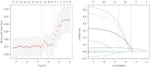
Fig.1
LASSO regression results"
Tab.4
Multivariate logistic regression analysis of aortic valve calcification"
| 变量 | OR(95%CI) | P值 |
|---|---|---|
| 年龄/岁 | 1.055(1.040 ~ 1.071) | < 0.001 |
| 身高/cm | 0.980(0.962 ~ 0.997) | 0.023 |
| LM钙化积分 | 1.002(1.000 ~ 1.003) | 0.076 |
| RCA钙化积分 | 1.000(0.999 ~ 1.001) | 0.411 |
| 总钙化积分 | 1.000(0.999 ~ 1.001) | 0.346 |
| RVEDd/cm | 1.719(1.102 ~ 2.692) | 0.017 |
| LVPW/cm | 1.909(0.921 ~ 4.088) | 0.087 |
| 高血压 | 1.402(0.981 ~ 2.023) | 0.067 |
| 他汀类药物 | 1.408(1.047 ~ 1.899) | 0.024 |
| TG/(mmol/L) | 0.932(0.806 ~ 1.060) | 0.311 |
| Lp(a)/(mg/L) | 1.001(1.000 ~ 1.001) | 0.123 |
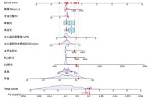
Fig.2
Nomogram for predicting aortic valve calcification in CT"
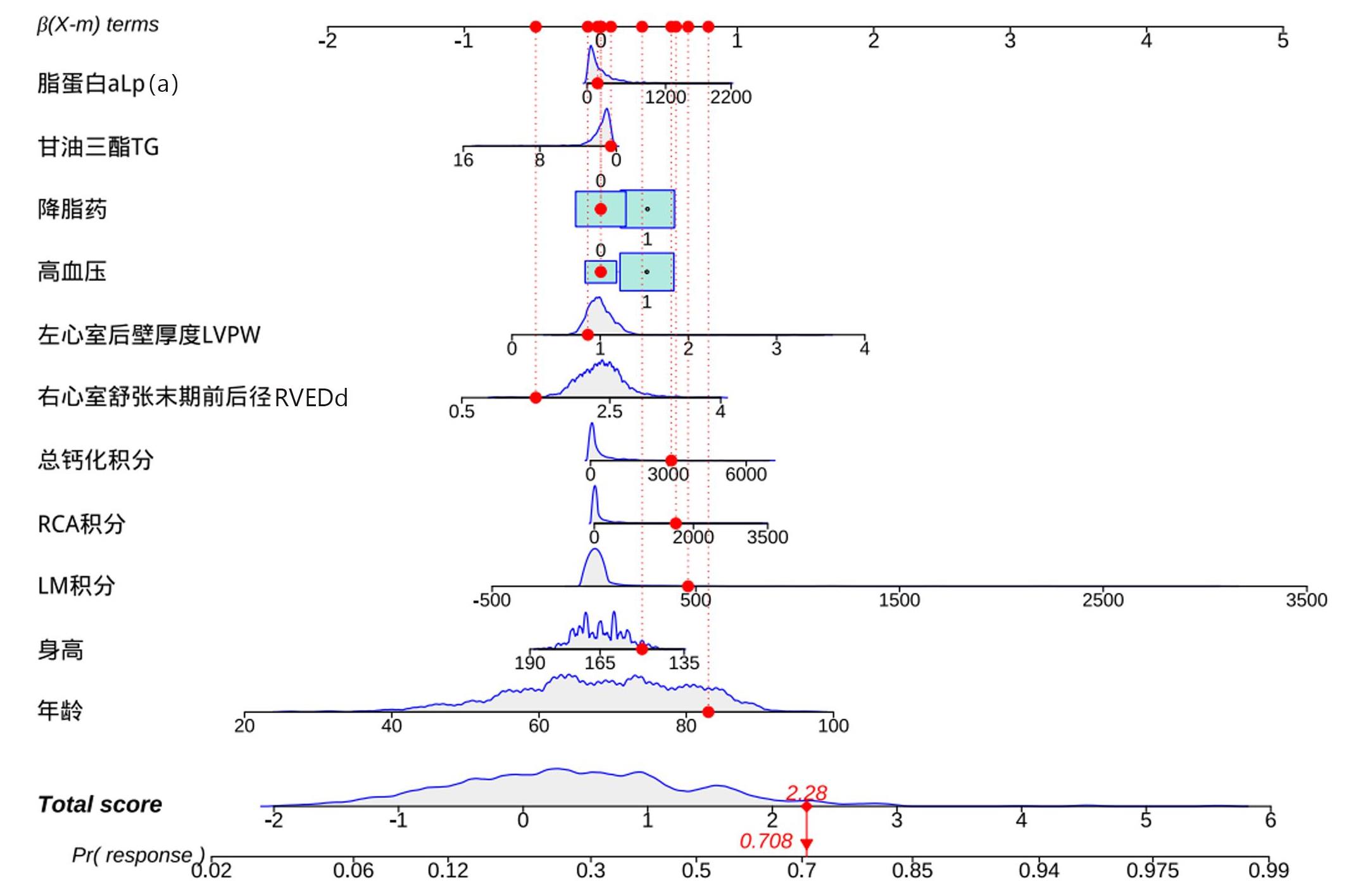
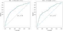
Fig.3
ROC curves of the training set (left) and the validation set (right)"
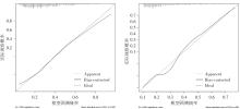
Fig.4
The training set (left), validation set (right), and the calibration curve of the nomogram"

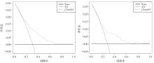
Fig.5
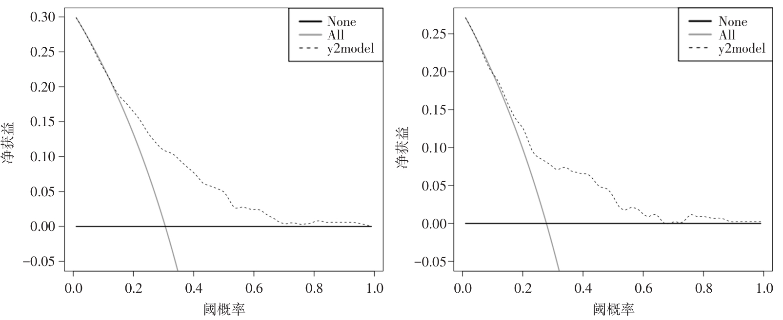
DCA curves of the training set (left) and the validation set (right)"
| [1] |
DEMER L L, TINTUT Y. Interactive and Multifactorial Mechanisms of Calcific Vascular and Valvular Disease[J]. Trends Endocrinol Metab, 2019, 30(9): 646-657.doi: 10.1016/j.tem. 2019. 06.001 .
doi: 10.1016/j.tem. 2019. 06.001 |
| [2] |
ONNIS C, VIRMANI R, KAWAI K, et al. Coronary Artery Calcification: Current Concepts and Clinical Implications[J]. Circulation, 2024, 149(3): 251-266. doi: 10.1161/CIRCULATIONAHA.123.065657 .
doi: 10.1161/CIRCULATIONAHA.123.065657 |
| [3] |
PAWADE T, SHETH T, GUZZETTI E, et al. Why and How to Measure Aortic Valve Calcification in Patients With Aortic Stenosis[J]. JACC Cardiovasc Imaging,2019, 12(9): 1835-1848. doi: 10.1016/j.jcmg.2019.01.045 .
doi: 10.1016/j.jcmg.2019.01.045 |
| [4] |
中华医学会放射学分会心胸学组,《中华放射学杂志》心脏冠状动脉多排CT临床应用指南写作专家组. 心脏冠状动脉CT血管成像技术规范化应用中国指南[J]. 中华放射学杂志, 2017, 51(10): 732-743. doi:10.3760/j.issn.1005-1201. 2017. 10.004 .
doi: 10.3760/j.issn.1005-1201. 2017. 10.004 |
| [5] |
MONCLA L M, BRIEND M, BOSSE Y, et al. Calcific aortic valve disease: Mechanisms, prevention and treatment[J]. Nat Rev Cardiol, 2023, 20(8):546-559.doi: 10.1038/s41569-023-00845-7 .
doi: 10.1038/s41569-023-00845-7 |
| [6] |
YU J, WANG Z, BAO Q, et al. Global burden of calcific aortic valve disease and attributable risk factors[J]. Front Cardiovasc Med, 2022, 9:1003233.doi: 10.3389/fcvm.2022.1003233 .
doi: 10.3389/fcvm.2022.1003233 |
| [7] |
LIU Z, DONG N, HUI H, et al. Endothelial cell-derived tetrahydrobiopterin prevents aortic valve calcification[J]. Eur Heart J,2022,43(17):1652-1664. doi: 10.1093/eurheartj/ehac037 .
doi: 10.1093/eurheartj/ehac037 |
| [8] |
CHENG S Q, LIU N F, FANG L J, et al. Factors predicting the occurrence of aortic valve calcification in patients with coronary artery calcification[J]. Acta Cardiol, 2022, 77(10): 910-917.doi: 10.1080/00015385.2022.2072053 .
doi: 10.1080/00015385.2022.2072053 |
| [9] |
KANAAN C N, LAYOUN H, KONDOLEON N P, et al. Comparison of CT acquired cardiac valvular calcification scores in hemodialysis and peritoneal dialysis patients undergoing open heart surgery[J]. Am Heart J Plus,2022, 25: 100234. doi: 10.1016/j.ahjo.2022.100234 .
doi: 10.1016/j.ahjo.2022.100234 |
| [10] |
ING S W, MOHLER III E R, PUTT M E, et al. Correlates of valvular ossification in patients with aortic valve stenosis[J]. Clin Transl Sci, 2009, 2(6):431-435.doi: 10.1111/j.1752-8062. 2009.00168.x .
doi: 10.1111/j.1752-8062. 2009.00168.x |
| [11] |
CHEN H Y, ENGERT J C, THANASSOULIS G. Risk factors for valvular calcification[J]. Curr Opin Endocrinol Diabetes Obes, 2019, 26(2):96-102.doi: 10.1097/MED.0000000000000471 .
doi: 10.1097/MED.0000000000000471 |
| [12] |
LIU Z, SU Z, LI W, et al. Global, regional, and national time trends in disability-adjusted life years, mortality, and variable risk factors of non-rheumatic calcified aortic valve disease, 1990-2019: An age-period-cohort analysis of the Global Burden of Disease 2019 study[J]. J Thorac Dis, 2023, 15(4):2079-2097. doi: 10.21037/jtd-23-480 .
doi: 10.21037/jtd-23-480 |
| [13] |
MESSAOUDI H, LEVESQUE T, PERZO N, et al. Subtotal Nephrectomy Associated with a High-Phosphate Diet in Rats Mimics the Development of Calcified Aortic Valve Disease Associated with Chronic Renal Failure[J]. J Clin Med,2023,12(4):1539. doi: 10.3390/jcm12041539 .
doi: 10.3390/jcm12041539 |
| [14] |
XINYU Z, DONGXIA M, YUE H, et al. Statins Accelerate Coronary Calcification and Reduce the Risk of Cardiovascular Events[J]. Cardiol Rev, 2023, 31(6):293-298.doi: 10.1097/CRD. 0000000000000438 .
doi: 10.1097/CRD. 0000000000000438 |
| [15] |
PURI R, NICHOLLS S J, SHAO M, et al. Impact of statins on serial coronary calcification[J]. J Am Coll Cardiol, 2015, 65(13):1273-1282. doi: 10.1016/j.jacc.2015.01.036 .
doi: 10.1016/j.jacc.2015.01.036 |
| [16] |
DYKUN I, LEHMANN N, KÄLSCH H, et al. Statin Medication Enhances Progression of Coronary Artery Calcification: The Heinz Nixdorf Recall Study[J]. J Am Coll Cardiol, 2016,68(19): 2123-2125. doi: 10.1016/j.jacc.2016.08.040 .
doi: 10.1016/j.jacc.2016.08.040 |
| [17] |
FLETCHER A J, SINGH T, SYED M B J, et al. Imaging aortic valve calcification: Significance, approach and implications[J]. Clin Radiol,2021,76(1):15-26. doi: 10.1016/j.crad. 2020. 04.007 .
doi: 10.1016/j.crad. 2020. 04.007 |
| [18] |
WANG C, WANG S, WANG Z, et al. Andrographolide regulates H3 histone lactylation by interfering with p300 to alleviate aortic valve calcification[J]. Br J Pharmacol,2024,181(12):1843-1856. doi: 10.1111/bph.16332 .
doi: 10.1111/bph.16332 |
| Viewed | ||||||
|
Full text |
|
|||||
|
Abstract |
|
|||||