

The Journal of Practical Medicine ›› 2026, Vol. 42 ›› Issue (1): 110-118.doi: 10.3969/j.issn.1006-5725.2026.01.014
• Chronic Disease Control • Previous Articles
Zhouzhan LUO1,Qingling HU1,Qiaofeng WANG1,Guolong LEI1,Mengyao TANG1,Chao PENG1,Yingying TANG2( )
)
Received:2025-10-11
Online:2026-01-10
Published:2026-01-14
Contact:
Yingying TANG
E-mail:13975141084@163.com
CLC Number:
Zhouzhan LUO,Qingling HU,Qiaofeng WANG,Guolong LEI,Mengyao TANG,Chao PENG,Yingying TANG. Value of left ventricular contrast echocardiography and three-dimensional echocardiography heart model parameters on predicting major adverse cardiovascular events in patients with coronary heart disease after percutaneous coronary intervention[J]. The Journal of Practical Medicine, 2026, 42(1): 110-118.
Tab.1
Comparison of 3D-HM parameters and MCE parameters between CHD patients and healthy volunteers"
| 组别 | 例数 | LVEDV/mL | LVESV/mL | LVEF/% | A/dB | Β/s-1 | A×β/(dB/s) | PSI |
|---|---|---|---|---|---|---|---|---|
| 冠心病患者 | 197 | 136.57 ± 16.26 | 69.47 ± 8.21 | 49.13 ± 2.17 | 8.29 ± 1.85 | 1.22 ± 0.19 | 10.11 ± 1.87 | 1.84 ± 0.43 |
| 健康志愿者 | 104 | 107.05 ± 12.67 | 38.68 ± 5.95 | 63.87 ± 2.19 | 12.47 ± 2.09 | 1.74 ± 0.32 | 21.70 ± 4.46 | 1.21 ± 0.27 |
| t值 | 16.108 | 33.831 | 55.863 | 17.813 | 17.671 | 31.620 | 13.588 | |
| P值 | < 0.001 | < 0.001 | < 0.001 | < 0.001 | < 0.001 | < 0.001 | < 0.001 |
Tab.2
Comparison of baseline data between the two groups of patients"
| 指标 | 发生MACE组(n = 26) | 未发生MACE组(n = 171) | χ2/t值 | P值 |
|---|---|---|---|---|
| 性别/[例(%)] | 0.061 | 0.805 | ||
| 男 | 15(57.69) | 103(60.23) | ||
| 女 | 11(42.31) | 68(39.77) | ||
| 年龄/岁 | 62.07 ± 10.17 | 59.18 ± 10.03 | 1.366 | 0.173 |
| 体质量指数/(kg/m2) | 22.94 ± 1.75 | 22.61 ± 1.82 | 0.866 | 0.388 |
| 发病至PCI时间/h | 3.67 ± 0.32 | 3.58 ± 0.29 | 1.454 | 0.148 |
| 静息心率/(次/min) | 76.05 ± 8.55 | 77.47 ± 8.61 | 0.784 | 0.434 |
| 脉压/mmHg | 61.07 ± 10.36 | 62.19 ± 10.44 | 0.510 | 0.611 |
| 吸烟史/[例(%)] | 12(46.15) | 69(40.35) | 0.314 | 0.575 |
| 饮酒史/[例(%)] | 5(19.23) | 39(22.81) | 0.166 | 0.683 |
| 心血管疾病家族史/[例(%)] | 15(57.69) | 75(43.86) | 1.740 | 0.187 |
| 高血压/[例(%)] | 12(46.15) | 60(35.09) | 1.192 | 0.275 |
| 糖尿病/[例(%)] | 11(42.31) | 55(32.16) | 1.042 | 0.307 |
| 高脂血症/[例(%)] | 10(38.46) | 40(23.39) | 2.706 | 0.100 |
| 病变部位/[例(%)] | 2.256 | 0.521 | ||
| 左主干 | 7(26.92) | 52(30.41) | ||
| 左前降支 | 6(23.08) | 44(25.73) | ||
| 左回旋支 | 6(23.08) | 49(28.65) | ||
| 右冠状动脉 | 7(26.92) | 26(15.21) | ||
| 病变支数/[例(%)] | 1.651 | 0.438 | ||
| 1支 | 7(26.92) | 63(36.84) | ||
| 2支 | 8(30.77) | 56(32.75) | ||
| > 2支 | 11(42.31) | 52(30.41) | ||
| 疾病临床分型/[例(%)] | 0.508 | 0.776 | ||
| 稳定型心绞痛 | 8(30.77) | 43(25.15) | ||
| 不稳定型心绞痛 | 8(30.77) | 63(36.84) | ||
| 急性心肌梗死 | 10(38.46) | 65(38.01) | ||
| 存活心肌节段/[例(%)] | 6.038 | 0.014 | ||
| < 4个 | 17(65.38) | 68(39.77) | ||
| ≥ 4个 | 9(34.62) | 103(60.23) | ||
| 介入治疗门-球时间≥ 90 min/[例(%)] | 1.803 | 0.179 | ||
| 是 | 11(42.31) | 50(29.24) | ||
| 否 | 15(57.69) | 121(70.76) | ||
| 支架置入数量/[例(%)] | 1.063 | 0.303 | ||
| ≤ 2个 | 14(53.85) | 110(64.33) | ||
| > 2个 | 12(46.15) | 61(35.67) | ||
| TG/(mmol/L) | 1.69 ± 0.50 | 1.54 ± 0.45 | 1.560 | 0.120 |
| TC/(mmol/L) | 4.41 ± 1.26 | 4.14 ± 1.18 | 1.077 | 0.283 |
| LDL-C/(mmol/L) | 2.27 ± 0.67 | 2.11 ± 0.64 | 1.180 | 0.239 |
| HDL-C/(mmol/L) | 1.20 ± 0.28 | 1.29 ± 0.37 | 1.189 | 0.236 |
| HbAlc/% | 7.05 ± 2.03 | 6.43 ± 1.89 | 1.543 | 0.124 |
| FBG/(mmol/L) | 8.57 ± 2.47 | 7.89 ± 2.12 | 1.490 | 0.138 |
| BNP/(ng/L) | 110.58 ± 32.25 | 99.42 ± 29.51 | 1.775 | 0.078 |
| cTnI/(μg/L) | 0.77 ± 0.23 | 0.71 ± 0.21 | 1.340 | 0.182 |
| CK-MB/(μg/L) | 6.61 ± 1.85 | 6.17 ± 1.76 | 1.180 | 0.240 |
| 术后用药情况/[例(%)] | ||||
| 阿司匹林 | 21(80.77) | 157(91.81) | 3.159 | 0.076 |
| P2Y12抑制剂 | 21(80.77) | 152(88.89) | 1.391 | 0.238 |
| β受体阻滞剂 | 20(76.92) | 148(86.55) | 1.666 | 0.197 |
Tab.3
Comparison of 3D-HM parameters at different time points between the two groups of patients with coronary heart disease x ± s"
| 组别 | LVEDV/mL | LVESV/mL | LVEF/% | |||
|---|---|---|---|---|---|---|
| 发生MACE组(n = 26) | 未发生MACE组(n = 171) | 发生MACE组(n = 26) | 未发生MACE组(n = 171) | 发生MACE组(n = 26) | 未发生MACE组(n = 171) | |
| 术前 | 141.97 ± 15.81 | 135.75 ± 16.22 | 72.08 ± 7.33 | 69.07 ± 8.28 | 47.23 ± 2.15 | 47.52 ± 2.18 |
| 术后1周 | 132.36 ± 14.30a | 123.45 ± 14.92ad | 65.34 ± 7.42a | 58.91 ± 6.82ad | 49.63 ± 3.22a | 51.28 ± 2.26ad |
| 术后3个月 | 125.85 ± 13.75ab | 116.64 ± 13.03abd | 57.47 ± 6.95ab | 48.27 ± 6.04abd | 52.33 ± 2.31ab | 56.62 ± 2.47abd |
| 术后6个月 | 118.51 ± 12.42abc | 110.93 ± 12.24abcd | 48.59 ± 6.30abc | 39.21 ± 5.17abcd | 55.06 ± 2.47abc | 59.65 ± 2.05abcd |
| F组间/P组间 | 28.590/< 0.001 | 98.080/< 0.001 | 6.940/< 0.001 | |||
| F时间/P时间 | 47.660/< 0.001 | 265.600/< 0.001 | 50.400/< 0.001 | |||
| F交互/P交互 | 0.211/0.889 | 4.456/0.004 | 2.710/< 0.001 | |||
Tab.4
Comparison of MCE parameters between the two groups of patients with coronary heart disease at different time points x ± s"
| 组别 | A/dB | β/s-1 | A×β/(dB/s) | PSI | ||||
|---|---|---|---|---|---|---|---|---|
| 发生MACE组(n = 26) | 未发生MACE组(n = 171) | 发生MACE组(n = 26) | 未发生MACE组(n = 171) | 发生MACE组(n = 26) | 未发生MACE组(n = 171) | 发生MACE组(n = 26) | 未发生MACE组(n = 171) | |
| 术前 | 8.15 ± 1.86 | 8.31 ± 1.85 | 1.20 ± 0.18 | 1.22 ± 0.19 | 9.79 ± 1.94 | 10.16 ± 1.86 | 1.94 ± 0.42 | 1.82 ± 0.43 |
| 术后1周 | 8.94 ± 1.90a | 10.05 ± 1.96ad | 1.29 ± 0.19a | 1.43 ± 0.24ad | 11.53 ± 2.53a | 14.37 ± 2.61ad | 1.79 ± 0.32a | 1.61 ± 0.30ad |
| 术后3个月 | 10.02 ± 1.94ab | 11.36 ± 1.99abd | 1.43 ± 0.22ab | 1.59 ± 0.29abd | 14.33 ± 3.01ab | 18.06 ± 3.85abd | 1.58 ± 0.29ab | 1.40 ± 0.28abd |
| 术后6个月 | 11.05 ± 2.01abc | 12.42 ± 2.07abcd | 1.57 ± 0.28abc | 1.73 ± 0.31abcd | 17.35 ± 3.97abc | 21.49 ± 4.22abcd | 1.44 ± 0.27abc | 1.22 ± 0.25abcd |
| F组间/P组间 | 23.170/< 0.001 | 19.170/< 0.001 | 66.150/< 0.001 | 26.430/< 0.001 | ||||
| F时间/P时间 | 53.590/< 0.001 | 49.310/< 0.001 | 143.300/< 0.001 | 49.890/< 0.001 | ||||
| F交互/P交互 | 1.892/0.129 | 1.551/0.200 | 6.152/0.001 | 0.367/0.777 | ||||
Tab.5
Multivariate logistic regression analysis of factors affecting poor prognosis after PCI in patients with coronary heart disease"
| 变量 | β | SE | Wald χ2 | OR | 95%CI | P值 |
|---|---|---|---|---|---|---|
| 高水平LVEDV | 0.874 | 0.492 | 3.156 | 2.396 | 0.914 ~ 6.286 | 0.076 |
| 高水平LVESV | 0.718 | 0.467 | 2.364 | 2.050 | 0.821 ~ 5.121 | 0.125 |
| 高水平LVEF | 0.965 | 0.263 | 13.463 | 0.381 | 0.228 ~ 0.638 | < 0.001 |
| 高水平A | -0.693 | 0.197 | 12.375 | 0.500 | 0.340 ~ 0.736 | < 0.001 |
| 高水平β | -0.652 | 0.184 | 12.556 | 0.521 | 0.363 ~ 0.747 | < 0.001 |
| 高水平A×β | -0.619 | 0.176 | 12.370 | 0.538 | 0.381 ~ 0.760 | < 0.001 |
| 高水平PSI | 0.855 | 0.236 | 13.125 | 2.351 | 1.481 ~ 3.734 | < 0.001 |
| 存活心肌节段< 4个 | 1.051 | 0.577 | 3.318 | 2.861 | 0.923 ~ 8.863 | 0.069 |
Tab.6
Predictive value of left ventricular contrast echocardiography and 3D-HM parameters alone and in combinationon the prognosis in patients withcoronary heart disease undergoing PCI"
| 指标 | 截点值 | AUC | 95%CI | P值 | 约登指数 | 敏感度/% | 特异度/% |
|---|---|---|---|---|---|---|---|
| LVEF | 49.05% | 0.647 | 0.510 ~ 0.785 | 0.016 | 0.426 | 65.38 | 77.19 |
| A | 8.99 dB | 0.697 | 0.567 ~ 0.827 | 0.001 | 0.464 | 69.23 | 77.19 |
| β | 1.32 s-1 | 0.676 | 0.546 ~ 0.805 | 0.004 | 0.429 | 69.23 | 73.68 |
| A×β | 11.76 dB/s | 0.681 | 0.556 ~ 0.807 | 0.003 | 0.364 | 61.54 | 74.85 |
| PSI | 1.75 | 0.709 | 0.599 ~ 0.819 | 0.001 | 0.414 | 65.38 | 76.02 |
| 联合 | 0.891 | 0.836 ~ 0.947 | < 0.001 | 0.687 | 96.15 | 72.51 |
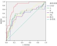
Fig.1
ROC curves of left ventricular contrast echocardiography and 3D-HM parameters alone and in combination at 1 week after PCI for predicting MACE after PCI in patients with coronary heart disease"
| [1] |
DUGGAN J P, PETERS A S, TRACHIOTIS G D, et al. Epidemiology of coronary artery disease[J]. Surg Clin North Am, 2022, 102(3): 499-516. doi: 10.1016/j.suc.2022.01.007 .
doi: 10.1016/j.suc.2022.01.007 |
| [2] |
刘庆爽, 杨志强, 贾崇富, 等. 负荷动态CT心肌灌注成像对疑似冠心病患者的预后价值[J]. 放射学实践, 2025, 40(3): 356-361. doi: 10.13609/j.cnki.1000-0313.2025.03.011 .
doi: 10.13609/j.cnki.1000-0313.2025.03.011 |
| [3] |
GRECO A, CAPODANNO D. Differences in coronary artery disease and outcomes of percutaneous coronary intervention with drug-eluting stents in women and men[J]. Expert Rev Cardiovasc Ther, 2021, 19(4): 301-312. doi: 10.1080/14779072. 2021. 1902806 .
doi: 10.1080/14779072. 2021. 1902806 |
| [4] |
张启贤, 高松原, 方舒, 等. 基于冠状动脉造影的微循环阻力指数对冠心病患者PCI术后的预后价值[J]. 中华心血管病杂志, 2025, 53(5): 505-513. doi: 10.3760/cma.j.cn112148-20250123-00065 .
doi: 10.3760/cma.j.cn112148-20250123-00065 |
| [5] |
SHAH B, SMILOWITZ N R, XIA Y, et al. Major Adverse cardiovascular events after colchicine administration before percutaneous coronary intervention: Follow-up of the colchicine-PCI trial[J]. Am J Cardiol, 2023, 204: 26-28. doi: 10.1016/j.amjcard.2023.07.029 .
doi: 10.1016/j.amjcard.2023.07.029 |
| [6] |
高玲, 刘金涛, 刘岩, 等. 高压氧联合心脏康复训练对冠心病PCI后患者临床疗效及MACE的影响[J]. 中华航海医学与高气压医学杂志, 2024, 31(5): 625-629. doi: 10.3760/cma.j.cn311847-20240130-00022 .
doi: 10.3760/cma.j.cn311847-20240130-00022 |
| [7] |
吴方芳, 陈建华, 宋钰, 等. 3D-HM联合2D-STI评价慢性肾脏病HFpEF患者左心房功能的应用价值[J]. 中国超声医学杂志, 2025, 41(4): 430-433. doi: 10.3969/j.issn.1002-0101. 2025. 04.021 .
doi: 10.3969/j.issn.1002-0101. 2025. 04.021 |
| [8] |
杨芳, 岳文胜, 于粒粒, 等. 三维超声心动图及心肌声学造影对缺血性二尖瓣反流患者左心室及乳头肌功能的评估[J]. 中国超声医学杂志, 2023, 39(1): 21-25. doi: 10.3969/j.issn. 1002-0101.2023.01.006 .
doi: 10.3969/j.issn. 1002-0101.2023.01.006 |
| [9] |
QI Y, GU R, XU J, et al. Index of microcirculatory resistance predicts long term cardiac systolic function in patients with STEMI undergoing primary PCI[J]. BMC Cardiovasc Disord, 2021, 21(1): 66. doi: 10.1186/s12872-021-01887-w .
doi: 10.1186/s12872-021-01887-w |
| [10] |
CAPDEVILLE S, GHOLSON B A, LINDNER J R. Contrast echocardiography for assessing myocardial perfusion[J]. Curr Cardiol Rep, 2023, 25(11): 1581-1587. doi: 10.1007/s11886-023-01970-y .
doi: 10.1007/s11886-023-01970-y |
| [11] |
中国康复医学会心脏介入治疗与康复专业委员会, 世界中医药学会联合会心脏康复专业委员会, 世界中医药学会联合会介入心脏病学专业委员会. 经皮冠状动脉介入术后中西医结合心脏康复专家共识[J]. 中国康复医学杂志, 2022, 37(11): 1517-1528. doi: 10.3969/j.issn.1001-1242.2022.11.013 .
doi: 10.3969/j.issn.1001-1242.2022.11.013 |
| [12] |
AMPONSAH D K, FEARON W F. Medical therapy alone, percutaneous coronary intervention, or coronary artery bypass grafting for treatment of coronary artery disease[J]. Annu Rev Med, 2025, 76(1): 267-281. doi: 10.1146/annurev-med-050423-085207 .
doi: 10.1146/annurev-med-050423-085207 |
| [13] |
刘梅颜, 张丽军, 鲍彦平, 等. 双心治疗对PCI术后冠心病患者发生心血管事件的影响及其经济学效益[J]. 中华医学杂志, 2025, 105(16): 1248-1255. doi: 10.3760/cma.j.cn112137-20250102-00018 .
doi: 10.3760/cma.j.cn112137-20250102-00018 |
| [14] |
ROLDAN P, RAVI S, HODOVAN J, et al. Myocardial contrast echocardiography assessment of perfusion abnormalities in hypertrophic cardiomyopathy[J]. Cardiovasc Ultrasound, 2022, 20(1): 23-25. doi: 10.1186/s12947-022-00293-2 .
doi: 10.1186/s12947-022-00293-2 |
| [15] |
包道日娜, 梁思颖, 朱天刚. 心脏声学造影对冠状动脉粥样硬化性心脏病并发症的诊断及心功能的评估[J]. 中华医学超声杂志(电子版), 2021, 18(5): 482-486. doi: 10.3877/cma.j.issn.1672-6448.2021.05.008 .
doi: 10.3877/cma.j.issn.1672-6448.2021.05.008 |
| [16] |
刘银萍, 吴秀琴, 许善玲, 等. 二维斑点追踪联合三维超声定量评价慢性肾脏病患者左心室收缩功能受损[J]. 中国超声医学杂志, 2024, 40(11): 1272-1276. doi: 10.3969/j.issn.1002-0101.2024.11.023 .
doi: 10.3969/j.issn.1002-0101.2024.11.023 |
| [17] |
代爽, 冯艳红. 全自动三维超声右心室定量软件评价系统性红斑狼疮患者右心室收缩功能[J]. 实用医学杂志, 2023, 39(5): 636-641. doi: 10.3969/j.issn.1006-5725.2023.05.019 .
doi: 10.3969/j.issn.1006-5725.2023.05.019 |
| [18] |
WANG J, YANG M, YANG Z, et al. Long-Term prognostic value of myocardial viability by myocardial contrast echocardiography in patients after acute myocardial infarction: A systematic review and meta-analysis[J]. Medicina (Kaunas), 2022, 58(10): 1429. doi: 10.3390/medicina58101429 .
doi: 10.3390/medicina58101429 |
| [19] |
GUERRA E, BERGAMASCHI L, TUTTOLOMONDO D, et al. Contrast stress echocardiography findings in myocardial bridging compared to normal coronary course, with and without coronary artery disease[J]. J Am Soc Echocardiogr, 2023, 36(10): 1092-1099. doi: 10.1016/j.echo.2023.06.008 .
doi: 10.1016/j.echo.2023.06.008 |
| [20] |
HUANG W, MORELLO M, GHOLSON B A, et al. Precision medicine in ischemic heart disease through point-of-care myocardial contrast echocardiography[J]. JACC Cardiovasc Imaging, 2024, 17(10): 1246-1251. doi: 10.1016/j.jcmg.2024.07.022 .
doi: 10.1016/j.jcmg.2024.07.022 |
| [21] |
黄琨, 张红利, 张春来, 等. 早期多巴酚丁胺负荷超声心动图监测预测急性心肌梗死患者不良心血管事件的价值[J]. 中国医学前沿杂志(电子版), 2025, 17(5): 55-62. doi: 10.12037/YXQY.2025.05-10 .
doi: 10.12037/YXQY.2025.05-10 |
| [22] |
WANG L, MA Y, JIN W, et al. Coronary microcirculation dysfunction evaluated by myocardial contrast echocardiography predicts poor prognosis in patients with ST-segment elevation myocardial infarction after percutaneous coronary intervention[J]. BMC Cardiovasc Disord, 2022, 22(1): 572. doi: 10.1186/s12872-022-02947-5 .
doi: 10.1186/s12872-022-02947-5 |
| [23] |
ZHANG R, LIANG S, ZHAO F, et al. Association between segmental noninvasive longitudinal strain and quantitative microvascular perfusion in ST-segment elevation myocardial infarction: Implications for clinical outcomes[J]. BMC Cardiovasc Disord, 2025, 25(1): 109. doi: 10.1186/s12872-025-04547-5 .
doi: 10.1186/s12872-025-04547-5 |
| [24] |
CHEN F, WENG W, YANG D, et al. Myocardial contrast echocardiography evaluation of coronary microvascular dysfunction to Predict MACEs in patients with heart failure with preserved ejection fraction follow-up[J]. BMC Cardiovasc Disord, 2024, 24(1): 496. doi: 10.1186/s12872-024-04173-7 .
doi: 10.1186/s12872-024-04173-7 |
| [25] |
LI L, HU N, LI L, et al. Low-dose dobutamine stress myocardial contrast echocardiography for evaluating myocardial microcirculation perfusion and predicting long-term prognosis in ST-segment elevation myocardial infarction after percutaneous coronary intervention[J]. J Cardiothorac Surg, 2025, 20(1): 125. doi: 10.1186/s13019-024-03216-6 .
doi: 10.1186/s13019-024-03216-6 |
| [26] |
LYU W Y, QIN C Y, WANG X T, et al. The application of myocardial contrast echocardiography in assessing microcirculation perfusion in patients with acute myocardial infarction after PCI[J]. BMC Cardiovasc Disord, 2022, 22(1): 233-235. doi: 10.1186/s12872-021-02404-9 .
doi: 10.1186/s12872-021-02404-9 |
| [27] |
ZHANG J, GUAN L, LI X, et al. Value of myocardial contrast echocardiography in detecting coronary microcirculatory dysfunction in ischemia with non-obstructive coronary artery disease[J]. Ultrasound Med Biol, 2023, 49(9): 2089-2094. doi: 10.1016/j.ultrasmedbio.2023.05.014 .
doi: 10.1016/j.ultrasmedbio.2023.05.014 |
| [28] |
汤勇, 罗裕, 颜艳, 等. 心肌声学造影对急性心肌梗死患者存活心肌及经皮冠状动脉介入术后临床预后的评估价值[J]. 心脑血管病防治, 2024, 24(9): 9-14. doi: 10.3969/j.issn.1009-816x.2024.09.003 .
doi: 10.3969/j.issn.1009-816x.2024.09.003 |
| Viewed | ||||||
|
Full text |
|
|||||
|
Abstract |
|
|||||