

The Journal of Practical Medicine ›› 2024, Vol. 40 ›› Issue (20): 2860-2866.doi: 10.3969/j.issn.1006-5725.2024.20.008
• Clinical Research • Previous Articles Next Articles
Menglu ZHU1,Fengjiao ZHANG2,Zhiqiang KANG2( )
)
Received:2024-06-28
Online:2024-10-25
Published:2024-11-05
Contact:
Zhiqiang KANG
E-mail:kzq9229@163.com
CLC Number:
Menglu ZHU,Fengjiao ZHANG,Zhiqiang KANG. Effects of telomere length and plasma AGEs on bone mineral density in type 2 diabetic patients[J]. The Journal of Practical Medicine, 2024, 40(20): 2860-2866.
Tab.1
Comparison of data across different groups"
| 项目 | 骨量正常组 | 骨量减少组 | 骨质疏松组 | F/H/χ2 值 | P值 |
|---|---|---|---|---|---|
| 年龄/岁 | 59.00(53.50,68.50) | 58.50(55.00,65.00) | 65.00(59.00,71.00)?# | 12.334 | 0.002 |
| 性别(男/女)/例 | 37/16 | 35/31 | 19/36 | 13.478 | 0.001 |
| 病程/a | 7.00(4.00,13.50) | 6.00(2.75,12.50) | 10.00(5.00,15.00) | 2.626 | 0.269 |
| BMI(x ± s)/(kg/m2) | 25.36 ± 2.93 | 25.21 ± 3.65 | 22.37 ± 2.84?# | 15.616 | < 0.001 |
| TG/(mmol/L) | 1.35(0.96,1.76) | 1.50(1.10,2.27) | 1.20(0.87,1.74) | 4.141 | 0.126 |
| TC(x ± s)/(mmol/L) | 4.39 ± 1.02 | 4.57 ± 1.36 | 4.12 ± 1.21 | 2.042 | 0.133 |
| HDL(x ± s)/(mmol/L) | 1.16 ± 0.36 | 1.09 ± 0.26 | 1.12 ± 0.29 | 0.772 | 0.464 |
| LDL(x ± s)/(mmol/L) | 2.46 ± 0.82 | 2.56 ± 0.99 | 2.09 ± 0.86?# | 4.543 | 0.012 |
| UA(x ± s)/(μmol/L) | 312.09 ± 69.71 | 294.67 ± 80.43 | 290.27 ± 84.02 | 1.174 | 0.312 |
| Cr/(μmol/L) | 62.10(53.65 | 54.75(46.98 | 56.90(48.10 | 8.666 | 0.013 |
| FPG/(mmol/L) | 7.35(5.73 | 7.01(5.86 | 6.93(5.99 | 0.912 | 0.634 |
| HbA1c/% | 7.70(6.80 | 7.30(6.35 | 7.80(6.50 | 0.504 | 0.777 |
| FPC/(μg/L) | 1.64(1.03 | 1.72(0.96 | 1.39(0.79 | 2.612 | 0.271 |
| 空腹胰岛素/(μg/L) | 6.74(4.28 | 6.80(3.87 | 5.13(3.07 | 4.335 | 0.114 |
| HOMA-IR | 2.07(1.42 | 2.08(1.17 | 1.71(1.13 | 2.350 | 0.309 |
| 25(OH)D3/(ng/mL) | 20.80(17.60 | 17.14(12.84 | 13.41(10.30 | 31.146 | < 0.001 |
| 股骨颈BMD | -0.50(-0.80 | -1.60(-1.90 | -2.60(-3.00 | 131.145 | < 0.001 |
| 全髋关节BMD(x ± s) | 0.03 ± 0.59 | -1.03 ± 0.48? | -2.14 ± 0.72?# | 176.951 | < 0.001 |
| 腰椎BMD | 0.20(-0.40 | -1.25(-1.70 | -2.70(-3.10 | 116.167 | < 0.001 |
| AGEs浓度/(ng/mL) | 20.11(17.61 | 21.91(19.77 | 26.93(23.96 | 47.171 | < 0.001 |
| CTX浓度/(pg/mL) | 0.95(0.43 | 1.14(0.58 | 1.79(1.19 | 27.157 | < 0.001 |
| PINP浓度/(ng/mL) | 29.38(26.66 | 43.23(32.66 | 58.20(53.78 | 75.042 | < 0.001 |
| 端粒长度 | 2.00(1.42 | 1.47(1.17 | 1.26(1.03 | 34.166 | < 0.001 |
Tab.2
Comparison of relevant indicators across different gender groups"
| 项目 | 男 | 女 | Z/t值 | P值 |
|---|---|---|---|---|
| 股骨颈BMD | -1.23 ± 0.94 | -1.82 ± 1.06 | 3.928 | < 0.001 |
| 全髋关节BMD | -0.82 ± 0.85 | -1.31 ± 1.16 | 3.219 | 0.002 |
| 腰椎BMD | -0.76 ± 1.42 | -1.58 ± 1.40 | 3.842 | < 0.001 |
| AGEs浓度[M(P25,P75)]/(ng/mL) | 21.86(18.34,25.26) | 24.85(21.62,27.65) | -4.388 | < 0.001 |
| 端粒长度 | 1.91 ± 1.24 | 1.58 ± 0.73 | -3.546 | 0.001 |
Tab.3
Correlation of various indicators with BMD at different sites and bone metabolism markers"
| 项目 | 股骨颈BMD | 全髋关节BMD | 腰椎BMD | CTX浓度 | PINP浓度 | |||||
|---|---|---|---|---|---|---|---|---|---|---|
| r | P值 | r | P值 | r | P值 | r | P值 | r | P值 | |
| 年龄 | -0.217 | 0.004* | -0.200 | 0.008* | -0.121 | 0.112 | 0.060 | 0.437 | 0.023 | 0.065 |
| 病程 | -0.054 | 0.479 | -0.011 | 0.887 | 0.007 | 0.929 | 0.013 | 0.865 | 0.024 | 0.753 |
| BMI | 0.318 | < 0.001** | 0.420 | < 0.001** | 0.352 | < 0.001** | -0.109 | 0.151 | -0.297 | < 0.001** |
| TG | 0.071 | 0.350 | 0.075 | 0.327 | 0.008 | 0.914 | 0.031 | 0.682 | 0.043 | 0.575 |
| TC | 0.129 | 0.090 | 0.090 | 0.240 | 0.071 | 0.352 | -0.070 | 0.357 | 0.047 | 0.542 |
| HDL | 0.110 | 0.148 | 0.109 | 0.152 | 0.059 | 0.438 | 0.028 | 0.717 | 0.123 | 0.105 |
| LDL | 0.154 | 0.042* | 0.071 | 0.354 | 0.120 | 0.115 | -0.081 | 0.291 | -0.019 | 0.800 |
| UA | 0.064 | 0.399 | 0.122 | 0.108 | 0.159 | 0.036* | 0.041 | 0.594 | -0.116 | 0.126 |
| Cr | 0.104 | 0.174 | 0.129 | 0.090 | 0.212 | 0.005* | -0.062 | 0.413 | -0.209 | 0.006* |
| FPG | -0.009 | 0.910 | -0.029 | 0.708 | -0.062 | 0.420 | 0.088 | 0.246 | 0.163 | 0.031* |
| HbA1c | 0.034 | 0.656 | -0.065 | 0.395 | 0.035 | 0.644 | -0.104 | 0.174 | 0.090 | 0.238 |
| FPC | 0.085 | 0.264 | 0.173 | 0.022* | 0.092 | 0.227 | 0.009 | 0.910 | -0.130 | 0.088 |
| 空腹胰岛素 | 0.150 | 0.049* | 0.222 | 0.003* | 0.147 | 0.053 | 0.029 | 0.700 | -0.160 | 0.034* |
| HOMA-IR | 0.114 | 0.134 | 0.172 | 0.023* | 0.106 | 0.164 | 0.071 | 0.349 | -0.073 | 0.338 |
| 25(OH)D3 | 0.392 | < 0.001** | 0.332 | < 0.001** | 0.345 | < 0.001** | -0.025 | 0.744 | -0.230 | 0.002* |
| AGEs | -0.401 | < 0.001** | -0.441 | < 0.001** | -0.375 | < 0.001** | 0.174 | 0.022* | 0.399 | < 0.001** |
| 端粒长度 | 0.332 | < 0.001** | 0.368 | < 0.001** | 0.386 | < 0.001** | -0.278 | < 0.001** | -0.299 | < 0.001** |
Tab.4
Multiple stepwise regression analysis of factorsaffecting Telomere Length"
| 变量 | b | S.E | t | P值 | β(95%CI) |
|---|---|---|---|---|---|
| Intercept | 1.74 | 0.62 | 2.81 | 0.006 | 1.74(0.53 ~ 2.96) |
| BMI | 0.04 | 0.02 | 1.60 | 0.112 | 0.04(-0.01 ~ 0.08) |
| TG | -0.23 | 0.07 | -3.27 | 0.001 | -0.23(-0.37 ~ -0.09) |
| 性别 | -0.33 | 0.16 | -2.13 | 0.035 | -0.33(-0.64 ~ -0.03) |
Tab.5
Multiple stepwise regression analysis of factors affecting AGEs"
| 变量 | b | S.E | t | P值 | β(95%CI) |
|---|---|---|---|---|---|
| Intercept | 31.74 | 3.82 | 8.32 | < 0.001 | 31.74(24.27 ~ 39.22) |
| BMI | -0.22 | 0.11 | -2.04 | 0.043 | -0.22(-0.44 ~ -0.01) |
| TG | -0.87 | 0.32 | -2.73 | 0.007 | -0.87(-1.49 ~ -0.25) |
| Cr | 0.01 | 0.00 | 1.96 | 0.052 | 0.01(0.00 ~ 0.02) |
| HbA1c | -0.40 | 0.20 | -1.97 | 0.051 | -0.40(-0.80 ~ -0.00) |
| 空腹胰岛素 | -0.59 | 0.14 | -4.19 | < 0.001 | -0.59(-0.87 ~ -0.31) |
| HOMA?IR | 1.26 | 0.31 | 3.99 | < 0.001 | 1.26(0.64 ~ 1.87) |
| 25(OH)D3 | -0.19 | 0.04 | -4.41 | < 0.001 | -0.19(-0.28 ~ -0.11) |
| 性别 | 2.64 | 0.76 | 3.50 | < 0.001 | 2.64(1.16 ~ 4.13) |
Tab.6
Multiple stepwise regression analysis of factors affecting femoral neck BMD"
| 变量 | b | S.E | t | P值 | β(95%CI) |
|---|---|---|---|---|---|
| Intercept | -1.13 | 0.84 | -1.35 | 0.179 | -1.13(-2.76 ~ 0.51) |
| BMI | 0.07 | 0.02 | 3.59 | < 0.001 | 0.07(0.03 ~ 0.10) |
| 25(OH)D3 | 0.03 | 0.01 | 3.17 | 0.002 | 0.03(0.01 ~ 0.04) |
| AGEs | -0.07 | 0.01 | -4.99 | < 0.001 | -0.07(-0.09 ~ -0.04) |
| 端粒 | 0.19 | 0.06 | 3.12 | 0.002 | 0.19(0.07 ~ 0.32) |
| 年龄 | -0.02 | 0.01 | -2.59 | 0.010 | -0.02(-0.04 ~ -0.01) |
Tab.7
Multiple stepwise regression analysis of factors affecting total hip BMD"
| 变量 | b | S.E | t | P值 | β(95%CI) |
|---|---|---|---|---|---|
| Intercept | -1.67 | 0.82 | -2.04 | 0.043 | -1.67(-3.26 ~ -0.07) |
| BMI | 0.10 | 0.02 | 5.37 | < 0.001 | 0.10(0.06 ~ 0.14) |
| 25(OH)D3 | 0.02 | 0.01 | 3.08 | 0.002 | 0.02(0.01 ~ 0.04) |
| AGEs | -0.06 | 0.01 | -4.30 | < 0.001 | -0.06(-0.08 ~ -0.03) |
| 端粒 | 0.22 | 0.06 | 3.70 | < 0.001 | 0.22(0.11 ~ 0.34) |
| 年龄 | -0.02 | 0.01 | -2.74 | 0.007 | -0.02(-0.04 ~ -0.01) |
Tab.8
Multiple stepwise regression analysis of factors affecting lumbar spine BMD"
| 变量 | b | S.E | t | P值 | β(95%CI) |
|---|---|---|---|---|---|
| Intercept | -4.25 | 1.02 | -4.16 | < 0.001 | -4.25(-6.25 ~ -2.25) |
| BMI | 0.11 | 0.03 | 3.94 | < 0.001 | 0.11(0.06 ~ 0.17) |
| Cr | 0.01 | 0.00 | 1.64 | 0.102 | 0.01(-0.00 ~ 0.01) |
| 25(OH)D3 | 0.04 | 0.01 | 3.19 | 0.002 | 0.04(0.01 ~ 0.06) |
| AGEs | -0.06 | 0.02 | -2.84 | 0.005 | -0.06(-0.09 ~ -0.02) |
| 端粒 | 0.35 | 0.09 | 3.79 | < 0.001 | 0.35(0.17 ~ 0.53) |
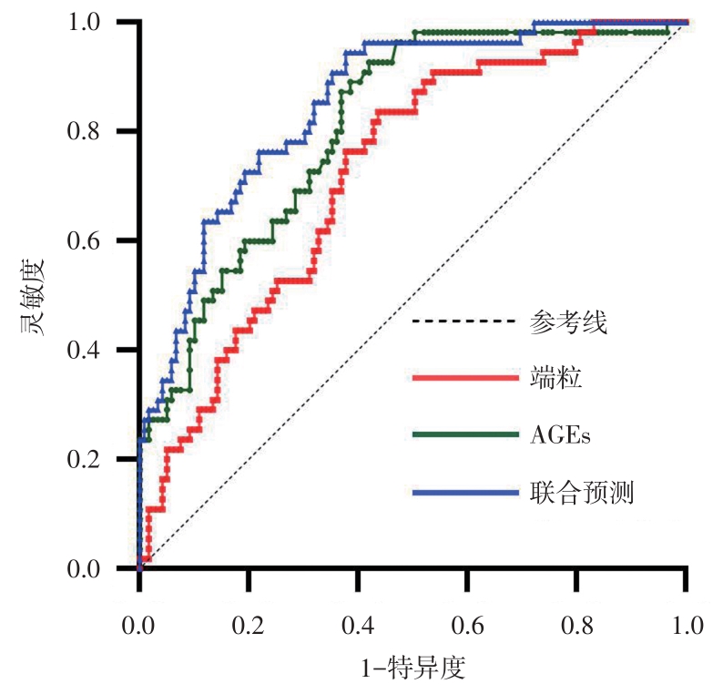
Tab.9
The Value of Telomere Length and AGEs in Predicting Osteoporosis in Patients with T2DM"
| 参数 | 端粒长度 | AGEs | 二者联合 |
|---|---|---|---|
| 曲线下面积 | 0.724 | 0.809 | 0.853 |
| 95%CI | 0.647 ~ 0.801 | 0.743 ~ 0.874 | 0.796 ~ 0.910 |
| P值 | < 0.001 | < 0.001 | < 0.001 |
| 阈值 | 1.569 | 22.270 ng/mL | 0.186 |
| 灵敏度 | 0.836 | 0.927 | 0.945 |
| 特异度 | 0.563 | 0.580 | 0.622 |
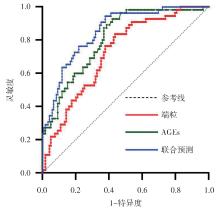
Fig.1
ROC Curve for Predicting Osteoporosis in T2DM Using Telomere Length and AGEs"
| 1 | 杨亚军,崔燎. FoxO/Wnt通路在氧化应激介导的骨质疏松中的调控机制[J]. 中国药理学通报,2013,29(1):27-30. |
| 2 |
WANG L, YU W, YIN X, et al. Prevalence of Osteoporosis and Fracture in China: The China Osteoporosis Prevalence Study[J]. JAMA Netw Open, 2021, 4(8):e2121106. doi:10.1001/jamanetworkopen.2021.21106
doi: 10.1001/jamanetworkopen.2021.21106 |
| 3 | 中华医学会骨质疏松和骨矿盐疾病分会,章振林. 原发性骨质疏松症诊疗指南(2022)[J]. 中国全科医学,2023,26(14):1671-1691. |
| 4 | SI Y, WANG C, GUO Y, et al. Prevalence of Osteoporosis in Patients with Type 2 Diabetes Mellitus in the Chinese Mainland: A Systematic Review and Meta-Analysis[J]. Iran J Public Health, 2019, 48(7): 1203-1214. |
| 5 |
TURNER K J, VASU V, GRIFFIN D K. Telomere Biology and Human Phenotype [J]. Cells, 2019, 8(1):73. doi:10.3390/cells8010073
doi: 10.3390/cells8010073 |
| 6 |
HERRMANN M, PUSCEDDU I, MARZ W, et al. Telomere biology and age-related diseases [J]. Clin Chem Lab Med, 2018, 56(8): 1210-1222. doi:10.1515/cclm-2017-0870
doi: 10.1515/cclm-2017-0870 |
| 7 |
CHENG F, CARROLL L, JOGLEKAR M V, et al. Diabetes, metabolic disease, and telomere length [J]. Lancet Diabetes Endocrinol, 2021, 9(2): 117-126. doi:10.1016/s2213-8587(20)30365-x
doi: 10.1016/s2213-8587(20)30365-x |
| 8 |
SNELSON M, LUCUT E, COUGHLAN M T. The Role of AGE-RAGE Signalling as a Modulator of Gut Permeability in Diabetes [J]. Int J Mol Sci, 2022, 23(3):1766. doi:10.3390/ijms23031766
doi: 10.3390/ijms23031766 |
| 9 |
DEO P, MCCULLOUGH C L, ALMOND T, et al. Dietary sugars and related endogenous advanced glycation end-products increase chromosomal DNA damage in WIL2-NS cells, measured using cytokinesis-block micronucleus cytome assay [J]. Mutagenesis, 2020, 35(2): 169-177. doi:10.1093/mutage/geaa002
doi: 10.1093/mutage/geaa002 |
| 10 | 陈晨,李莉. 晚期糖基化终产物及其受体在新疆维吾尔族2型糖尿病性骨质疏松症中的作用研究[J]. 中国社区医师,2020,36(32):22-23. |
| 11 |
CAWTHON R M. Telomere measurement by quantitative PCR [J]. Nucleic Acids Res, 2002, 30(10): e47. doi:10.1093/nar/30.10.e47
doi: 10.1093/nar/30.10.e47 |
| 12 |
RHARASS T, LUCAS S. High Glucose Level Impairs Human Mature Bone Marrow Adipocyte Function Through Increased ROS Production [J]. Front Endocrinol (Lausanne), 2019, 10: 607. doi:10.3389/fendo.2019.00607
doi: 10.3389/fendo.2019.00607 |
| 13 |
BERGAMINI C M, GAMBETTI S, DONDI A D, et al. Oxygen, reactive oxygen species and tissue damage [J]. Curr Pharm Des, 2004, 10(14): 1611-1626. doi:10.2174/1381612043384664
doi: 10.2174/1381612043384664 |
| 14 |
ZHAO F, GUO L, WANG X, et al. Correlation of oxidative stress-related biomarkers with postmenopausal osteoporosis: a systematic review and meta-analysis [J]. Arch Osteoporos, 2021, 16(1): 4. doi:10.1007/s11657-020-00854-w
doi: 10.1007/s11657-020-00854-w |
| 15 |
PERRONE A, GIOVINO A, BENNY J, et al. Advanced Glycation End Products (AGEs): Biochemistry, Signaling, Analytical Methods, and Epigenetic Effects [J]. Oxid Med Cell Longev, 2020, 2020: 3818196. doi:10.1155/2020/3818196
doi: 10.1155/2020/3818196 |
| 16 |
FISHMAN S L, SONMEZ H, BASMAN C, et al. The role of advanced glycation end-products in the development of coronary artery disease in patients with and without diabetes mellitus: a review [J]. Mol Med, 2018, 24(1): 59. doi:10.1186/s10020-018-0060-3
doi: 10.1186/s10020-018-0060-3 |
| 17 |
SUZUKI A, YABU A, NAKAMURA H. Advanced glycation end products in musculoskeletal system and disorders [J]. Methods, 2022, 203: 179-186. doi:10.1016/j.ymeth.2020.09.012
doi: 10.1016/j.ymeth.2020.09.012 |
| 18 |
SAKAMOTO E, KIDO J I, TAKAGI R, et al. Advanced glycation end-product 2 and Porphyromonas gingivalis lipopolysaccharide increase sclerostin expression in mouse osteocyte-like cells [J]. Bone, 2019, 122: 22-30. doi:10.1016/j.bone.2019.02.001
doi: 10.1016/j.bone.2019.02.001 |
| 19 |
LLABRE J E, SROGA G E, TICE M J L, et al. Induction and rescue of skeletal fragility in a high-fat diet mouse model of type 2 diabetes: An in vivo and in vitro approach [J]. Bone, 2022, 156: 116302. doi:10.1016/j.bone.2021.116302
doi: 10.1016/j.bone.2021.116302 |
| 20 |
GE W, JIE J, YAO J, et al. Advanced glycation end products promote osteoporosis by inducing ferroptosis in osteoblasts [J]. Mol Med Rep, 2022, 25(4):140. doi:10.3892/mmr.2022.12656
doi: 10.3892/mmr.2022.12656 |
| 21 |
HEIN G, WIEGAND R, LEHMANN G, et al. Advanced glycation end-products pentosidine and N epsilon-carboxymethyllysine are elevated in serum of patients with osteoporosis [J]. Rheumatology (Oxford), 2003, 42(10): 1242-1246. doi:10.1093/rheumatology/keg324
doi: 10.1093/rheumatology/keg324 |
| 22 |
YAVUZ D G, APAYDIN T. Skin autofluorescence Is associated With low bone mineral density in type 2 diabetic patients [J]. J Clin Densitom, 2022, 25(3): 373-379. doi:10.1016/j.jocd.2021.11.010
doi: 10.1016/j.jocd.2021.11.010 |
| 23 |
AHMAD S, KHAN M S, AKHTER F, et al. Glycoxidation of biological macromolecules: a critical approach to halt the menace of glycation [J]. Glycobiology, 2014, 24(11): 979-990. doi:10.1093/glycob/cwu057
doi: 10.1093/glycob/cwu057 |
| 24 |
BARNES R P, FOUQUEREL E, OPRESKO P L. The impact of oxidative DNA damage and stress on telomere homeostasis [J]. Mech Ageing Dev, 2019, 177: 37-45. doi:10.1016/j.mad.2018.03.013
doi: 10.1016/j.mad.2018.03.013 |
| 25 |
CHAKRAVARTI D, LABELLA K A, DEPINHO R A. Telomeres: history, health, and hallmarks of aging [J]. Cell, 2021, 184(2): 306-322. doi:10.1016/j.cell.2020.12.028
doi: 10.1016/j.cell.2020.12.028 |
| 26 |
WANG J, DONG X, CAO L, et al. Association between telomere length and diabetes mellitus: A meta-analysis [J]. J Int Med Res, 2016, 44(6): 1156-1173. doi:10.1177/0300060516667132
doi: 10.1177/0300060516667132 |
| 27 |
TAMAYO M, MOSQUERA A, REGO J I, et al. Differing patterns of peripheral blood leukocyte telomere length in rheumatologic diseases [J]. Mutat Res, 2010, 683(1-2): 68-73. doi:10.1016/j.mrfmmm.2009.10.010
doi: 10.1016/j.mrfmmm.2009.10.010 |
| 28 |
VALDES A M, RICHARDS J B, GARDNER J P, et al. Telomere length in leukocytes correlates with bone mineral density and is shorter in women with osteoporosis [J]. Osteoporos Int, 2007, 18(9): 1203-1210. doi:10.1007/s00198-007-0357-5
doi: 10.1007/s00198-007-0357-5 |
| 29 | FRAGKIADAKI P, NIKITOVIC D, KALLIANTASI K, et al. Telomere length and telomerase activity in osteoporosis and osteoarthritis [J]. Exp Ther Med, 2020, 19(3): 1626-1632. |
| 30 |
GRUBER H J, SEMERARO M D, RENNER W, et al. Telomeres and Age-Related Diseases [J]. Biomedicines, 2021, 9(10):1355. doi:10.3390/biomedicines9101335
doi: 10.3390/biomedicines9101335 |
| 31 |
PIGNOLO R J, LAW S F, CHANDRA A. Bone Aging, Cellular Senescence, and Osteoporosis [J]. JBMR Plus, 2021, 5(4): e10488. doi:10.1002/jbm4.10488
doi: 10.1002/jbm4.10488 |
| Viewed | ||||||
|
Full text |
|
|||||
|
Abstract |
|
|||||