实用医学杂志 ›› 2025, Vol. 41 ›› Issue (14): 2143-2151.doi: 10.3969/j.issn.1006-5725.2025.14.004

• 专题报道:乳腺癌 • 上一篇    下一篇

术前腋窝超声正常的临床T1—2 N0乳腺癌患者发生前哨淋巴结转移的预测模型

丘海,归奕飞(),刘媛   

  1. 柳州市工人医院普通外科四病区(乳腺) (广西 柳州 545005 )
  • 收稿日期:2025-04-09 出版日期:2025-07-25 发布日期:2025-07-29
  • 通讯作者: 归奕飞 E-mail:guiyifei@hotmail.com
  • 基金资助:
    广西壮族自治区卫生和计划生育委员会计划课题(Z20170911)

Predictors of sentinel lymph node metastasis in clinical T1⁃2 N0 breast cancer patients with preoperatively normal axillary ultrasound

Hai QIU,Yifei GUI(),Yuan. LIU   

  1. Fourth Ward (Breast) of General Surgery,Liuzhou Workers' Hospital,Liuzhou 545005,Guangxi,China
  • Received:2025-04-09 Online:2025-07-25 Published:2025-07-29
  • Contact: Yifei GUI E-mail:guiyifei@hotmail.com

摘要:

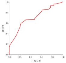
目的 建立多因素预测模型,筛选术前腋窝超声(axillary ultrasound, AUS)正常的临床T1—2N0患者发生前哨淋巴结(sentinel lymph node, SLN)转移的高危人群,为腋窝分期的个体化管理策略提供参考。 方法 回顾性分析柳州市工人医院普通外科四病区(乳腺)2018年1月至2023年12月收治的427例浸润性乳腺癌患者的临床病理资料及超声检查数据,进行单因素相关性分析和多因素logistic回归分析,从而确定影响SLN转移的独立危险因素,然后采用ROC曲线评估模型的准确性和预测价值。 结果 本研究共纳入427例术前AUS评估正常的临床T1—2 N0女性乳腺癌患者,其中47例(11.0%)患者探及正常腋窝淋巴结,余380例(89.0%)患者腋窝超声均未见报告肿大淋巴结。78例(18.3%)患者经术后病理证实SLN转移,单因素分析显示SLN转移与雌激素受体(estrogen receptor, ER)、肿瘤最大径、肿瘤位置、脉管癌栓和神经侵犯相关(P < 0.05),而多因素分析显示肿瘤位于外上象限(OR = 4.118, 95%CI: 1.349 ~ 12.571)、肿瘤最大径> 2 cm(OR = 2.246, 95%CI: 1.252 ~ 4.029)、脉管癌栓(OR = 4.477, 95%CI: 2.207 ~ 9.081)和神经侵犯(OR = 3.013, 95%CI: 1.573 ~ 5.771)是影响SLN转移的独立危险因素(P < 0.05)。超声下腋窝淋巴结的阴性或阳性、最短径、数目与SLN转移数目无关(P ≥ 0.05),而与病理淋巴结(pathological node, pN)分期有关(P < 0.05)。1 ~ 2枚SLN转移患者的超声下腋窝淋巴结的阴性或阳性、最长径、最短径、数目与ALN转移数目、pN分期变化均无关(P ≥ 0.05)。根据预测模型绘制ROC曲线的AUC值为0.702(95%CI: 0.651 ~ 0.749, P < 0.000 1),该模型预测的敏感度为78.21%,特异度为59.12%。 结论 肿瘤位于外上象限、肿瘤最大径> 2 cm、脉管癌栓和神经侵犯是术前AUS正常的临床T1-T2N0女性乳腺癌患者发生SLN转移的显著危险因素。另外,单从对pN分期的影响来说,对于cT1—2 N0且1 ~ 2枚SLN转移的绝大部分患者,豁免腋窝淋巴结清扫术(ALND)是可行的,但尚不能从超声腋窝淋巴结的特征预测与腋窝淋巴结(ALN)转移、pN分期的相关性。

关键词: 乳腺癌, 前哨淋巴结转移, 预测因素, 腋窝超声, 预测模型

Abstract:

Objective To develop a multivariate predictive nomogram to identify high-risk cohorts for sentinel lymph node (SLN) metastasis among cT1-2N0 breast cancer patients with preoperatively normal axillary ultrasound (AUS), thereby providing a reference for personalized axillary management. Methods A retrospective analysis was conducted on the clinicopathological and ultrasonographic data of 427 patients diagnosed with invasive breast cancer who received treatment at Ward 4 (Breast Unit), Department of General Surgery, Liuzhou Workers' Hospital, between January 2018 and December 2023. Univariate correlation analysis and multivariate logistic regression analysis were employed to identify independent risk factors associated with SLN metastasis. The accuracy and predictive performance of the nomogram were assessed using receiver operating characteristic (ROC) curve analysis. Results Our study enrolled 427 women diagnosed with clinical T1-2N0 breast cancer who underwent preoperative AUS with normal findings. Among these patients, 47 cases (11.0%) exhibited sonographically normal axillary lymph nodes, whereas 380 cases (89.0%) showed non-visualized lymph nodes. SLN metastasis, confirmed by postoperative pathological examination, was identified in 78 patients (18.3%). Univariate analysis revealed that estrogen receptor (ER) status, maximal tumor diameter, tumor location, lymphovascular invasion (LVI), and perineural invasion (PNI) were significantly associated with the presence of SLN metastasis (P < 0.05). Multivariate logistic regression analysis further identified the following independent high-risk factors for SLN metastasis: tumor location in the upper outer quadrant (OR = 4.118, 95% CI = 1.349 ~ 12.571), tumor size greater than 2 cm (OR = 2.246, 95% CI = 1.252 ~ 4.029), presence of LVI (OR = 4.477, 95% CI = 2.207 ~ 9.081), and presence of PNI (OR = 3.013, 95% CI = 1.573 ~ 5.771) (all P < 0.05). Ultrasonographic features of axillary lymph nodes—including their positivity status, short-axis diameter, and numerical count—did not show a statistically significant association with the SLN metastatic burden (P ≥ 0.05). However, these features demonstrated a statistically significant correlation with the pathological nodal stage (pN-stage) classification (P < 0.05). In patients with 1 ~ 2 positive sentinel lymph nodes, sonographic characteristics of axillary lymph nodes (including status, maximum diameter, minimum diameter, and numerical count) did not exhibit a significant association with either axillary lymph node metastatic burden or pN-stage classification (all P ≥ 0.05). The area under the receiver operating characteristic curve (AUC) for the predictive nomogram was 0.702 (95% CI: 0.651 ~ 0.749, P < 0.0001), with a sensitivity of 78.21% and specificity of 59.12%. Conclusions Tumor location in the upper outer quadrant, tumor size greater than 2 cm, LVI, and PNI were identified as significant independent risk factors for SLN metastasis among patients with clinical T1-2N0 breast cancer who underwent preoperative AUS with normal findings. Furthermore, from the perspective of pN-stage stratification, omission of axillary lymph node dissection (ALND) appears to be clinically feasible for the majority of cT1-2N0 patients with 1 ~ 2 metastatic SLNs. However, preoperative ultrasonographic characteristics of lymph nodes demonstrate limited predictive value for axillary lymph node (ALN) metastatic burden or pN-stage progression.

Key words: breast cancer, sentinel lymph node metastasis, predictors, axillary ultrasonography, predictive nomogram

中图分类号: