实用医学杂志 ›› 2026, Vol. 42 ›› Issue (15): 2847-2852.doi: 10.3969/j.issn.1006-5725.2026.15.022

• 论著·机制与实践 • 上一篇    

磁共振质子密度脂肪分数评估肝脏脂肪含量的空间分布特征

王梓华1,2,吴红珍3,谭俊杰1,2,曾雪伟1,2()   

  1. 1.广州中医药大学第八临床医学院 (广东 佛山 528000 )
    2.佛山市中医院医学影像科 (广东 佛山 528000 )
    3.广州市第一人民医院放射科 (广东 广州 510180 )
  • 收稿日期:2026-05-18 修回日期:2026-06-11 接受日期:2026-06-16 出版日期:2026-08-10 发布日期:2026-08-13
  • 通讯作者: 曾雪伟 E-mail:81842829@qq.com
  • 基金资助:
    佛山市自筹经费类科技创新项目(2520001002860)

Spatial distribution characteristics of liver fat content evaluated by magnetic resonance imaging proton density fat fraction

Zihua WANG1,2,Hongzhen WU3,Junjie TAN1,2,Xuewei ZENG1,2()   

  1. 1.The Eighth Clinical Medical College of Guangzhou University of Chinese Medicine,Foshan 528000,Guangdong,China
    2.Department of Radiology,Foshan Hospital of Traditional Chinese Medicine,Foshan 528000,Guangdong,China
    3.Department of Radiology,Guangzhou First People′s Hospital,Guangzhou 510180,Guangdong,China
  • Received:2026-05-18 Revised:2026-06-11 Accepted:2026-06-16 Online:2026-08-10 Published:2026-08-13
  • Contact: Xuewei ZENG E-mail:81842829@qq.com

摘要:

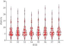
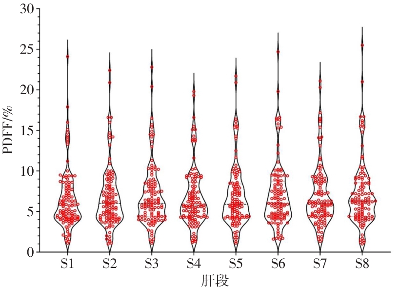
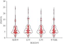
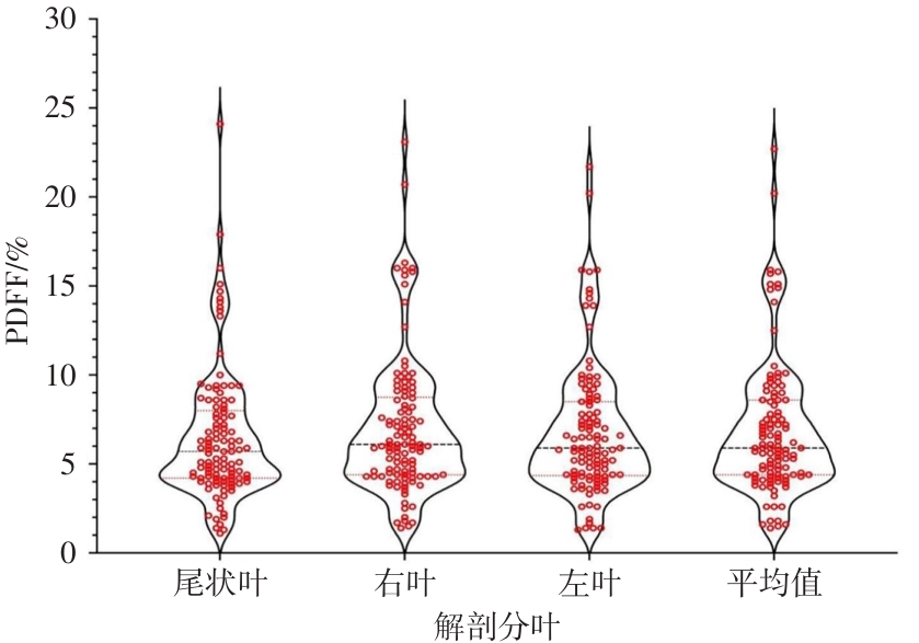
目的 旨在通过MRI质子密度脂肪分数(PDFF)比较肝脏各段与各叶之间脂肪含量的差异,明确肝脏脂肪含量的空间分布特征,为临床肝脏脂肪精准定量提供依据。 方法 回顾性纳入113例患者,收集肝脏MRI检查及生化检查资料并测定肝脏PDFF值。采用Friedman秩和检验结合Bonferroni事后比较分析肝段、肝叶脂肪含量差异。使用Spearman相关系数分析全肝PDFF与生化指标的相关性。 结果 各段之间的PDFF值总体差异有统计学意义(P < 0.05)。两两比较显示,S1与S2 ~ S8、S2与S6 ~ S8、S4与S7 ~ S8,以及尾状叶与左叶、右叶及全肝平均值之间,右叶与左叶、全肝平均值之间的PDFF值差异均有统计学意义(均校正后P < 0.05)。相关性分析显示,全肝PDFF平均值与糖化血红蛋白、甘油三酯呈正相关,与载脂蛋白A呈负相关。 结论 肝脏脂肪含量存在明显空间异质性,不同肝叶及肝段的脂肪含量分布存在显著差异。此外,PDFF与部分糖脂代谢指标存在相关性,可为临床评估提供参考。

关键词: 脂肪肝, 磁共振, 脂肪定量, 化学位移成像, 质子密度脂肪分数

Abstract:

Objective The aims of this study were to compare the differences in fat content between liver segments and liver lobes by utilizing magnetic resonance imaging proton density fat fraction (PDFF), thereby clarifying the spatial distribution characteristics of liver fat content and providing evidence for the clinical accurate quantitative evaluation of liver fat. Methods A total of 113 patients were retrospectively included. Liver magnetic resonance imaging (MRI) and biochemical data were gathered to ascertain hepatic PDFF values. The Friedman rank-sum test, in combination with the Bonferroni post-hoc test, was employed to analyze the disparities in fat content among liver segments and lobes. The Spearman correlation coefficient was utilized to investigate the correlation between whole-liver PDFF and biochemical indicators. Results There were significant differences in PDFF values among liver segments (P < 0.05). Pair-wise comparisons indicated significant differences in PDFF values between S1 and S2 - S8, between S2 and S6 - S8, and between S4 and S7 - S8. Additionally, there were significant differences between the caudate lobe, left lobe, right lobe, and the whole-liver mean, as well as between the right lobe, left lobe, and the whole-liver mean (all adjusted P < 0.05). Correlation analysis revealed that the average whole-liver PDFF was weakly positively correlated with glycated hemoglobin and triglycerides, and weakly negatively correlated with apolipoprotein A. Conclusions There is evident spatial heterogeneity in liver fat content, and the distribution of fat content varies significantly among different liver lobes and segments. Moreover, PDFF is correlated with certain glycolipid metabolism indicators, which can offer a reference for clinical evaluation.

Key words: fatty liver, magnetic resonance imaging, fat quantification, chemical shift imaging, proton density fat fraction

中图分类号: