

The Journal of Practical Medicine ›› 2025, Vol. 41 ›› Issue (3): 330-338.doi: 10.3969/j.issn.1006-5725.2025.03.004
• Basic Research • Previous Articles
Lu ZHENG1,Haohao ZHANG2,Feifei WU1,Jiaqi GUO1,Youqin WANG1,Ruimin HAO1,Lihui FENG1,Yan. LI1( )
)
Received:2024-10-06
Online:2025-02-10
Published:2025-02-19
Contact:
Yan. LI
E-mail:liyanweiwei@126.com
CLC Number:
Lu ZHENG,Haohao ZHANG,Feifei WU,Jiaqi GUO,Youqin WANG,Ruimin HAO,Lihui FENG,Yan. LI. Protective effect of exenatide on oxidative stress in hypothalamus of diabetes mice and its mechanism[J]. The Journal of Practical Medicine, 2025, 41(3): 330-338.
Tab.1
Comparison of body weight and glycolipid metabolism indexes in each group"
| 组别 | 体质量/g | FBG/(mmol/L) | TC/(mmol/L) | TG/(mmol/L) | FFA/(mmol/L) | Ins/(mIU/L) | HOMA-IR |
|---|---|---|---|---|---|---|---|
| NC组 | 30.35 ± 3.34 | 5.70 ± 0.93 | 2.36 ± 0.72 | 0.50 ± 1.17 | 0.41 ± 0.12 | 2.51 ± 0.12 | 0.64 ± 0.13 |
| T2DM组 | 35.32 ± 2.65? | 14.15 ± 2.24? | 4.99 ± 0.39? | 1.73 ± 0.24? | 0.83 ± 0.08? | 5.56 ± 0.45? | 3.52 ± 0.78? |
| T2DM+Exe组 | 31.45 ± 2.00# | 9.50 ± 0.85?# | 3.97 ± 0.28?# | 0.88 ± 0.10?# | 0.34 ± 0.14# | 2.65 ± 0.13# | 1.12 ± 0.10?# |
| F值 | 10.33 | 32.45 | 34.99 | 61.67 | 25.93 | 153.34 | 44.83 |
| P值 | < 0.05 | < 0.05 | < 0.05 | < 0.05 | < 0.05 | < 0.05 | < 0.05 |
Fig.1
Inflammatory factors and adipokines in each group"
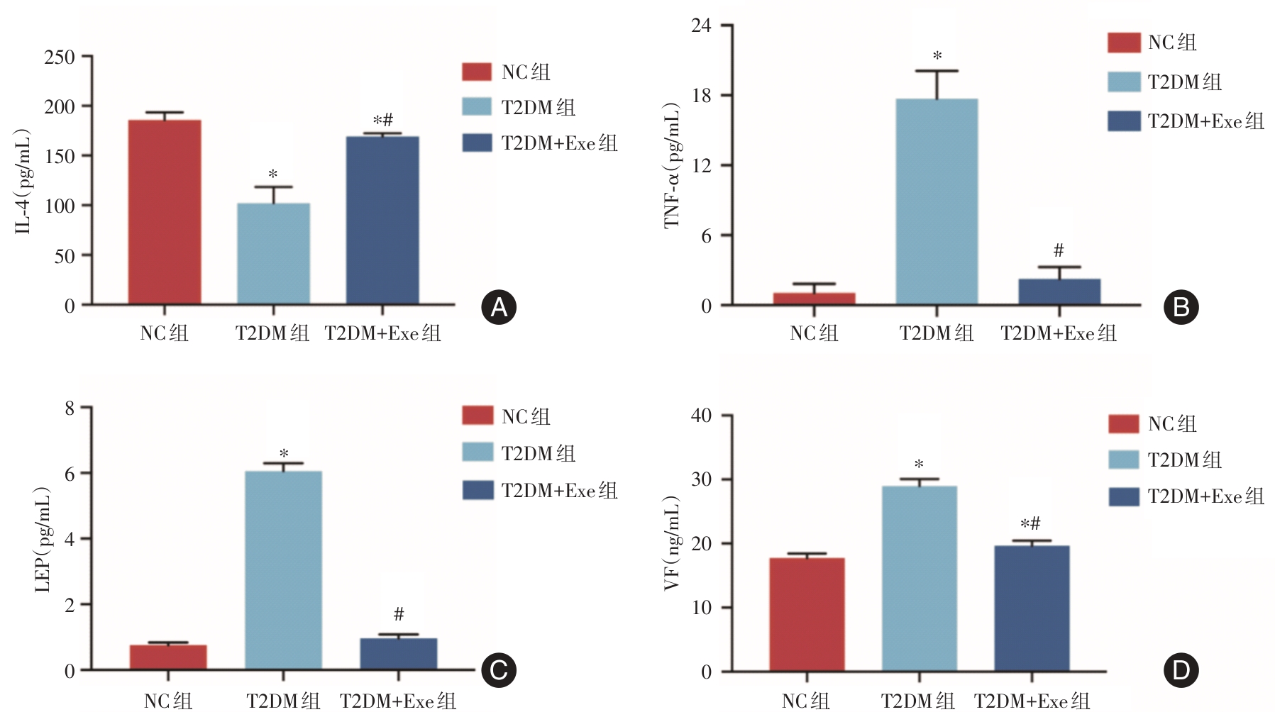
Tab.2
Inflammatory and adipokine factors in each group"
| 组别 | IL-4/ (pg/mL) | TNF-α/ (pg/mL) | LEP/ (pg/mL) | Visfitin/ (ng/mL) |
|---|---|---|---|---|
| NC组 | 185.74 ± 7.75 | 1.04 ± 0.79 | 0.76 ± 0.08 | 17.75 ± 0.72 |
| T2DM组 | 102.13 ± 16.29? | 17.71 ± 2.38? | 6.06 ± 0.24? | 28.98 ± 1.09? |
| T2DM+Exe组 | 169.04 ± 3.44?# | 2.25 ± 1.02# | 0.96 ± 0.12# | 19.66 ± 0.79*# |
| F值 | 87.014 | 176.09 | 1764.931 | 233.229 |
| P值 | < 0.05 | < 0.05 | < 0.05 | < 0.05 |
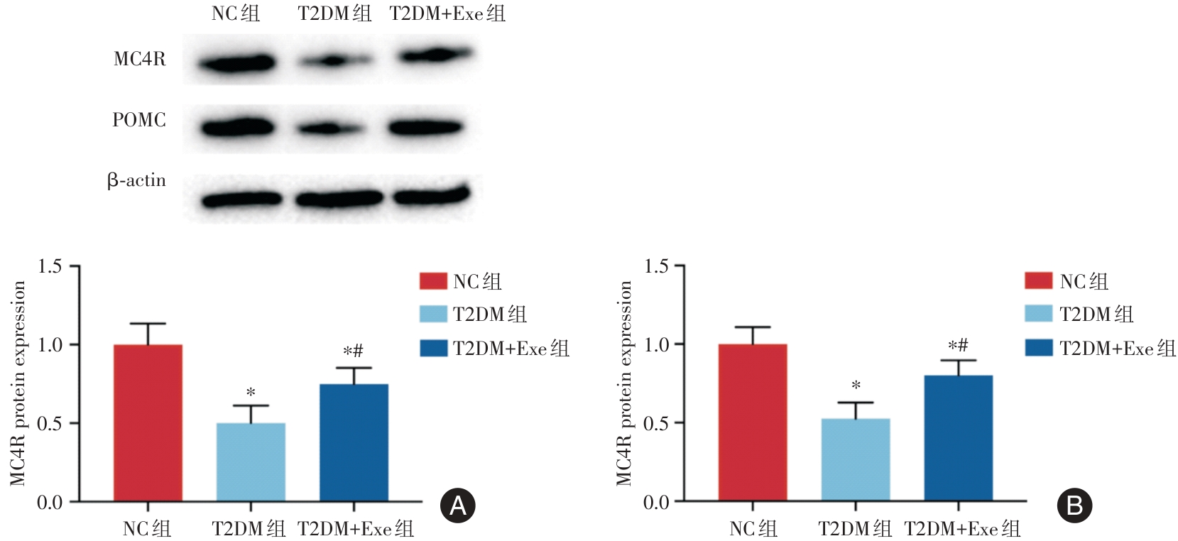
Tab.3
Expression of MC4R and POMC protein in hypothalamus of mice in each group"
| 组别 | MC4R蛋白水平 | POMC蛋白水平 |
|---|---|---|
| NC组 | 1.00 ± 0.13 | 1.00 ± 0.11 |
| T2DM组 | 0.50 ± 0.11? | 0.53 ± 0.10? |
| T2DM+Exe组 | 0.75 ± 0.10?# | 0.80 ± 0.10?# |
| F值 | 27.613 | 32.647 |
| P值 | < 0.05 | < 0.05 |
Fig.2
Expression of MC4R and POMC protein in hypothalamus of mice in each group"
Tab.4
Oxidative stress levels of hypothalamic mitochondria in each group"
| 组别 | ROS/(MFI) | MDA/(nmol/mgprot) | SOD/(U/mgprot) |
|---|---|---|---|
| NC组 | 1.00 ± 0.13 | 3.22 ± 0.40 | 13.42 ± 2.30 |
| T2DM组 | 2.15 ± 0.39? | 6.56 ± 1.98? | 4.94 ± 1.50? |
| T2DM+Exe组 | 1.24 ± 0.37# | 4.35 ± 1.85# | 7.80 ± 1.32?# |
| F值 | 21.726 | 11.041 | 35.8 |
| P值 | < 0.05 | < 0.05 | < 0.05 |

Fig.3
Oxidative stress levels of hypothalamic mitochondria in each group"
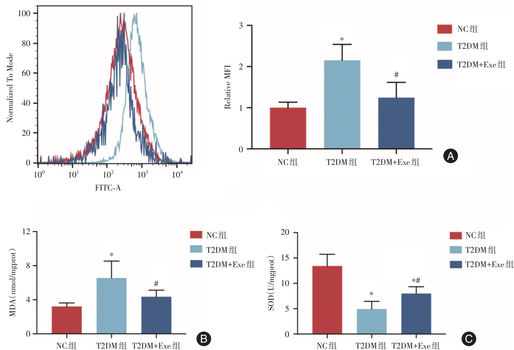
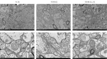
Fig.4
Morphological changes of hypothalamic mitochondria in each group"

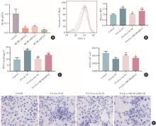
Fig.5
The activity of antioxidant enzymes and lipid deposition in each group after transfection"
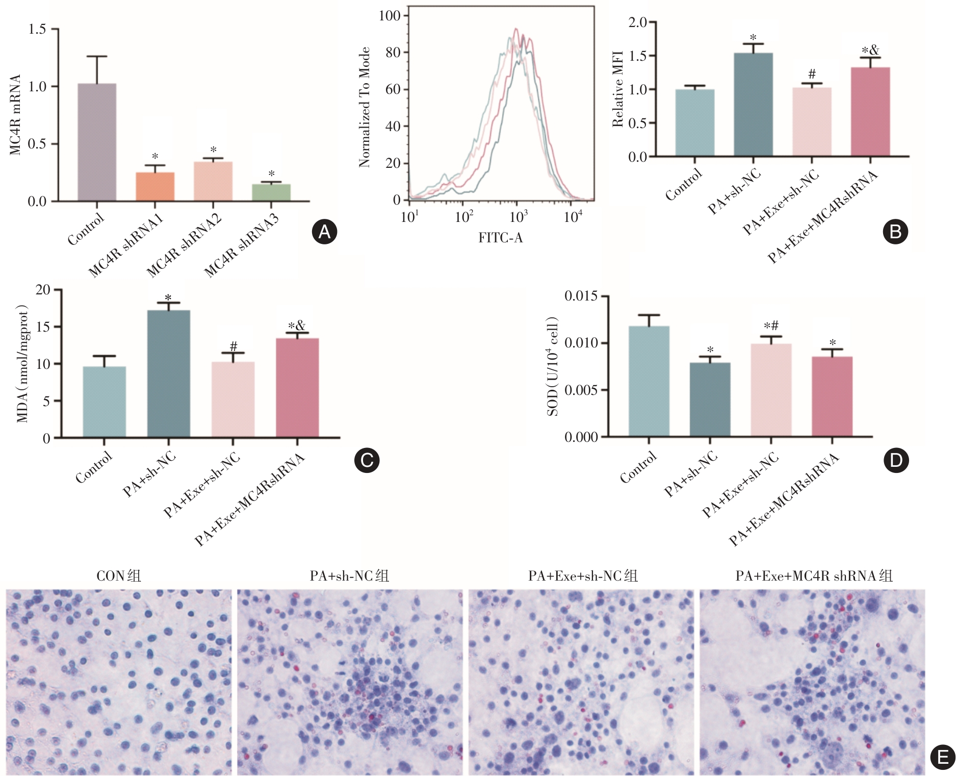
Tab.5
Efficiency of MC4R shRNA transfection in GT1-7 cells"
| 组别 | MC4R mRNA |
|---|---|
| CON组 | 1.03 ± 0.30 |
| MC4R shRNA1组 | 0.25 ± 0.06? |
| MC4R shRNA2组 | 0.35 ± 0.03?* |
| MC4R shRNA3组 | 0.15 ± 0.02? |
| F值 | 18.397 |
| P值 | < 0.05 |
Tab.6
The activity of antioxidant enzymes and lipid deposition in each group after transfection"
| 组别 | ROS/ (MFI) | MDA/ (nmol/mgprot) | SOD/ (U/104cell) |
|---|---|---|---|
| CON组 | 1.00 ± 0.05 | 9.64 ± 1.41 | 0.0118 ± 0.001 |
| PA+sh-NC组 | 1.54 ± 0.13? | 17.24 ± 1.01? | 0.0079 ± 0.0006? |
| PA+Exe+sh-NC组 | 1.03 ± 0.06# | 10.26 ± 1.21# | 0.010 ± 0.0007?# |
| PA+Exe+MC4RshRNA组 | 1.33 ± 0.15?& | 13.46 ± 0.72?& | 0.009 ± 0.0007? |
| F值 | 29.264 | 48.683 | 12.147 |
| P值 | < 0.05 | < 0.05 | < 0.05 |
| 1 | NCD Risk Factor Collaboration (NCD-RisC).Worldwide trends in diabetes prevalence and treatment from 1990 to 2022: A pooled analysis of 1108 population-representative studies with 141 million participants [J]. Lancet, 2024, 404(10467): 2077-2093. |
| 2 |
PANG D, YANG C, LUO Q, et al. Soy isoflavones improve the oxidative stress induced hypothalamic inflammation and apoptosis in high fat diet-induced obese male mice through PGC1-alpha pathway [J]. Aging (Albany NY), 2020, 12(9): 8710-8727. doi:10.18632/aging.103197
doi: 10.18632/aging.103197 |
| 3 |
JO D, YOON G, SONG J. Role of Exendin-4 in Brain Insulin Resistance, Mitochondrial Function, and Neurite Outgrowth in Neurons under Palmitic Acid-Induced Oxidative Stress [J]. Antioxidants (Basel), 2021, 10(1):78. doi:10.3390/antiox10010078
doi: 10.3390/antiox10010078 |
| 4 |
MALONE J I, HANSEN B C. Does obesity cause type 2 diabetes mellitus (T2DM)?Or is it the opposite? [J]. Pediatr Diabetes, 2019, 20(1): 5-9. doi:10.1111/pedi.12787
doi: 10.1111/pedi.12787 |
| 5 | 马刚,孙家忠. GLP-1 RAs联合恩格列净对2型糖尿病患者的治疗效果及对胰岛素抵抗的影响[J]. 实用医学杂志,2020,36(18):2500-2504. |
| 6 |
DE SOUZA CORDEIRO L M, ELSHEIKH A, DEVISETTY N, et al. Hypothalamic MC4R regulates glucose homeostasis through adrenaline-mediated control of glucose reabsorption via renal GLUT2 in mice [J]. Diabetologia, 2021, 64(1): 181-194. doi:10.1007/s00125-020-05289-z
doi: 10.1007/s00125-020-05289-z |
| 7 |
POLENI P E, AKIEDA-ASAI S, KODA S, et al. Possible involvement of melanocortin-4-receptor and AMP-activated protein kinase in the interaction of glucagon-like peptide-1 and leptin on feeding in rats [J]. Biochem Biophys Res Commun, 2012, 420(1): 36-41. doi:10.1016/j.bbrc.2012.02.109
doi: 10.1016/j.bbrc.2012.02.109 |
| 8 |
TANAKA T, MASUZAKI H, YASUE S, et al. Central melanocortin signaling restores skeletal muscle AMP-activated protein kinase phosphorylation in mice fed a high-fat diet [J]. Cell Metab, 2007, 5(5): 395-402. doi:10.1016/j.cmet.2007.04.004
doi: 10.1016/j.cmet.2007.04.004 |
| 9 |
GOKUL P R, APPERLEY L, PARKINSON J, et al. Semaglutide, A Long-Acting GLP-1 Analogue, for the management of early onset obesity due to MC4R defect-A case report [J]. Horm Res Paediatr, 2024. doi: 10.1159/000537921 .
doi: 10.1159/000537921 |
| 10 |
LAMBADIARI V, PAVLIDIS G, KOUSATHANA F, et al. Effects of 6-month treatment with the glucagon like peptide-1 analogue liraglutide on arterial stiffness, left ventricular myocardial deformation and oxidative stress in subjects with newly diagnosed type 2 diabetes [J]. Cardiovasc Diabetol, 2018, 17(1): 8. doi:10.1186/s12933-017-0646-z
doi: 10.1186/s12933-017-0646-z |
| 11 |
YAGISHITA Y, URUNO A, FUKUTOMI T, et al. Nrf2 Improves Leptin and Insulin Resistance Provoked by Hypothalamic Oxidative Stress [J]. Cell Rep, 2017, 18(8): 2030-2044. doi:10.1016/j.celrep.2017.01.064
doi: 10.1016/j.celrep.2017.01.064 |
| 12 |
LIN M H, CHENG P C, HSIAO P J, et al. The GLP-1 receptor agonist exenatide ameliorates neuroinflammation, locomotor activity, and anxiety-like behavior in mice with diet-induced obesity through the modulation of microglial M2 polarization and downregulation of SR-A4 [J]. Int Immunopharmacol, 2023, 115: 109653. doi:10.1016/j.intimp.2022.109653
doi: 10.1016/j.intimp.2022.109653 |
| 13 |
STENLID R, CERENIUS S Y, WEN Q, et al. Adolescents with obesity treated with exenatide maintain endogenous GLP-1, reduce DPP-4, and improve glycemic control [J]. Front Endocrinol (Lausanne), 2023, 14: 1293093. doi:10.3389/fendo.2023.1293093
doi: 10.3389/fendo.2023.1293093 |
| 14 |
BAI C, WANG Y, NIU Z, et al. Exenatide improves hepatocyte insulin resistance induced by different regional adipose tissue[J]. Front Endocrinol (Lausanne), 2022, 13: 1012904. doi:10.3389/fendo.2022.1012904
doi: 10.3389/fendo.2022.1012904 |
| 15 |
XU Q, ZHANG X, LI T, et al. Exenatide regulates Th17/Treg balance via PI3K/Akt/FoxO1 pathway in db/db mice [J]. Mol Med, 2022, 28(1): 144. doi:10.1186/s10020-022-00574-6
doi: 10.1186/s10020-022-00574-6 |
| 16 |
XU F, CAO H, CHEN Z, et al. Short-term GLP-1 receptor agonist exenatide ameliorates intramyocellular lipid deposition without weight loss in ob/ob mice [J]. Int J Obes (Lond), 2020, 44(4): 937-947. doi:10.1038/s41366-019-0513-y
doi: 10.1038/s41366-019-0513-y |
| 17 |
ADRIAENSSENS A E, BIGGS E K, DARWISH T, et al. Glucose-Dependent Insulinotropic Polypeptide Receptor-Expressing Cells in the Hypothalamus Regulate Food Intake [J]. Cell Metab, 2019, 30(5):987-996.e6. doi:10.1016/j.cmet.2019.07.013
doi: 10.1016/j.cmet.2019.07.013 |
| 18 |
HUANG Z, LIU L, ZHANG J, et al. Glucose-sensing glucagon-like peptide-1 receptor neurons in the dorsomedial hypothalamus regulate glucose metabolism [J]. Sci Adv, 2022, 8(23): eabn5345. doi:10.1126/sciadv.abn5345
doi: 10.1126/sciadv.abn5345 |
| 19 |
HAVEL P J, HAHN T M, SINDELAR D K, et al. Effects of streptozotocin-induced diabetes and insulin treatment on the hypothalamic melanocortin system and muscle uncoupling protein 3 expression in rats [J]. Diabetes, 2000, 49(2): 244-252. doi:10.2337/diabetes.49.2.244
doi: 10.2337/diabetes.49.2.244 |
| 20 |
DONG Y, CARTY J, GOLDSTEIN N, et al. Time and metabolic state-dependent effects of GLP-1R agonists on NPY/AgRP and POMC neuronal activity in vivo [J]. Mol Metab, 2021, 54: 101352. doi:10.1016/j.molmet.2021.101352
doi: 10.1016/j.molmet.2021.101352 |
| 21 |
SPEZANI R, MARINHO T S, REIS T S, et al. Cotadutide (GLP-1/Glucagon dual receptor agonist) modulates hypothalamic orexigenic and anorexigenic neuropeptides in obese mice [J]. Peptides, 2024, 173: 171138. doi:10.1016/j.peptides.2023.171138
doi: 10.1016/j.peptides.2023.171138 |
| 22 |
GONZÁLEZ P, LOZANO P, ROS G, et al. Hyperglycemia and Oxidative Stress: An Integral, Updated and Critical Overview of Their Metabolic Interconnections [J]. Int J Mol Sci, 2023, 24(11):9352. doi:10.3390/ijms24119352
doi: 10.3390/ijms24119352 |
| 23 |
XIA B, DING J, LI Q, et al. Loganin protects against myocardial ischemia-reperfusion injury by modulating oxidative stress and cellular apoptosis via activation of JAK2/STAT3 signaling [J]. Int J Cardiol, 2024, 395: 131426. doi:10.1016/j.ijcard.2023.131426
doi: 10.1016/j.ijcard.2023.131426 |
| 24 |
PANDEY S, MANGMOOL S, MADREITER-SOKOLOWSKI C T, et al. Exendin-4 protects against high glucose-induced mitochondrial dysfunction and oxidative stress in SH-SY5Y neuroblastoma cells through GLP-1 receptor/Epac/Akt signaling [J]. Eur J Pharmacol, 2023, 954: 175896. doi:10.1016/j.ejphar.2023.175896
doi: 10.1016/j.ejphar.2023.175896 |
| 25 |
WANG Y, FANG N, WANG Y, et al. Activating MC4R Promotes Functional Recovery by Repressing Oxidative Stress-Mediated AIM2 Activation Post-spinal Cord Injury [J]. Mol Neurobiol, 2024, 61(8):6101-6118. doi:10.1007/s12035-024-03936-9
doi: 10.1007/s12035-024-03936-9 |
| 26 |
ZHANG H H, LIU J, QIN G J, et al. Melanocortin 4 Receptor Activation Attenuates Mitochondrial Dysfunction in Skeletal Muscle of Diabetic Rats [J]. J Cell Biochem, 2017, 118(11): 4072-4079. doi:10.1002/jcb.26062
doi: 10.1002/jcb.26062 |
| Viewed | ||||||
|
Full text |
|
|||||
|
Abstract |
|
|||||