

The Journal of Practical Medicine ›› 2024, Vol. 40 ›› Issue (23): 3367-3372.doi: 10.3969/j.issn.1006-5725.2024.23.014
• Medical Examination and Clinical Diagnosis • Previous Articles
Liping CHEN1,Juyu LUO1,Zhangyan PENG1,Xiulan WU1( ),Yuhong YANG1,Lianyan SHI1,Xiaoyun LI2,Ling. WANG3
Received:2024-08-14
Online:2024-12-10
Published:2024-12-16
Contact:
Xiulan WU
E-mail:741903320@qq.com
CLC Number:
Liping CHEN,Juyu LUO,Zhangyan PENG,Xiulan WU,Yuhong YANG,Lianyan SHI,Xiaoyun LI,Ling. WANG. Correlation between the ratio of tumor volume to uterine volume and the expression of Ki⁃67 and p16 protein in tissues with the pathological features and recurrence of endometrial carcinoma[J]. The Journal of Practical Medicine, 2024, 40(23): 3367-3372.
Tab.1
Comparison of the expression of T/U, Ki-67 protein, and p16 protein"
| 组别 | 例数 | T/U | Ki-67蛋白 | p16蛋白 | |||||
|---|---|---|---|---|---|---|---|---|---|
| ≥ 0.18 | < 0.18 | 阳性 | 阴性 | 阳性 | 阴性 | ||||
| 复发组 | 28 | 17(60.71) | 11(39.29) | 15(53.57) | 13(46.43) | 12(42.86) | 16(57.14) | ||
| 非复发组 | 122 | 44(36.07) | 78(63.93) | 40(32.79) | 82(67.21) | 78(63.93) | 44(36.07) | ||
| χ2值 | 5.734 | 4.236 | 4.215 | ||||||
| P值 | 0.017 | 0.040 | 0.040 | ||||||
Tab. 2
Association between the expression of T / U, Ki-67 protein and p16 protein and the pathological features"
| 因素 | T/U ≥ 0.18(n = 61) | T/U < 0.18(n = 89) | χ2值 | P值 | Ki-67阳性(n = 55) | Ki-67阴性(n = 95) | χ2值 | P值 | p16阳性(n = 90) | p16阴性(n = 60) | χ2值 | P值 |
|---|---|---|---|---|---|---|---|---|---|---|---|---|
| 年龄 | 2.049 | 0.152 | 2.926 | 0.087 | 1.703 | 0.192 | ||||||
| ≥ 50岁 | 42(68.85) | 51(57.3) | 39(70.91) | 54(56.84) | 52(57.78) | 41(68.33) | ||||||
| < 50岁 | 19(31.15) | 38(42.7) | 16(29.09) | 41(43.16) | 38(42.22) | 19(31.67) | ||||||
| FIGO分期 | 11.594 | 0.001 | 8.422 | 0.004 | 4.768 | 0.029 | ||||||
| Ⅰ+Ⅱ期 | 27(44.26) | 64(71.91) | 25(45.45) | 66(69.47) | 61(67.78) | 30(50) | ||||||
| Ⅲ期 | 34(55.74) | 25(28.09) | 30(54.55) | 29(30.53) | 29(32.22) | 30(50) | ||||||
| 病理学分化程度 | 21.988 | < 0.001 | 4.587 | 0.032 | 6.459 | 0.011 | ||||||
| 高+中分化 | 14(22.95) | 55(61.8) | 19(34.55) | 50(52.63) | 49(54.44) | 20(33.33) | ||||||
| 低分化 | 47(77.05) | 34(38.2) | 36(65.45) | 45(47.37) | 41(45.56) | 40(66.67) | ||||||
| 淋巴结转移 | 2.691 | 0.101 | 6.964 | 0.008 | 8.378 | 0.004 | ||||||
| 是 | 43(70.49) | 51(57.3) | 42(76.36) | 52(54.74) | 48(53.33) | 46(76.67) | ||||||
| 否 | 18(29.51) | 38(42.7) | 13(23.64) | 43(45.26) | 42(46.67) | 14(23.33) | ||||||
| 肌层浸润深度 | 2.215 | 0.137 | 3.704 | 0.054 | 2.79 | 0.095 | ||||||
| ≥ 1/2 | 37(60.66) | 43(48.31) | 35(63.64) | 45(47.37) | 43(47.78) | 37(61.67) | ||||||
| < 1/2 | 24(39.34) | 46(51.69) | 20(36.36) | 50(52.63) | 47(52.22) | 23(38.33) | ||||||
| 累及宫颈 | 0.762 | 0.382 | 1.869 | 0.172 | 3.32 | 0.068 | ||||||
| 是 | 13(21.31) | 14(15.73) | 13(23.64) | 14(14.74) | 12(13.33) | 15(25) | ||||||
| 否 | 48(78.69) | 75(84.27) | 42(76.36) | 81(85.26) | 78(86.67) | 45(75) |
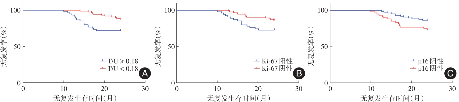
Fig.1
Association between T/U, Ki?67 protein expression, p16 protein expression and recurrence after surgery in patientswith endometrial cancer"
Tab. 3
Multivariate analysis of recurrence after endometrial cancer surgery"
| 因素 | β | SE | Walds | P值 | OR | 95%CI |
|---|---|---|---|---|---|---|
| FIGO分期 | 0.582 | 0.243 | 5.736 | 0.002 | 1.790 | 1.112 ~ 2.881 |
| 淋巴结转移 | 0.401 | 0.186 | 4.648 | 0.044 | 1.493 | 1.037 ~ 2.150 |
| 肌层浸润深度 | 0.397 | 0.171 | 5.390 | 0.018 | 1.487 | 1.064 ~ 2.080 |
| T/U | 0.518 | 0.207 | 6.262 | < 0.001 | 1.679 | 1.119 ~ 2.519 |
| Ki-67蛋白 | 0.476 | 0.225 | 4.476 | 0.046 | 1.610 | 1.036 ~ 2.502 |
| p16蛋白 | -0.500 | 0.268 | 3.481 | 0.097 | 0.607 | 0.359 ~ 1.026 |
| 常数项 | 1.409 | 0.840 | 2.814 | 0.125 | 4.092 | 0.789 ~ 21.230 |
| 1 |
KARPEL H C, SLOMOVITZ B, COLEMAN R L, et al. Treatment options for molecular subtypes of endometrial cancer in 2023[J]. Curr Opin Obstet Gynecol, 2023,35(3):270-278. doi:10.1097/gco.0000000000000855
doi: 10.1097/gco.0000000000000855 |
| 2 |
TRONCONI F, NERO C, GIUDICE E, et al. Advanced and recurrent endometrial cancer: State of the art and future perspectives[J]. Crit Rev Oncol Hematol, 2022,180(8):e103851. doi:10.1016/j.critrevonc.2022.103851
doi: 10.1016/j.critrevonc.2022.103851 |
| 3 |
CLARKE M A, LONG B J, SHERMAN M E, et al. Risk assessment of endometrial cancer and endometrial intraepithelial neoplasia in women with abnormal bleeding and implications for clinical management algorithms[J]. Am J Obstet Gynecol, 2020,223(4):e549. doi:10.1016/j.ajog.2020.03.032
doi: 10.1016/j.ajog.2020.03.032 |
| 4 |
MOAR K, PANT A, SAINI V, et al. Potential biomarkers in endometrial cancer: A narrative review[J]. Biomarkers,2023,28(4):358-371. doi:10.1080/1354750x.2023.2179114
doi: 10.1080/1354750x.2023.2179114 |
| 5 |
HOIVIK E A. Using an MRI-based radiomics model to predict recurrence of endometrial cancer: A step towards meeting a key clinical need[J]. Eur Radiol, 2023,33(8):5812-5813. doi:10.1007/s00330-023-09764-0
doi: 10.1007/s00330-023-09764-0 |
| 6 |
KULINCZAK M, SROMEK M, PANEK G, et al. Endometrial Cancer-Adjacent Tissues Express Higher Levels of Cancer-Promoting Genes than the Matched Tumors[J]. Genes (Basel), 2022,13(9):1611-1613. doi:10.3390/genes13091611
doi: 10.3390/genes13091611 |
| 7 |
林培培,周晓冬,宋伟. 子宫内膜癌肿瘤体积与子宫体积比值联合肿瘤ADC值对病理分级的预测价值[J]. 实用医学杂志, 2023, 39(23):3071-3075. doi:10.3969/j.issn.1006-5725.2023.23.008
doi: 10.3969/j.issn.1006-5725.2023.23.008 |
| 8 |
KARPEL H, SLOMOVITZ B, COLEMAN R L, et al. Biomarker-driven therapy in endometrial cancer[J]. Int J Gynecol Cancer, 2023,33(3):343-350. doi:10.1136/ijgc-2022-003676
doi: 10.1136/ijgc-2022-003676 |
| 9 |
WONG R W, CHEUNG A N Y. Predictive and prognostic biomarkers in female genital tract tumours: An update highlighting their clinical relevance and practical issues[J]. Pathology, 2024, 56(2):214-227. doi:10.1016/j.pathol.2023.10.013
doi: 10.1016/j.pathol.2023.10.013 |
| 10 | 周琦,吴小华,刘继红,等. 子宫内膜癌诊断与治疗指南(第四版)[J]. 中国实用妇科与产科杂志, 2018, 34(8):880-886. |
| 11 | GHALIB FARHOOD R, AL-HUMAIRI I ABD ALI. Immunohistochemical Study of Ki-67 in Hyperplastic and Endometrium Carcinoma: A Comparative Study[J]. Arch Razi Inst,2022,77(1):229-234. |
| 12 |
邓茜,俞文英. 子宫内膜癌的临床病理学进展[J]. 现代实用医学,2021,33(5):565-568. doi:10.3969/j.issn.1671-0800.2021.05.002
doi: 10.3969/j.issn.1671-0800.2021.05.002 |
| 13 |
VOLINSKY-FREMOND S, HOREWEG N, ANDANI S, et al. Prediction of recurrence risk in endometrial cancer with multimodal deep learning [J]. Nat Med,2024,30(7):1962-1973. doi:10.1038/s41591-024-02993-w
doi: 10.1038/s41591-024-02993-w |
| 14 |
BRUNO V, BETTI M, D'AMBROSIO L, et al. Machine learning endometrial cancer risk prediction model: Integrating guidelines of European Society for Medical Oncology with the tumor immune framework[J]. Int J Gynecol Cancer,2023,33(11):1708-1714. doi:10.1136/ijgc-2023-004671
doi: 10.1136/ijgc-2023-004671 |
| 15 |
DE VITIS L A, MULTINU F. Advancing endometrial cancer management in the era of molecular classification: Insights into pattern of recurrence[J]. Int J Gynecol Cancer, 2024,34(5):667-668. doi:10.1136/ijgc-2024-005527
doi: 10.1136/ijgc-2024-005527 |
| 16 |
LABAN M, EL-SWAIFY S T, ALI S H, et al. The Prediction of Recurrence in Low-Risk Endometrial Cancer: Is It Time for a Paradigm Shift in Adjuvant Therapy?[J]. Reprod Sci, 2022,29(4):1068-1085. doi:10.1007/s43032-021-00565-8
doi: 10.1007/s43032-021-00565-8 |
| 17 |
RIEDINGER C J, PATTERSON J M, BACKES F J, et al. The contemporary presentation and diagnosis of endometrial cancer recurrence: When, where, and how?[J]. Gynecol Oncol, 2022,167(2):174-180. doi:10.1016/j.ygyno.2022.09.014
doi: 10.1016/j.ygyno.2022.09.014 |
| 18 |
OAKNIN A, BOSSE T J, CREUTZBERG C L, et al. Endometrial cancer: ESMO Clinical Practice Guideline for diagnosis, treatment and follow-up[J]. Ann Oncol,2022,33(9):860-877. doi:10.1016/j.annonc.2022.05.009
doi: 10.1016/j.annonc.2022.05.009 |
| 19 | 朱争艳,王彩霞,梁菲梅,等. 早期高危子宫内膜癌患者术后辅助治疗与预后的关系[J]. 中华肿瘤防治杂志,2023,30(2):99-103. |
| 20 |
MAHESHWARI E, NOUGARET S, STEIN E B, et al. Update on MRI in Evaluation and Treatment of Endometrial Cancer[J]. Radiographics,2022,42(7):2112-2130. doi:10.1148/rg.220070
doi: 10.1148/rg.220070 |
| 21 | 孔伟,余裕珍,王康,等. 术前MRI影像组学模型预测子宫内膜癌风险分层[J]. 中国医学影像技术, 2023, 39(12):1857-1861. |
| 22 |
DING S X, SUN Y F, MENG H, et al. Radiomics model based on multi-sequence MRI for preoperative prediction of ki-67 expression levels in early endometrial cancer[J]. Sci Rep, 2023,13(1):e22052. doi:10.1038/s41598-023-49540-0
doi: 10.1038/s41598-023-49540-0 |
| 23 |
YASUTAKE N, YAMAMOTO H, KUGA R, et al. Immunohistochemical p16 overexpression and Rb loss correlate with high-risk human papillomavirus infection in endocervical adenocarcinomas[J]. Histopathology, 2024,84(7):1178-1191. doi:10.1111/his.15169
doi: 10.1111/his.15169 |
| 24 | KALKAN H E, AKMAN L, SERIN G, et al. The usefulness of p16 and COX-2 expression on the prediction of progression to endometrial cancer[J]. Histol Histopathol, 2024,39(5):565-571. |
| 25 |
YANG Z, YANG X, LIU X, et al. Clinical characteristics and prognostic characterization of endometrial carcinoma: A comparative analysis of molecular typing protocols[J]. BMC Cancer,2023,23(1):243-245. doi:10.1186/s12885-023-10706-8
doi: 10.1186/s12885-023-10706-8 |
| 26 | 张继屏,范锶丝,江洁丽,等. Ki67、突变型P53、突变型P16、CK7与宫颈病复发相关性临床研究[J]. 大医生,2023,8(22):102-105. |
| [1] | Shaojin LI,Shipeng. ZHENG. Relevant preoperative imaging pathological features and tumor markers serve as predictive indicators for the risk of sentinel lymph node metastasis in breast cancer [J]. The Journal of Practical Medicine, 2024, 40(17): 2418-2424. |
| [2] | Lingyu FANG,Jinghua HU,Junfeng WEN,Shiqi HAN,Yali WANG,Lulan PU,Jingjia LI,Yi YANG,Shishan DENG,Lingmi HOU,Fangfang. ZHOU. Expression and significance of ubiquitin⁃specific proteases 20 and hypoxia inducible factor⁃1α in breast cancer [J]. The Journal of Practical Medicine, 2024, 40(16): 2270-2276. |
| [3] | Fei CHAI,Zhenwen CHEN,Hongyan LIU,Su LI,Yuanyuan GONG,Yingying. ZHAO. Expression level of serum UBE2C and TRIM27 in patients with endometrial cancer and their correlation with pathological parameters [J]. The Journal of Practical Medicine, 2024, 40(13): 1808-1813. |
| [4] |
AI Yongbiao, HUANG Jun, YUAN Jie, LI Wenfang..
Androgen receptor expression in early triple negative breast cancer and its association with the clinicopath⁃ ological features and prognosis [J]. The Journal of Practical Medicine, 2023, 39(8): 975-979. |
| [5] |
JIN Bei, CHENG Cheng, ZHUANG Hongjie, OUYANG Xiaojun, LI Jie, CHEN Lizhi, JIANG Xiaoyun..
The clinicopathological features and long ⁃ term prognosis inlupus nephritis children of different gender [J]. The Journal of Practical Medicine, 2023, 39(8): 997-1001. |
| [6] | Peipei LIN,Xiaodong ZHOU,Wei. SONG. Prediction value of tumor volume to uterine volume ratio combined with tumor ADC value in pathological grading of endometrial carcinoma [J]. The Journal of Practical Medicine, 2023, 39(23): 3071-3075. |
| [7] | Chongfeng SUN,Ping YANG,Jishuai HOU,Zouyu ZHAO,Panpan. YU. Expression and prognosis of STT3A and STT3B in epithelial ovarian cancer [J]. The Journal of Practical Medicine, 2023, 39(16): 2062-2070. |
| [8] |
CHEN Rui, XIONG Zhuang, CHEN Renfu..
Expression of interleukin 38 in renal cell carcinoma and its correlation with clinicopathological features [J]. The Journal of Practical Medicine, 2022, 38(12): 1506-1511. |
| [9] |
WANG Saifeng, SHI Wei, XIE Xianjing..
RBM5 knockout in endometrial cancer cells and its effect [J]. The Journal of Practical Medicine, 2022, 38(10): 1203-1207. |
| [10] |
LI Song, DONG Lili, SUN Li, LI Gang, YUAN Shuang, WANG Xuebo, WANG Ke.
Effect of MicroRNA⁃191 on proliferation and invasion of endometrial cancer cells through targeted regula⁃ tion of TET1 [J]. The Journal of Practical Medicine, 2021, 37(8): 1014-1018. |
| [11] |
SU Bo, YAN Zhenyu, HAO Shanghui, SUN Li, HU Ping, GAO Yi..
Association of expression of IL⁃33 with clinic⁃pathological features and prognosis in patients with cervical cancer tissues [J]. The Journal of Practical Medicine, 2021, 37(17): 2214-2218. |
| [12] |
SU Bo, YAN Zhenyu, HAO Shanghui, SUN Li, HU Ping, GAO Yi..
Association of expression of IL⁃33 with clinic⁃pathological features and prognosis in patients with cervical cancer tissues [J]. The Journal of Practical Medicine, 2021, 37(17): 2214-2218. |
| Viewed | ||||||
|
Full text |
|
|||||
|
Abstract |
|
|||||