

The Journal of Practical Medicine ›› 2024, Vol. 40 ›› Issue (17): 2418-2424.doi: 10.3969/j.issn.1006-5725.2024.17.011
• Clinical Research • Previous Articles Next Articles
Shaojin LI,Shipeng. ZHENG( )
)
Received:2023-12-29
Online:2024-09-10
Published:2024-09-13
Contact:
Shipeng. ZHENG
E-mail:1147966825@qq.com
CLC Number:
Shaojin LI,Shipeng. ZHENG. Relevant preoperative imaging pathological features and tumor markers serve as predictive indicators for the risk of sentinel lymph node metastasis in breast cancer[J]. The Journal of Practical Medicine, 2024, 40(17): 2418-2424.
Tab.1
Comparison of training set and validation set features"
| 特征 | 训练集(n = 174) | 验证集(n = 58) | t/Z/χ 2 值 | P值 |
|---|---|---|---|---|
| 年龄(x ± s)/岁 | 49.3 ± 9.96 | 49.21 ± 11.72 | 0.058 | 0.954 |
| BMI(x ± s)/(kg/㎡) | 24.79 ± 4.22 | 24.34 ± 3.31 | 0.744 | 0.458 |
| 可触及性 | 0.232 | 0.630 | ||
| 可触及 | 117(67.2) | 37(63.8) | ||
| 未触及 | 57(32.8) | 21(36.2) | ||
| 已绝经 | 114(65.5) | 35(60.3) | 0.507 | 0.477 |
| CEA[M(P25,P75)]/(ng/mL) | 1.03(0.67,1.74) | 1.01(0.62,1.50) | -0.497 | 0.619 |
| CA125[M(P25,P75)]/(U/mL) | 9.34(7.35,14.13) | 9.06(6.97,15.53) | -0.316 | 0.752 |
| CA153[M(P25,P75)]/(U/mL) | 6.58(4.44,10.75) | 6.67(4.55,10.13) | -0.026 | 0.979 |
| CA199[M(P25,P75)]/(U/mL) | 6.58(4.22,10.23) | 6.31(4.34,10.17) | -0.434 | 0.664 |
| CA724[M(P25,P75)]/(U/mL) | 1.50(0.60,2.36) | 1.50(0.53,2.01) | -0.893 | 0.372 |
| 病理类型 | 1.037 | 0.595 | ||
| 浸润性导管癌 | 143(82.2) | 49(84.5) | ||
| 浸润性小叶癌 | 3(1.7) | 0(0) | ||
| 其他 | 28(16.1) | 9(15.5) | ||
| 组织学分级 | 2.130 | 0.345 | ||
| Ⅰ | 3(1.7) | 3(5.2) | ||
| Ⅱ | 121(69.5) | 40(69.0) | ||
| Ⅲ | 50(28.7) | 15(25.9) | ||
| 分子分型 | 3.144 | 0.370 | ||
| Luminal A | 22(12.6) | 8(13.8) | ||
| Luminal B | 117(67.2) | 43(74.1) | ||
| HER2过表达 | 14(8.0) | 1(1.7) | ||
| 三阴性 | 21(12.1) | 6(10.3) | ||
| ER阳性 | 137(78.7) | 51(87.9) | 2.393 | 0.122 |
| PR阳性 | 126(72.4) | 42(72.4) | 0.000 | 1.000 |
| HER2阳性 | 34(19.5) | 9(15.5) | 0.466 | 0.495 |
| Ki67高表达 | 147(84.5) | 43(74.1) | 3.140 | 0.076 |
| 脉管浸润 | 46(26.4) | 14(24.1) | 0.120 | 0.729 |
| 超声[肿瘤最大径(x ± s)/mm] | 19.69 ± 8.01 | 19.37 ± 8.67 | 0.261 | 0.795 |
| 超声(肿瘤回声均匀) | 13(7.5) | 6(10.3) | 0.172 | 0.678 |
| 超声(肿瘤边界清晰) | 19(10.9) | 9(15.5) | 0.866 | 0.352 |
| 超声(有肿瘤血流信号) | 122(70.1) | 37(63.8) | 0.806 | 0.369 |
| 钼靶(有钙化) | 83(47.7) | 29(50.0) | 0.092 | 0.762 |
| 超声(有ALN回声) | 73(42.0) | 25(43.1) | 0.024 | 0.878 |
| 超声(ALN分界清晰) | 35(20.1) | 11(19.0) | 0.036 | 0.849 |
| 超声(有ALN血流信号) | 59(33.9) | 23(39.7) | 0.629 | 0.428 |
| SLN转移 | 66(37.9) | 25(43.1) | 0.488 | 0.485 |
Tab.2
Univarate analysis of baseline characteristics of the training set"
| 特征 | SLN阳性(n = 66) | SLN阴性(n = 108) | t/Z/χ 2 值 | P值 |
|---|---|---|---|---|
| 可触及性 | ||||
| 可触及 | 56(47.9) | 61(52.1) | 14.965 | < 0.001 |
| 未触及 | 10(17.5) | 47(82.5) | ||
| BMI(x ± s)/(kg/m2) | 25.67 ± 4.73 | 24.25 ± 3.8 | 2.176 | 0.031 |
| CA153[M(P25,P75)]/(U/mL) | 8.70(5.35,14.01) | 5.47(4.01,9.51) | -3.503 | < 0.001 |
| CA199[M(P25,P75)]/(U/mL) | 8.19(4.70,12.32) | 6.00(3.78,10.05) | -2.132 | 0.033 |
| 脉管浸润 | 24(52.2) | 22(47.8) | 5.388 | 0.020 |
| 超声[肿瘤最大径(x ± s)/mm] | 21.32 ± 8.42 | 18.7 ± 7.6 | 2.117 | 0.036 |
| 超声(肿瘤血流信号) | 53(43.4) | 69(56.6) | 5.267 | 0.022 |
| 钼靶(钙化) | 39(47.0) | 44(53.0) | 5.529 | 0.019 |
| 超声(ALN回声) | 43(58.9) | 30(41.1) | 23.497 | < 0.001 |
| 超声(ALN分界清晰) | 24(68.6) | 11(31.4) | 17.471 | < 0.001 |
| 超声(ALN血流信号) | 39(66.1) | 20(33.9) | 30.090 | < 0.001 |
Tab.3
Multivariate logistic regression analysis for predicting SLN metastasis"
| 因素 | OR值 | 95%CI | P值 |
|---|---|---|---|
| 可触及性 | 4.511 | 1.698 ~ 11.987 | 0.003 |
| BMI | 1.072 | 0.985 ~ 1.168 | 0.109 |
| CA153 | 1.065 | 1.003 ~ 1.130 | 0.039 |
| CA199 | 1.012 | 0.953 ~ 1.075 | 0.692 |
| 脉管浸润 | 1.749 | 0.749 ~ 4.086 | 0.197 |
| 超声(肿瘤最大径,mm) | 0.978 | 0.927 ~ 1.031 | 0.404 |
| 超声(肿瘤血流信号) | 2.147 | 0.854 ~ 5.394 | 0.104 |
| 钼靶(钙化) | 2.202 | 1.014 ~ 4.783 | 0.046 |
| 超声(ALN回声) | 0.428 | 0.092 ~ 1.994 | 0.280 |
| 超声(ALN分界) | 0.320 | 0.101 ~ 1.017 | 0.053 |
| 超声(ALN血流信号) | 7.427 | 1.720 ~ 32.076 | 0.007 |
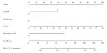
Fig.1
Nomogram for predicting SLN positivity"
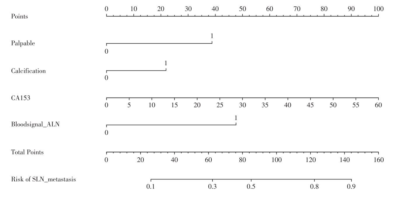
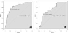
Fig.2
ROC curves for nomogram in training and validation sets"
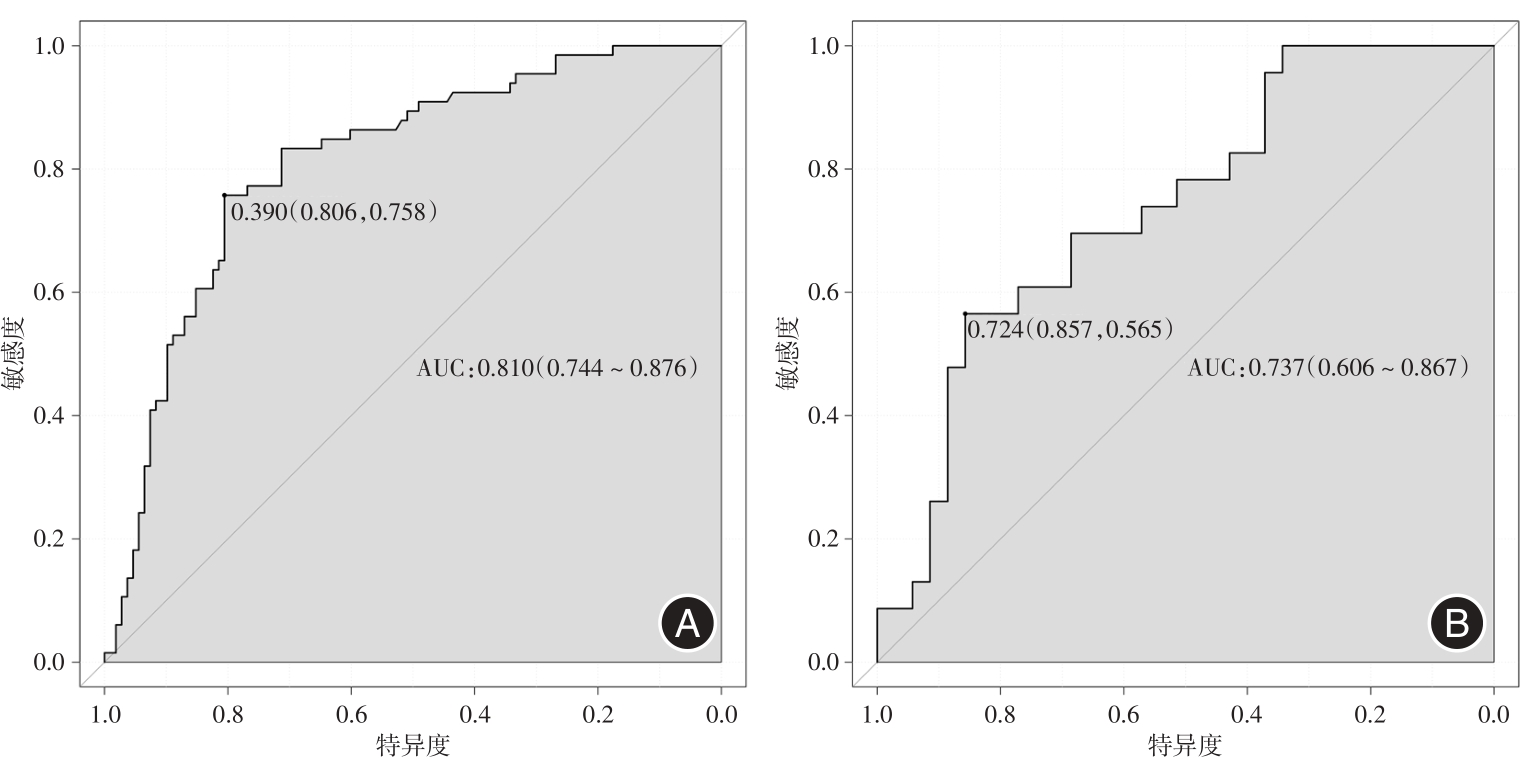
Tab.4
Diagnostic efficacy of ROC for training and validation sets"
| 指标 | AUC | 95%CI | 敏感度(%) | 特异度(%) | 截断值 |
|---|---|---|---|---|---|
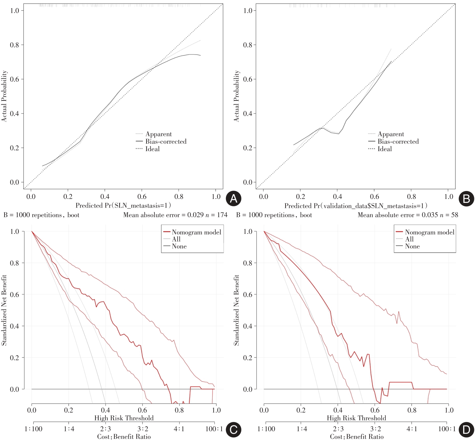
| 训练集 | 0.810 | 0.744 ~ 0.876 | 75.8 | 80.6 | 0.390 |
| 验证集 | 0737 | 0.606 ~ 0.867 | 56.5 | 85.7 | 0.724 |
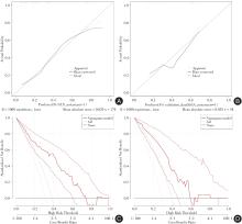
Fig.3
Calibration curve and decision curve analysis of training set and validation set"
| 1 |
SUNG H, FERLAY J, SIEGEL R L,et al. Global Cancer Statistics 2020: GLOBOCAN Estimates of Incidence and Mortality Worldwide for 36 Cancers in 185 Countries[J]. CA Cancer J Clin, 2021,71(3):209-249. doi:10.3322/caac.21660
doi: 10.3322/caac.21660 |
| 2 |
ARNOLD M, MORGAN E, RUMGAY H,et al. Current and future burden of breast cancer: Global statistics for 2020 and 2040[J]. Breast,2022,66:15-23. doi:10.1016/j.breast.2022.08.010
doi: 10.1016/j.breast.2022.08.010 |
| 3 |
VERONESI P, CORSO G. Standard and controversies in sentinel node in breast cancer patients[J]. Breast,2019,48():S53-S56. doi:10.1016/s0960-9776(19)31124-5
doi: 10.1016/s0960-9776(19)31124-5 |
| 4 |
QIU S Q, ZHANG G J, JANSEN L, et al. Evolution in sentinel lymph node biopsy in breast cancer[J]. Crit Rev Oncol Hematol,2018,123:83-94. doi:10.1016/j.critrevonc.2017.09.010
doi: 10.1016/j.critrevonc.2017.09.010 |
| 5 |
MANCA G, RUBELLO D, TARDELLI E,et al. Sentinel Lymph Node Biopsy in Breast Cancer: Indications, Contraindications, and Controversies[J]. Clin Nucl Med,2016,41(2):126-133. doi:10.1097/rlu.0000000000000985
doi: 10.1097/rlu.0000000000000985 |
| 6 | GOLDHIRSCH A, WINER E P, COATES A S, et al. Personalizing the treatment of women with early breast cancer: highlights of the St Gallen International Expert Consensus on the Primary Therapy of Early Breast Cancer 2013[J]. Ann Oncol,2013,24(9):2206-2223. |
| 7 |
张强,牛连杰, 黄涛,等.早期乳腺癌520例前哨淋巴结转移关联性分析及预测研究[J]. 中华肿瘤防治杂志, 2020, 27 (22): 1850-1854. doi:10.16073/j.cnki.cjcpt.2020.22.13
doi: 10.16073/j.cnki.cjcpt.2020.22.13 |
| 8 |
VAN LA PARRA R F, FRANCISSEN C M, PEER P G,et al. Assessment of the Memorial Sloan-Kettering Cancer Center nomogram to predict sentinel lymph node metastases in a Dutch breast cancer population[J]. Eur J Cancer, 2013,49(3):564-571. doi:10.1016/j.ejca.2012.04.025
doi: 10.1016/j.ejca.2012.04.025 |
| 9 |
ZHU L, JIN L, LI S, et al.Which nomogram is best for predicting non-sentinel lymph node metastasis in breast cancer patients? A meta-analysis[J]. Breast Cancer Res Treat,2013,137(3):783-795. doi:10.1007/s10549-012-2360-6
doi: 10.1007/s10549-012-2360-6 |
| 10 |
LALE A, YUR M, ÖZGÜL H,et al. Predictors of non-sentinel lymph node metastasis in clinical early stage (cT1-2N0) breast cancer patients with 1-2 metastatic sentinel lymph nodes[J]. Asian J Surg,2020,43(4):538-549. doi:10.1016/j.asjsur.2019.07.019
doi: 10.1016/j.asjsur.2019.07.019 |
| 11 |
LEONARDI M C, ARROBBIO C, GANDINI S,et al. Predictors of positive axillary non-sentinel lymph nodes in breast cancer patients with positive sentinel lymph node biopsy after neoadjuvant systemic therapy[J]. Radiother Oncol,2021,163:128-135. doi:10.1016/j.radonc.2021.08.013
doi: 10.1016/j.radonc.2021.08.013 |
| 12 |
QIU Y, ZHANG X, WU Z,et al. MRI-Based Radiomics Nomogram: Prediction of Axillary Non-Sentinel Lymph Node Metastasis in Patients With Sentinel Lymph Node-Positive Breast Cancer[J]. Front Oncol,2022,12:811347. doi:10.3389/fonc.2022.811347
doi: 10.3389/fonc.2022.811347 |
| 13 |
FONG W, TAN L, TAN C, et al.Predicting the risk of axillary lymph node metastasis in early breast cancer patients based on ultrasonographic-clinicopathologic features and the use of nomograms: a prospective single-center observational study[J]. Eur Radiol,2022,32(12):8200-8212. doi:10.1007/s00330-022-08855-8
doi: 10.1007/s00330-022-08855-8 |
| 14 |
GAO Y, LUO Y, ZHAO C, et al. Nomogram based on radiomics analysis of primary breast cancer ultrasound images: prediction of axillary lymph node tumor burden in patients[J]. Eur Radiol,2021,31(2):928-937. doi:10.1007/s00330-020-07181-1
doi: 10.1007/s00330-020-07181-1 |
| 15 |
LI X, YANG L, JIAO X. Development and Validation of a Nomogram for Predicting Axillary Lymph Node Metastasis in Breast Cancer[J]. Clin Breast Cancer,2023,23(5):538-545. doi:10.1016/j.clbc.2023.04.002
doi: 10.1016/j.clbc.2023.04.002 |
| 16 |
暴珞宁, 王瑛, 陈东, 等. 超声影像组学标签预测乳腺癌前哨淋巴结转移的价值[J]. 实用医学杂志, 2021, 37 (15): 2007-2011. doi:10.3969/j.issn.1006-5725.2021.15.020
doi: 10.3969/j.issn.1006-5725.2021.15.020 |
| 17 |
LIU M, MAO N, MA H, et al. Pharmacokinetic parameters and radiomics model based on dynamic contrast enhanced MRI for the preoperative prediction of sentinel lymph node metastasis in breast cancer[J]. Cancer Imaging, 2020,20(1):65. doi:10.1186/s40644-020-00342-x
doi: 10.1186/s40644-020-00342-x |
| 18 |
MATHIS K L, HOSKIN T L, BOUGHEY J C, et al. Palpable presentation of breast cancer persists in the era of screening mammography[J]. J Am Coll Surg,2010,210(3):314-318. doi:10.1016/j.jamcollsurg.2009.12.003
doi: 10.1016/j.jamcollsurg.2009.12.003 |
| 19 |
AZAM S, ERIKSSON M, SJÖLANDER A,et al. Mammographic microcalcifications and risk of breast cancer[J]. Br J Cancer,2021,125(5):759-765. doi:10.1038/s41416-021-01459-x
doi: 10.1038/s41416-021-01459-x |
| 20 |
胡仰玲, 曾辉, 何子龙, 等. 钙化型乳腺癌的分子分型特点及其预后分析[J]. 实用医学杂志, 2020, 36 (10): 1354-1359. doi:10.3969/j.issn.1006-5725.2020.10.017
doi: 10.3969/j.issn.1006-5725.2020.10.017 |
| 21 |
XIONG J, ZUO W, WU Y, et al.Ultrasonography and clinicopathological features of breast cancer in predicting axillary lymph node metastases[J]. BMC Cancer,2022,22(1):1155. doi:10.1186/s12885-022-10240-z
doi: 10.1186/s12885-022-10240-z |
| 22 |
WANG X F, ZHANG G C, ZUO Z C, et al. A novel nomogram for the preoperative prediction of sentinel lymph node metastasis in breast cancer[J]. Cancer Med,2023,12(6):7039-7050. doi:10.1002/cam4.5503
doi: 10.1002/cam4.5503 |
| 23 |
DARWISH I A, WANI T A, KHALIL N Y, et al. Novel automated flow-based immunosensor for real-time measurement of the breast cancer biomarker CA15-3 in serum[J]. Talanta,2012,97:499-504. doi:10.1016/j.talanta.2012.05.005
doi: 10.1016/j.talanta.2012.05.005 |
| 24 |
WANG W, XU X, TIAN B, et al. The diagnostic value of serum tumor markers CEA, CA19-9, CA125, CA15-3, and TPS in metastatic breast cancer[J]. Clin Chim Acta,2017,470:51-55. doi:10.1016/j.cca.2017.04.023
doi: 10.1016/j.cca.2017.04.023 |
| 25 |
冯霖, 何真, 王欣欣. 超声BI-RADS分级联合血清CA153、CEA、ALP检测在乳腺癌早期诊断及腋窝淋巴结转移预测中的应用[J]. 中国医学创新, 2023, 20 (28): 146-152. doi:10.3969/j.issn.1674-4985.2023.28.034
doi: 10.3969/j.issn.1674-4985.2023.28.034 |
| 26 |
LUO J, XIAO J, YANG Y, et al. Strategies for five tumour markers in the screening and diagnosis of female breast cancer[J]. Front Oncol,2023,12:1055855. doi:10.3389/fonc.2022.1055855
doi: 10.3389/fonc.2022.1055855 |
| [1] | Zixu SONG,Guangzheng ZHU,Chenxu GUO,Jiaqi WU,Ligong ZHANG,Jun. QIAN. Expression of SLC35A2 and PFDN2 in breast cancer and its relationship with clinical observables and prognosis [J]. The Journal of Practical Medicine, 2024, 40(4): 496-502. |
| [2] | Yinghua ZENG,Wenji LI,Li. ZHENG. The Impact of the timing of initial dressing change following PICC catheterization on postoperative breast cancer patients [J]. The Journal of Practical Medicine, 2024, 40(19): 2772-2777. |
| [3] | Che CHEN,Dehong LUO,Huangfei YU,Qin ZHANG,Xiaochi HU,Shenghua YU,Yajun. LI. Clinical Application of automatic delineation in whole breast radiotherapy with simultaneous integrated boost to the medial tumor beds [J]. The Journal of Practical Medicine, 2024, 40(17): 2406-2411. |
| [4] | Lingyu FANG,Jinghua HU,Junfeng WEN,Shiqi HAN,Yali WANG,Lulan PU,Jingjia LI,Yi YANG,Shishan DENG,Lingmi HOU,Fangfang. ZHOU. Expression and significance of ubiquitin⁃specific proteases 20 and hypoxia inducible factor⁃1α in breast cancer [J]. The Journal of Practical Medicine, 2024, 40(16): 2270-2276. |
| [5] |
AI Yongbiao, HUANG Jun, YUAN Jie, LI Wenfang..
Androgen receptor expression in early triple negative breast cancer and its association with the clinicopath⁃ ological features and prognosis [J]. The Journal of Practical Medicine, 2023, 39(8): 975-979. |
| [6] | Xiran SHI,Heng WANG,Libing HE,Zhiqiang QIU,Hongjian LI,Xiaoxue. XU. Advance in predicting lymph node metastasis of breast cancer by multimodal MRI [J]. The Journal of Practical Medicine, 2023, 39(22): 2861-2865. |
| [7] | Yadan CHE,Lixia. LI. Reaserch progress on small molecule anti⁃angiogenic drugs for advanced breast cancer [J]. The Journal of Practical Medicine, 2023, 39(22): 2866-2871. |
| [8] | Zhaowei ZHUANG,Wumei YUAN,Zuodong REN,Shangfei LI,Minggui CHEN,Yan. ZENG. Expression and significance of granzyme B and perforin in peripheral blood of patients with breast cancer [J]. The Journal of Practical Medicine, 2023, 39(22): 2872-2877. |
| [9] | Haiying LIU,Feng CHEN,Jia. YAO. Suppression of miR-767-5p expression of inhibits breast cancer cell proliferation, migration, invasion and EMT through down-regulating IGF1 [J]. The Journal of Practical Medicine, 2023, 39(22): 2878-2884. |
| [10] | Zhenghua ZHANG,Rongfu GONG,Wen. FANG. FGF2 gene knockout inhibits MCF⁃7 cell proliferation, motility and invasion while promotes cell apoptosis [J]. The Journal of Practical Medicine, 2023, 39(22): 2885-2890. |
| [11] | Yan LV,Xu ZHENG,Yanyan HAN,Shan GAO,Chong LI,Qiang. GENG. Effect of CCN5 gene knock⁃down on the proliferation of MCF⁃7 breast cancer cells and its mechanism [J]. The Journal of Practical Medicine, 2023, 39(22): 2898-2902. |
| [12] | Yao MENG,Zhao LIU,Jing ZHANG,Changxiao. ZHAO. Comparison of curative effect and prognosis of preserving nipple and areola in patients with breast cancer by different reconstruction operations [J]. The Journal of Practical Medicine, 2023, 39(22): 2903-2908. |
| [13] | Yuling LI,Jianlin YANG,Zhi CUI,Jing WANG,Yafeng LV,Chunyu. CAO. Effect of BMI-1 inhibitor PTC-596 on proliferation, cell cycle, and apoptosis of human breast cancer MCF-7cells [J]. The Journal of Practical Medicine, 2023, 39(18): 2317-2322. |
| [14] | Yin LIN,Dongdong ZOU,Yilong WU,Min LIN,Tuo. YANG. Comparison of endoscopic features between colorectal sessile serrated lesions and hyperplastic polyps and establishment of predictive model [J]. The Journal of Practical Medicine, 2023, 39(17): 2258-2264. |
| [15] | Cuixia HUANG,Yaqian ZHANG,Aiping YANG,Ruihanqiu LIU,Yan LU. Exploring the mechanism of using andrographolide to inhibit triple negative breast cancer based on Hippo/YAP signaling pathway [J]. The Journal of Practical Medicine, 2023, 39(16): 2050-2056. |
| Viewed | ||||||
|
Full text |
|
|||||
|
Abstract |
|
|||||