实用医学杂志 ›› 2025, Vol. 41 ›› Issue (6): 877-881.doi: 10.3969/j.issn.1006-5725.2025.06.016

• 医学检查与临床诊断 • 上一篇    下一篇

彩色多普勒联合实时剪切波弹性成像在诊断血管性勃起功能障碍中的应用价值

申佳琦1,2,康彧2(),南绪红1,沙晓溪2   

  1. 1.成都中医药大学医学与生命科学学院 (四川 成都 611137 )
    2.成都中医药大学附属医院超声医学科 (四川 成都 610075 )
  • 收稿日期:2024-12-07 出版日期:2025-03-25 发布日期:2025-03-31
  • 通讯作者: 康彧 E-mail:mofang5474@163.com
  • 基金资助:
    国家中医药管理局基金项目(JDZX2015218)

The application value of color doppler ultrasound combined with real⁃time shear wave elastography in the diagnosis of vascular erectile dysfunction

Jiaqi SHEN1,2,Yu KANG2(),Xuhong NAN1,Xiaoxi. SHA2   

  1. School of Medicine and Life Sciences,Chengdu University of Traditional Chinese Medicine,Chengdu 611137,Sichuan,China; *Department of Ultrasound Medicine,Affiliated Hospital of Chengdu University of Traditional Chinese Medicine,Chengdu 610075,Sichuan,China
  • Received:2024-12-07 Online:2025-03-25 Published:2025-03-31
  • Contact: Yu KANG E-mail:mofang5474@163.com

摘要:

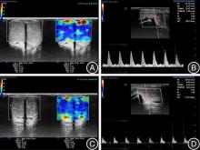
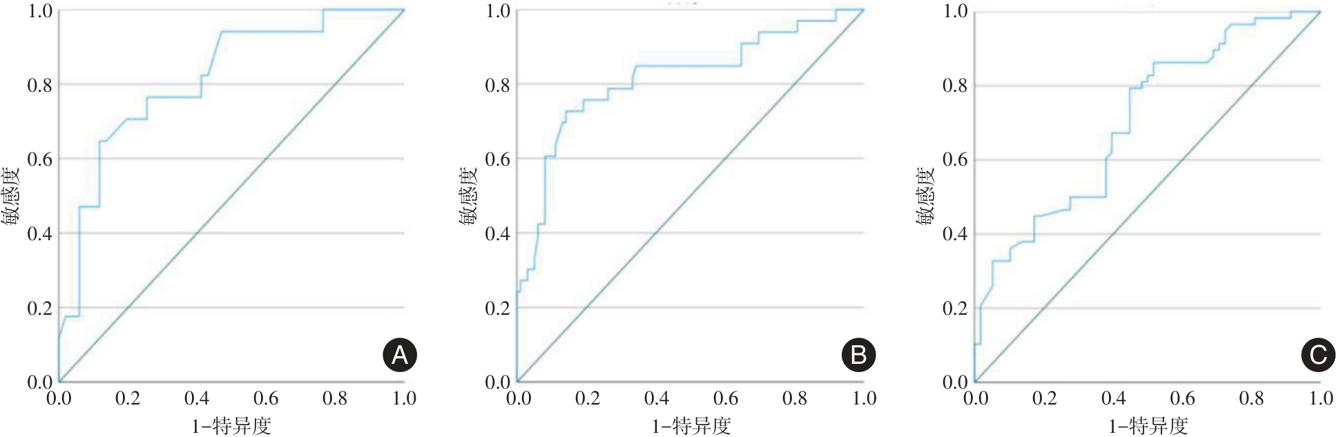
目的 探讨实时剪切波弹性成像(SWE)在血管性勃起功能障碍(ED)诊断及预测彩色多普勒血流显像(CDFI)检查时机中的应用价值。 方法 纳入因ED就诊并行血管活性药物阴茎海绵体内注射(ICI)的患者,经CDFI诊断分为动脉性ED组17例、静脉性ED组33例和非血管性ED组29例。采用SWE技术检测ED患者ICI前疲软状态及ICI诱导勃起后4个时间点阴茎海绵体平均杨氏模量值(E值),分析各组E值的差异。 结果 ICI前疲软状态下动脉性、静脉性及非血管性ED组患者阴茎海绵体E值比较差异无统计学意义(P > 0.05),各组疲软状态阴茎海绵体E值均高于ICI诱导勃起后4个时间点均值,差异均有统计学意义(P < 0.01)。ICI诱导勃起后各组4个时间点阴茎海绵体E值的均值组间比较差异有统计学意义(P < 0.01)。ROC曲线分析结果显示ICI后E值诊断动脉性、静脉性和非血管性ED的AUC分别为0.814、0.770、0.711,截断值分别为9.98、8.16、7.06 kPa。在ICI诱导勃起后,CDFI联合SWE截断值诊断不同类型ED,动脉性ED组及静脉性ED组检测时间缩短,差异有统计学意义(P < 0.01)。 结论 运用SWE技术检测血管活性药物ICI诱导勃起后海绵体E值,可以区分不同类型ED,与CDFI联合应用有助于缩短检查时间。

关键词: 勃起功能障碍, 实时剪切波弹性成像, 彩色多普勒血流显像, 诊断

Abstract:

Objective To assess the utility of real?time shear wave elastography (SWE) in diagnosing vascular erectile dysfunction (ED) and to predict the optimal timing for color Doppler flow imaging (CDFI) examination. Methods Patients diagnosed with ED who received intracavernosal injection (ICI) of vasoactive drugs were recruited and categorized based on CDFI findings into three groups: arterial ED (n = 17), venous ED (n = 33), and non?vascular ED (n = 29). SWE technology was utilized to measure the average Young's modulus (E value) of the corpus cavernosum in these patients, both in the flaccid state prior to ICI and at four time points following ICI?induced erection. Subsequently, the differences in E values among the three groups were analyzed. Results There was no significant difference in the E value of the corpus cavernosum in the flaccid state among the arterial, venous, and non?vascular ED groups before ICI (P > 0.05). However, the E value in the flaccid state for each group was significantly higher than the mean E values observed at the four time points after ICI?induced erection (P < 0.01). Additionally, the mean E values at these four time points post?ICI were also statistically significant (P < 0.01). ROC curve analysis revealed that the AUC for diagnosing arterial, venous, and non?vascular ED using the E value after ICI were 0.814, 0.770, and 0.711, respectively, with corresponding cutoff values of 9.98, 8.16 and 7.06 kPa. The combined use of CDFI and SWE cutoff values following ICI?induced erection significantly shortened the detection time for both arterial and venous ED groups (P < 0.01). Conclusions SWE can accurately measure the E value of the corpus cavernosum following erection induced by the vasoactive drug ICI, thereby facilitating the differentiation of various types of ED. Additionally, when combined with CDFI, this technique can reduce the time required for examination.

Key words: erectile dysfunction, real-time shear wave elastography, color doppler flow imaging, diagnosis

中图分类号: