

The Journal of Practical Medicine ›› 2026, Vol. 42 ›› Issue (13): 2284-2290.doi: 10.3969/j.issn.1006-5725.2026.13.003
• Feature Reports:Cardiovascular diseases • Previous Articles
Wentao QU1,Yuanlin LEI2,Zheng YANG2( ),Nanding WANG2,Liping FAN1
Received:2026-04-23
Online:2026-07-10
Published:2026-07-14
Contact:
Zheng YANG
E-mail:283565626@qq.com
CLC Number:
Wentao QU,Yuanlin LEI,Zheng YANG,Nanding WANG,Liping FAN. Diagnostic value of myocardial contrast echocardiography combined with 2D speckle tracking echocardiography for early quantitative assessment of HFpEF[J]. The Journal of Practical Medicine, 2026, 42(13): 2284-2290.
Fig.1
The MCE perfusion curves of myocardial segment in the normal group"
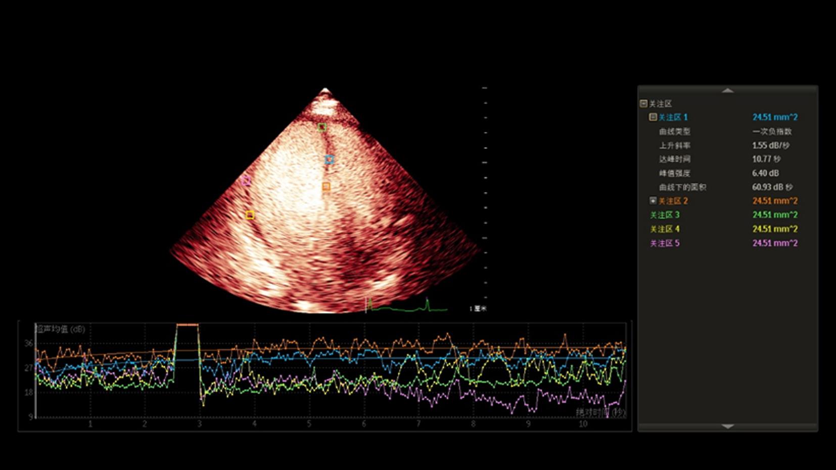
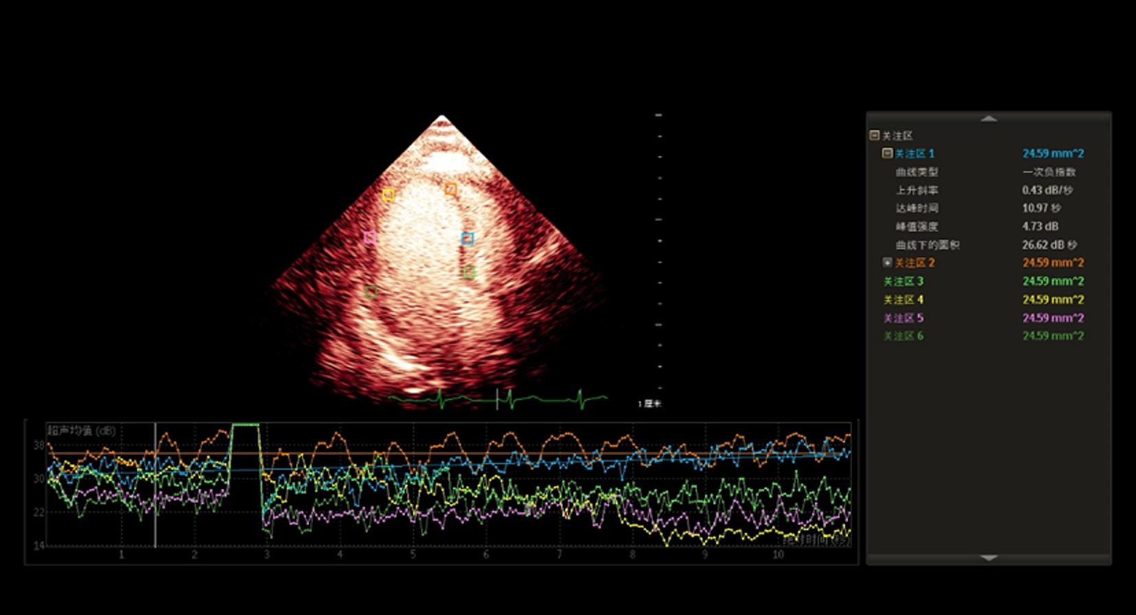
Fig.2
The MCE perfusion curves of myocardial segment in patients with heart failure with preserved ejection fraction"
Tab.1
Comparison of general clinical data between the two groups"
| 参数 | HFpEF组(n = 80) | 对照组(n = 60) | t/χ2值 | P值 |
|---|---|---|---|---|
| 年龄/岁 | 62.8 ± 5.3 | 63.4 ± 6.2 | 0.61 | 0.54 |
| 性别/[例(%)] | 0.03 | 0.71 | ||
| 男 | 36(45.0) | 27(45.0) | ||
| 女 | 44(55.0) | 33(55.0) | ||
| 心率/(次/min) | 66.4 ± 6.2 | 65.3 ± 5.8 | 1.06 | 0.29 |
| BMI/(kg/m2) | 25.6 ± 3.6 | 22.8 ± 2.8 | 5.17 | 0.00 |
| 收缩压/mmHg | 136.6 ± 13.2 | 120.4 ± 12.3 | 7.39 | 0.00 |
| 舒张压/mmHg | 90.2 ± 14.4 | 76.6 ± 9.3 | 6.38 | 0.00 |
| 糖尿病/[例(%)] | 42(52.5) | 17(28.3) | 9.03 | 0.00 |
| 高脂血症/[例(%)] | 34(42.5) | 24(40.0) | 0.09 | 0.86 |
| 肌酐/(μmol/L) | 66.6 ± 16.6 | 65.6 ± 17.8 | 0.35 | 0.73 |
| BNP/(pg/mL) | 282.8 ± 59.2 | 138.3 ± 32.3 | 17.08 | 0.00 |
Tab.2
Comparison of echocardiographic parameter between the two group"
| 参数 | HFpEF组 | 对照组 | t值 | P值 |
|---|---|---|---|---|
| LVEF/% | 62.80 ± 7.30 | 63.40 ± 6.20 | 0.51 | 0.61 |
| LVEDD/mm | 45.54 ± 4.53 | 44.87 ± 5.25 | 0.81 | 0.43 |
| IVST/mm | 12.23 ± 0.78 | 9.78 ± 1.12 | 15.25 | 0.00 |
| LAD/mm | 39.14 ± 3.10 | 34.31 ± 2.44 | 10.20 | 0.00 |
| PWT/mm | 8.65 ± 1.38 | 8.23 ± 1.24 | 1.86 | 0.06 |
| LAVI/(mL/m2) | 26.89 ± 2.77 | 23.58 ± 3.08 | 6.64 | 0.00 |
| E/A | 0.74 ± 0.23 | 0.72 ± 0.25 | 0.49 | 0.62 |
| E/e′ | 15.61 ± 4.56 | 11.71 ± 1.95 | 6.28 | 0.00 |
| LVMI/(g/m2) | 108.77 ± 14.23 | 95.45 ± 12.65 | 5.74 | 0.00 |
| GLS/% | 19.87 ± 2.78 | 20.01 ± 3.26 | 0.27 | 0.78 |
| LAS-s/% | 20.96 ± 4.72 | 33.73 ± 5.58 | 14.37 | 0.00 |
| LAS-a/% | 12.28 ± 3.51 | 16.10 ± 2.86 | 7.07 | 0.00 |
| A/dB | 5.76 ± 0.78 | 9.42 ± 0.64 | 29.62 | 0.00 |
| Β/(dB/s) | 0.72 ± 0.13 | 1.87 ± 0.26 | 31.44 | 0.00 |
| A×β/(dB2/s) | 4.45 ± 1.02 | 15.22 ± 0.95 | 63.65 | 0.00 |
Tab.3
Multivariate logistic regression analysis of the factors influencing HFpEF"
| 因素 | 回归系数 | 标准误 | Wald χ2 | P值 | OR | 95%CI |
|---|---|---|---|---|---|---|
| 收缩压 | 0.845 | 0.426 | 1.152 | 0.245 | 2.328 | 0.803 ~ 2.416 |
| 舒张压 | 0.972 | 0.671 | 1.246 | 0.124 | 2.643 | 0.822 ~ 2.869 |
| 糖尿病 | 0.890 | 0.235 | 1.524 | 0.302 | 2.435 | 0.786 ~ 2.754 |
| BMI | 1.023 | 0.651 | 3.246 | 0.072 | 2.781 | 0.965 ~ 2.951 |
| LAS-s | -0.933 | 0.442 | 4.367 | 0.013 | 0.393 | 0.327 ~ 0.872 |
| LAS-a | -0.581 | 0.274 | 6.772 | 0.028 | 0.559 | 0.389 ~ 0.742 |
| BNP | 1.014 | 0.235 | 6.254 | 0.034 | 2.756 | 1.037 ~ 2.926 |
| E/e' | 0.656 | 0.329 | 2.865 | 0.082 | 1.927 | 0.964 ~ 2.122 |
| LAVI | 1.109 | 0.487 | 3.862 | 0.042 | 3.031 | 1.021 ~ 3.764 |
| LVMI | 1.327 | 0.573 | 3.533 | 0.095 | 3.769 | 0.957 ~ 3.877 |
| A | -0.782 | 0.523 | 5.457 | 0.031 | 0.457 | 0.421 ~ 0.845 |
| β | -0.624 | 0.245 | 2.632 | 0.035 | 0.535 | 0.522 ~ 0.735 |
| A×β | -0.865 | 0.449 | 7.243 | 0.022 | 0.421 | 0.371 ~ 0.904 |
Tab.4
LAS-s, LAS-a, A×β, A, β for predicting the effect of HFpEF"
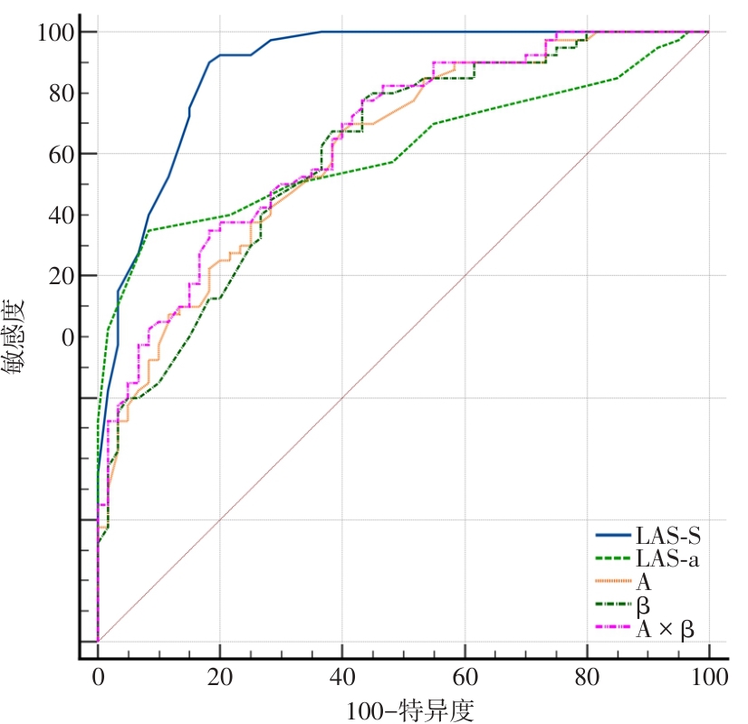
| 指标 | 最佳截断值 | AUC | 敏感度/% | 特异度/% | 95%CI |
|---|---|---|---|---|---|
| LAS-s | 28% | 0.939 | 95.2 | 81.2 | 0.886 ~ 0.973 |
| LAS-a | 12% | 0.806 | 67.5 | 91.6 | 0.734 ~ 0.878 |
| A | 8.2 dB | 0.804 | 68.7. | 75.0 | 0.748 ~ 0.882 |
| β | 1.04 dB/s | 0.802 | 83.7 | 61.7. | 0.729 ~ 0.866 |
| A×β | 7.2 dB2/s | 0.827 | 67.1 | 81.7 | 0.725 ~ 0.862 |
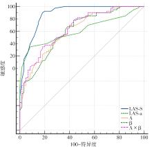
Fig.3
ROC curve analysis of MCE combined with 2D-STE"
| [1] |
UPADHYA B, ROSE G A, STACEY R B, et al.The role of echocardiography in the diagnosis of heart failure with preserved ejection fraction[J]. Heart Fail Rev, 2025,30(5):899-922. doi: 10.1007/s10741-025-10516-z .
doi: 10.1007/s10741-025-10516-z |
| [2] |
CAMPBELL P, RUTTEN F H, LEE M M, et al. Heart failure with preserved ejection fraction: Everything the clinician needs to know[J]. Lancet, 2024,403(10431):1083-1092. doi: 10.1016/S0140-6736(23)02756-3 .
doi: 10.1016/S0140-6736(23)02756-3 |
| [3] |
ALIZADEHASL A, MOKHAYERI M, SOHANI Z, et al. A Comprehensive Review of Two-Dimensional Speckle-Tracking Echocardiography in Assessing Right and Left Ventricular Function in Diabetic Patients[J]. Clin Cardiol, 2025,48(5):e70153. doi: 10.1002/clc.70153 .
doi: 10.1002/clc.70153 |
| [4] |
DE VET C M, NICHTING T J, FRANSEN A F, et al. Two-dimensional fetal speckle tracking; A learning curve study for offline strain analysis[J]. PLoS One, 2024,19(11):e0310307. doi: 10.1371/journal.pone.0310307 .
doi: 10.1371/journal.pone.0310307 |
| [5] |
CAPDEVILLE S, GHOLSON B A, LINDNER J R. Contrast Echocardiography for Assessing Myocardial Perfusion [J]. Curr Cardiol Rep, 2023,25(11):1581-1587. doi: 10.1007/s11886-023-01970-y .
doi: 10.1007/s11886-023-01970-y |
| [6] |
LI M, ZENG D, FEI H, et al. Automatic Myocardial Contrast Echocardiography Image Quality Assessment Using Deep Learning: Impact on Myocardial Perfusion Evaluation[J]. Ultrasound Med Biol, 2023,49(10):2247-2255. doi: 10.1016/j.ultrasmedbio .
doi: 10.1016/j.ultrasmedbio |
| [7] |
徐刚强,胡召锁,汪龙,等. 血清渗透压、N-端脑利钠肽前体、同型半胱氨酸在冠心病合并高血压患者发生急性心力衰竭早期诊断的应用价值[J]. 实用医学杂志,2026,42(6):1018-1023. doi: 10.3969/j.issn.1006-5725.2026.06.014
doi: 10.3969/j.issn.1006-5725.2026.06.014 |
| [8] |
伍鑫,丁戈琦,李禄丰,等. 血清可溶性生长刺激表达因子2,氨基末端B型脑钠肽前体及超声心动图参数与急性心肌梗死后心力衰竭患者心肌重塑的关系[J]. 实用医学杂志, 2025, 41(19):3034-3040. doi:10.3969/j.issn.1006-5725.2025.19.011 .
doi: 10.3969/j.issn.1006-5725.2025.19.011 |
| [9] |
YAO L, TA S, WANG J, et al. Myocardial perfusion improvement and mechanism after percutaneous intramyocardial septal radiofrequency ablation in obstructive hypertrophic cardiomyopathy: A study of myocardial contrast echocardiography[J]. Int J Cardiovasc Imaging, 2024,40(7):1483-1492. doi: 10.1007/s10554-024-03126-7 .
doi: 10.1007/s10554-024-03126-7 |
| [10] |
SUN Q, GÜVEN B, WAGG C S, et al.Mitochondrial fatty acid oxidation is the major source of cardiac adenosine triphosphate production in heart failure with preserved ejection fraction[J]. Cardiovasc Res. 2024, 120(4):360-371. doi: 10.1093/cvr/cvae006 .
doi: 10.1093/cvr/cvae006 |
| [11] |
BELMONTE M, FOÀ A, PAOLISSO P, et al. Coronary Physiopathology and Microcirculation Working Group of the Italian Society of Cardiology (SIC). Coronary microvascular dysfunction beyond the spectrum of chronic coronary syndromes[J]. Prog Cardiovasc Dis, 2024,87:73-82. doi: 10.1016/j.pcad.2024.10.006 .
doi: 10.1016/j.pcad.2024.10.006 |
| [12] |
SMATI H, SELLKE F W, BOURQUE J M, et al. Coronary Microvascular Dysfunction: A Guide for Clinicians[J]. Am J Med, 2024,137(9):810-817. doi: 10.1016/j.amjmed .
doi: 10.1016/j.amjmed |
| [13] |
INOUE K, OBOKATA M. Clinical utility of the left atrial strain analysis[J]. J Echocardiogr, 2025,23(3):145-155. doi: 10.1007/s12574-025-00695-x .
doi: 10.1007/s12574-025-00695-x |
| [14] |
KIM H, LEE H J, KIM I C, et al. Perfusion Abnormality in Adenosine Stress Myocardial Contrast Echocardiography in Intermediate-Risk Patients With Chest Pain[J]. J Clin Ultrasound, 2026,54(4):845-853. doi: 10.1002/jcu.70137 .
doi: 10.1002/jcu.70137 |
| [15] |
SEVERINO P, D'AMATO A, PROSPERI S, et al. Coronary microcirculation in myocardial ischemia: A genetic perspective[J]. J Mol Cell Cardiol, 2025,203:67-75. doi: 10.1016/j.yjmcc .
doi: 10.1016/j.yjmcc |
| [16] |
REN Z, XU F, WEI Z. Predictive value of NLR, PLR and MPVLR for recent major cardiovascular adverse events in elderly patients with heart failure[J]. Am J Transl Res, 2025,17(7):4964-4975. doi: 10.62347/XQNT2355 .
doi: 10.62347/XQNT2355 |
| [17] |
ROLDAN P, RAVI S, HODOVAN J, et al. Myocardial contrast echocardiography assessment of perfusion abnormalities in hypertrophic cardiomyopathy[J]. Cardiovasc Ultrasound, 2022, 20(1):23. doi: 10.1186/s12947-022-00293-2 .
doi: 10.1186/s12947-022-00293-2 |
| [18] |
CHUNYAO L, RUIHAN J, KAI L, et al. Quantitative Assessment of Myocardial Perfusion in Physiological and Pathological Hypertrophy Using Myocardial Contrast Echocardiography[J]. Clin Cardiol, 2026,49(5):e70285. doi: 10.1002/clc.70285 .
doi: 10.1002/clc.70285 |
| [19] |
GAO Y, LI B, MA Y, et al. Early detection of left atrial function alterations across the heart failure spectrum by feature tracking-cardiac magnetic resonance[J]. Eur Radiol, 2025,35(8):4786-4797. doi: 10.1007/s00330-025-11453-z .
doi: 10.1007/s00330-025-11453-z |
| [20] |
CHEN F, WENG W, YANG D, et al. Myocardial contrast echocardiography evaluation of coronary microvascular dysfunction to Predict MACEs in patients with heart failure with preserved ejection fraction follow-up [J]. BMC Cardiovasc Disord, 2024,24(1):496. doi: 10.1186/s12872-024-04173-7 .
doi: 10.1186/s12872-024-04173-7 |
| [21] |
YAN C, MA J, YAN T, et al. Research Progress in Imaging Evaluation of Myocardial Microcirculation[J]. Int J Gen Med, 2025, 18:6055-6061. doi: 10.2147/IJGM.S547970 .
doi: 10.2147/IJGM.S547970 |
| [22] |
MARKLEY R, DEL BUONO M G, MIHALICK V, et al. Abnormal left ventricular subendocardial perfusion and diastolic function in women with obesity and heart failure and preserved ejection fraction[J]. Int J Cardiovasc Imaging, 2023, 39(4):811-819. doi: 10.1007/s10554-022-02782-x .
doi: 10.1007/s10554-022-02782-x |
| [23] |
YANG Y, QIN D, LI C, et al. Prevalence and prognostic significance of reduced myocardial perfusion reserve in diabetic heart failure with preserved ejection fraction using quantitative perfusion cardiac magnetic resonance[J]. Eur Radiol, 2025, 35(9):5537-5550. doi: 10.1007/s00330-025-11474-8 .
doi: 10.1007/s00330-025-11474-8 |
| [24] |
THORP E B, FILIPP M. Contributions of Inflammation to Cardiometabolic Heart Failure with Preserved Ejection Fraction[J]. Annu Rev Pathol, 2025,20(1):143-167. doi: 10.1146/annurev-pathmechdis-111523-023405 .
doi: 10.1146/annurev-pathmechdis-111523-023405 |
| [25] |
ZHANG J, GUAN L, LI X, et al. Value of Myocardial Contrast Echocardiography in Detecting Coronary Microcirculatory Dysfunction in Ischemia With Non-obstructive Coronary Artery Disease[J]. Ultrasound Med Biol, 2023, 49(9):2089-2094. doi: 10.1016/j.ultrasmedbio .
doi: 10.1016/j.ultrasmedbio |
| [26] |
WAHYULAKSANA G, WEI L, VOORNEVELD J, et al. Assessment of Coronary Microcirculation with High Frame-Rate Contrast-Enhanced Echocardiography[J]. Ultrasound Med Biol, 2025,51(3):585-591. doi: 10.1016/j.ultrasmedbio .
doi: 10.1016/j.ultrasmedbio |
| Viewed | ||||||
|
Full text |
|
|||||
|
Abstract |
|
|||||
