

The Journal of Practical Medicine ›› 2026, Vol. 42 ›› Issue (5): 861-868.doi: 10.3969/j.issn.1006-5725.2026.05.018
• Treatise: Clinical Practice • Previous Articles
Liang CAO,Fang ZOU,Changhong ZHANG,Kailun XU,Jianqing ZHAO,Jingqi LI,Jianhua LIU( ),Zhanhong TANG
Received:2025-10-23
Online:2026-03-10
Published:2026-03-09
Contact:
Jianhua LIU
E-mail:15530396730@163.com
CLC Number:
Liang CAO,Fang ZOU,Changhong ZHANG,Kailun XU,Jianqing ZHAO,Jingqi LI,Jianhua LIU,Zhanhong TANG. Mechanism of CREG regulating PINK1/Parkin to promote mitophagy in sepsis-induced acute lung injury[J]. The Journal of Practical Medicine, 2026, 42(5): 861-868.
Tab.1
Gene primer sequences"
| 基因 | 方向 | 引物序列 |
|---|---|---|
| CREG | F | 5'-GAGGAAGAGAGGTGCAGGTG-3' |
| R | 5'-CATTGCTGTCCTCGACTGAA-3' | |
| Parkin | F | 5'-TGCAGCATTATTAGCCACTTCTT-3' |
| R | 5'-TTCGGCTATCATTAACTATCACAA-3' | |
| PINK1 | F | 5'-CACAATGAGCCAGGAGCTGGT-3' |
| R | 5'-GCTTGGGACCTCTCTTGGATTT-3' | |
| GAPDH | F | 5'-CTTTGGTATCGTGGAAGGACTC-3' |
| R | 5'-GTAGAGGCAGGGATGATGTTCT-3' |
Tab.2
Comparison of MH-S cell activity after treatment with different concentrations of LPS (490 nm, OD value)"
| LPS | 12 h | 24 h | 48 h |
|---|---|---|---|
| 1 μg/mL | 0.340 ± 0.07 | 0.590 ± 0.14a | 0.702 ± 0.18ab |
| 5 μg/mL | 0.592 ± 0.12① | 0.811 ± 0.15①a | 1.100 ± 0.24①ab |
| 10 μg/mL | 0.309 ± 0.10①② | 0.366 ± 0.17①②a | 0.449 ± 0.22①②ab |
| 15 μg/mL | 0.213 ± 0.11①③ | 0.238 ± 0.15①③a | 0.403 ± 0.24①③ab |
| F时间/组间/相互 | 19.630/38.170/1.791 | ||
| P时间/组间/相互 | < 0.001/< 0.001/0.116 | ||
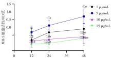
Fig.1
shows the cell viability of each group detected by CCK-8"

Tab.3
Effects of LPS on CREG protein and mRNA in MH-S cells"
| LPS | CREG蛋白 | CREG mRNA |
|---|---|---|
| 1 μg/mL | 0.36 ± 0.10 | 0.40 ± 0.07 |
| 5 μg/mL | 0.22 ± 0.06①② | 0.26 ± 0.05①② |
| 10 μg/mL | 0.41 ± 0.09①② | 0.50 ± 0.09①② |
| 15 μg/mL | 0.40 ± 0.11① | 0.52 ± 0.13① |
| F值 | 5.462 | 10.470 |
| P值 | 0.007 | 0.001 |
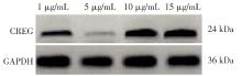
Fig.2
Effects of different concentrations of LPS on CREG protein"
Tab.4
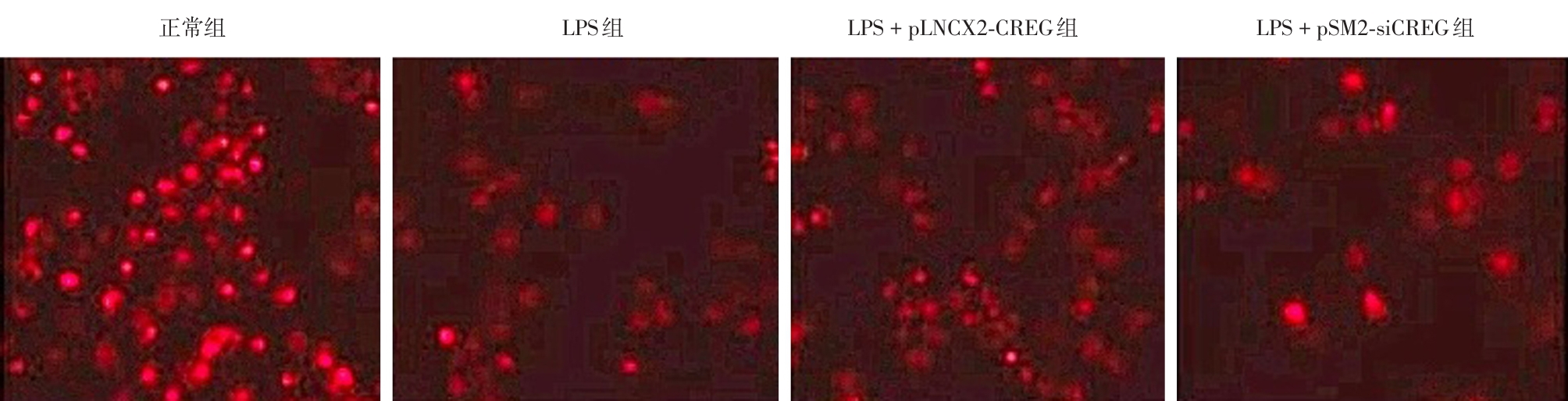
Comparison of lysosome quantities in each group"
| 组别 | 溶酶体数量 |
|---|---|
| 正常组 | 51.90 ± 3.70 |
| LPS组 | 40.28 ± 3.18? |
| LPS + pLNCX2-CREG组 | 46.81 ± 3.55?# |
| LPS + pSM2-siCREG组 | 32.76 ± 3.63?#? |
| F值 | 33.230 |
| P值 | < 0.001 |

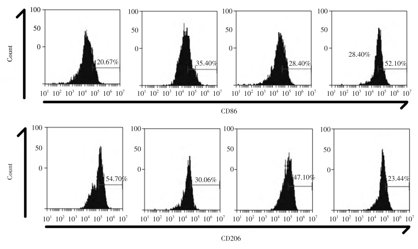
Fig.3
Number of lysosomes in each group (Red fluorescent probe for changing lysosomes, × 100)"
Tab.5
Comparison of CD86 and CD206 expressions in each group of cells"
| 组别 | CD86 | CD206 |
|---|---|---|
| 正常组 | 20.67 ± 3.50 | 54.70 ± 6.30 |
| LPS组 | 35.40 ± 4.18? | 30.06 ± 4.58? |
| LPS + pLNCX2-CREG组 | 28.40 ± 3.61?# | 47.10 ± 6.06?# |
| LPS + pSM2-siCREG组 | 52.10 ± 5.17?#? | 23.44 ± 4.80?#? |
| F值 | 62.000 | 42.120 |
| P值 | < 0.001 | < 0.001 |
Fig.4
shows the expression of CD86 and CD206 detected by flow cytometry"
Tab.6
Comparison of levels of IL-1β, IL-6, IL-10, CRP, ROS, MDA and SOD in each group"
| 组别 | IL-1β/(pg/mL) | IL-6/(pg/mL) | IL-10/(μg/L) | CRP/(ng/L) | ROS/(U/mg) | MDA/(nmol/mg) | SOD/(U/mg) |
|---|---|---|---|---|---|---|---|
| 正常组 | 201.60 ± 46.80 | 154.77 ± 30.15 | 3.84 ± 0.54 | 69.33 ± 10.54 | 6.21 ± 1.69 | 1.16 ± 0.15 | 4.18 ± 0.77 |
| LPS组 | 545.80 ± 100.02? | 510.79 ± 89.30? | 4.48 ± 0.68? | 196.51 ± 23.68? | 18.33 ± 3.50? | 3.31 ± 0.50? | 2.18 ± 0.33? |
| LPS + pLNCX2-CREG组 | 365.80 ± 80.44?# | 394.15 ± 81.10?# | 5.69 ± 0.71?# | 106.38 ± 22.40?# | 12.70 ± 2.96?# | 2.40 ± 0.49?# | 3.47 ± 0.48?# |
| LPS + pSM2-siCREG组 | 760.80 ± 120.69?#? | 626.33 ± 114.70?#? | 3.39 ± 0.50?#? | 287.11 ± 41.60?#? | 23.99 ± 4.12?#? | 4.28 ± 0.61?#? | 1.04 ± 0.22?#? |
| F值 | 41.700 | 34.060 | 15.880 | 132.800 | 34.100 | 47.920 | 47.380 |
| P值 | < 0.001 | < 0.001 | < 0.001 | < 0.001 | < 0.001 | < 0.001 | < 0.001 |
Tab.7
Comparison of CREG, Parkin, and PINK1 proteins and mRNA in each group"
| 组别 | CREG蛋白 | Parkin蛋白 | PINK1蛋白 | CREG mRNA | Parkin mRNA | PINK1 mRNA |
|---|---|---|---|---|---|---|
| 正常组 | 0.70 ± 0.09 | 0.65 ± 0.07 | 0.40 ± 0.06 | 0.84 ± 0.10 | 0.70 ± 0.11 | 0.54 ± 0.09 |
| LPS组 | 0.22 ± 0.06? | 0.36 ± 0.08? | 0.15 ± 0.03? | 0.26 ± 0.05? | 0.36 ± 0.08? | 0.28 ± 0.06? |
| LPS + pLNCX2-CREG组 | 0.50 ± 0.08?# | 0.52 ± 0.10?# | 0.29 ± 0.05?# | 0.61 ± 0.08?# | 0.51 ± 0.12?# | 0.40 ± 0.07?# |
| LPS + pSM2-siCREG组 | 0.14 ± 0.05?#? | 0.19 ± 0.03?#? | 0.08 ± 0.01?#? | 0.18 ± 0.03?#? | 0.21 ± 0.06?#? | 0.20 ± 0.04?#? |
| F值 | 77.510 | 42.880 | 69.180 | 115.000 | 28.870 | 28.970 |
| P值 | < 0.001 | < 0.001 | < 0.001 | < 0.001 | < 0.001 | < 0.001 |
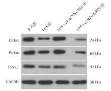
Fig.5
Electrophoresis images of CREG, Parkin and PINK1 proteins in each group"

| [1] |
周颖,蒋大军,田勇,等. 抑制TRAF6调节炎症和自噬改善脓毒症小鼠的心肌损伤和心功能[J]. 实用医学杂志,2024,40(5):608-614.doi:10.3969/j.issn.1006-5725.2024.05.004 .
doi: 10.3969/j.issn.1006-5725.2024.05.004 |
| [2] |
CHIU C, LEGRAND M. Epidemiology of sepsis and septic shock[J]. Curr Opin Anaesthesiol, 2021,34(2):71-76. doi: 10.1097/ACO.0000000000000958 .
doi: 10.1097/ACO.0000000000000958 |
| [3] |
HU Q, ZHANG S, YANG Y, et al. Extracellular vesicles in the pathogenesis and treatment of acute lung injury[J]. Mil Med Res, 2022,9(1):61. doi: 10.1186/s40779-022-00417-9 .
doi: 10.1186/s40779-022-00417-9 |
| [4] |
LI N, LIU B, XIONG R, et al. HDAC3 deficiency protects against acute lung injury by maintaining epithelial barrier integrity through preserving mitochondrial quality control[J]. Redox Biol, 2023, 63:102746. doi: 10.1016/j.redox.2023.102746 .
doi: 10.1016/j.redox.2023.102746 |
| [5] |
LI J, YANG D, LI Z, et al. PINK1/Parkin-mediated mitophagy in neurodegenerative diseases[J]. Ageing Res Rev,2023, 84:101817. doi: 10.1016/j.arr.2022.101817 .
doi: 10.1016/j.arr.2022.101817 |
| [6] |
CHEN H, LIN H, DONG B, et al. Hydrogen alleviates cell damage and acute lung injury in sepsis via PINK1/Parkin-mediated mitophagy[J]. Inflamm Res,2021,70(8):915-930. doi: 10.1007/s00011-021-01481-y .
doi: 10.1007/s00011-021-01481-y |
| [7] | 孙鸣宇,韩雅玲,闫承慧. E1A激活基因阻遏子通过稳定溶酶体抑制巨噬细胞炎症反应[C]. 北京:中国心脏大会 2014: 28. |
| [8] |
TIAN X, YAN C, HAN Y. Cellular Repressor of E1A-stimulated Genes, A New Potential Therapeutic Target for Atherosclerosis[J]. Curr Drug Targets, 2017,18(15):1800-1804. doi: 10.2174/1389450117666161026111250 .
doi: 10.2174/1389450117666161026111250 |
| [9] | 汪洁. MicroRNA-31对E1A激活基因阻遏子基因表达及血管平滑肌细胞表型转化的调控作用研究[D]. 西安:第四军医大学,2013. |
| [10] |
华天桢,汪海涛,魏淑婷,等. 脓毒症小鼠巨核细胞程序性死亡及对产血小板能力、凝血功能的影响[J]. 实用医学杂志,2025,41(15):2325-2335.doi:10.3969/j.issn.1006-5725. 2025. 15.006 .
doi: 10.3969/j.issn.1006-5725. 2025. 15.006 |
| [11] |
MOHSIN M, ZAKI A, TABASSUM G, et al. Urolithin-A supplementation alleviates sepsis-induced acute lung injury by reducing mitochondrial dysfunction and modulating macrophage polarization[J]. Mitochondrion,2025, 84:102047. doi: 10.1016/j.mito.2025.102047 .
doi: 10.1016/j.mito.2025.102047 |
| [12] |
WANG Z, WANG Z. The role of macrophages polarization in sepsis-induced acute lung injury[J]. Front Immunol, 2023, 14:1209438. doi: 10.3389/fimmu.2023.1209438 .
doi: 10.3389/fimmu.2023.1209438 |
| [13] |
JIAO Y, ZHANG T, ZHANG C, et al. Exosomal miR-30d-5p of neutrophils induces M1 macrophage polarization and primes macrophage pyroptosis in sepsis-related acute lung injury[J]. Crit Care, 2021,25(1):356. doi: 10.1186/s13054-021-03775-3 .
doi: 10.1186/s13054-021-03775-3 |
| [14] |
YE R, WEI Y, LI J, et al. Plasma-derived extracellular vesicles prime alveolar macrophages for autophagy and ferroptosis in sepsis-induced acute lung injury[J]. Mol Med, 2025, 31(1):40. doi: 10.1186/s10020-025-01111-x .
doi: 10.1186/s10020-025-01111-x |
| [15] |
WANG W B, LI J T, et al. Combination of pseudoephedrine and emodin ameliorates LPS-induced acute lung injury by regulating macrophage M1/M2 polarization through the VIP/cAMP/PKA pathway[J]. Chin Med,2022, 17(1):19. doi: 10.1186/s13020-021-00562-8 .
doi: 10.1186/s13020-021-00562-8 |
| [16] |
WANG C, MA C, GONG L, et al. Macrophage Polarization and Its Role in Liver Disease[J]. Front Immunol,2021, 12:803037. doi: 10.3389/fimmu.2021.803037 .
doi: 10.3389/fimmu.2021.803037 |
| [17] |
JIANG L, YANG D, ZHANG Z, et al. Elucidating the role of Rhodiola rosea L. in sepsis-induced acute lung injury via network pharmacology: Emphasis on inflammatory response, oxidative stress, and the PI3K-AKT pathway[J]. Pharm Biol, 2024,62(1):272-284. doi: 10.1080/13880209.2024.2319117 .
doi: 10.1080/13880209.2024.2319117 |
| [18] |
BIAN Z, CAI J, SHEN D F, et al. Cellular repressor of E1A-stimulated genes attenuates cardiac hypertrophy and fibrosis[J]. J Cell Mol Med, 2009,13(7):1302-1313. doi: 10.1111/j.1582-4934.2008.00633.x .
doi: 10.1111/j.1582-4934.2008.00633.x |
| [19] |
DUAN Y, LIU S, TAO J, et al. Cellular repressor of E1A stimulated genes enhances endothelial monolayer integrity[J]. Mol Biol Rep,2013, 40(6):3891-900. doi: 10.1007/s11033-012-2373-6 .
doi: 10.1007/s11033-012-2373-6 |
| [20] |
SUN M, TIAN X, LIU Y, et al. Cellular repressor of E1A-stimulated genes inhibits inflammation to decrease atherosclerosis in ApoE(-/-) mice[J]. J Mol Cell Cardiol, 2015, 86:32-41. doi: 10.1016/j.yjmcc.2015.07.001 .
doi: 10.1016/j.yjmcc.2015.07.001 |
| [21] |
宋钰. 参附注射液对脓毒症心肌损伤中线粒体自噬的影响[D]. 天津:天津中医药大学,2024.doi:10.27368/d.cnki.gtzyy. 2024.000058 .
doi: 10.27368/d.cnki.gtzyy. 2024.000058 |
| [22] |
叶莹莹. 溶酶体相关膜蛋白1在CXCL10-CXCR3轴调控巨噬细胞极化及肺损伤中的作用研究[D]. 蚌埠:蚌埠医学院,2023. doi:10.26925/d.cnki.gbbyc.2023.000330 .
doi: 10.26925/d.cnki.gbbyc.2023.000330 |
| [23] |
XIAO Z, LONG J, ZHANG J, et al. Administration of protopine prevents mitophagy and acute lung injury in sepsis[J]. Front Pharmacol, 2023, 14:1104185. doi: 10.3389/fphar. 2023. 1104185 .
doi: 10.3389/fphar. 2023. 1104185 |
| [24] |
孙鸣宇,闫承慧,田孝祥,等.CREG促进小鼠腹腔巨噬细胞溶酶体发生及溶酶体组织蛋白酶表达[J].现代生物医学进展,2016,16(8):1424-1427+1471.doi:10.13241/j.cnki.pmb. 2016. 08.006 .
doi: 10.13241/j.cnki.pmb. 2016. 08.006 |
| [25] |
ZHANG Y, XING D, LIU Y, et al. CREG1 attenuates intervertebral disc degeneration by alleviating nucleus pulposus cell pyroptosis via the PINK1/Parkin-related mitophagy pathway[J]. Int Immunopharmacol, 2025, 147:113974. doi: 10.1016/j.intimp. 2024.113974 .
doi: 10.1016/j.intimp. 2024.113974 |
| [1] | Weiyan LIAO,Qian ZHAO,Zeyu CHEN,Yue XUAN,Shengtao XIONG,Donglin LI,Xiao WANG. Mechanism of tauroursodeoxycholic acid improving mitochondrial autophagy and alleviating oxygen-glucose deprivation myocardial cell injury by regulating TBC1D15 [J]. The Journal of Practical Medicine, 2026, 42(2): 220-229. |
| [2] | Zhen ZHANG,Donghao WANG,Yang LV. Diagnostic value of skeletal muscle ultrasonography combined with bioelectrical impedance analysis in acquired weakness in the intensive care unit of patients with tumor sepsis [J]. The Journal of Practical Medicine, 2026, 42(1): 21-28. |
| [3] | Ziling ZENG,Xing WANG,Hongmei TANG,Zhibin WANG,Ning MA,Yuejiao LI,Xiaoyun WANG,Xiefang YUAN,Guofeng XU,Qiaoqiao WANG,Wen ZHANG,Jiayao DUAN,Yun ZHANG. House dust mite⁃induced autophagy affects airway epithelial barrier function through β⁃catenin⁃Snail signaling pathway [J]. The Journal of Practical Medicine, 2025, 41(9): 1309-1318. |
| [4] | Xu ZHOU,Ranran LU,Fangli REN,Xiaoyu PENG,Xinling. YANG. Effect of EGCG on MPTP⁃induced Parkinson′s model mice via autophagy⁃lysosomal pathway [J]. The Journal of Practical Medicine, 2025, 41(8): 1097-1104. |
| [5] | Songbai WU,Yao. DAI. Predictive value of multiligand proteoglycan⁃1, vascular endothelial⁃calmodulin and neuron⁃specific enolase in sepsis⁃associated encephalopathy in elderly patients [J]. The Journal of Practical Medicine, 2025, 41(8): 1111-1116. |
| [6] | Tong DUAN,Qi WU,Hui. LIU. Correlation between iron death⁃related gene expression level and myocardial dysfunction and prognosis in sepsis [J]. The Journal of Practical Medicine, 2025, 41(6): 846-851. |
| [7] | Zhizhou XIAO,Ying HUANG,Huawei. BIAN. To investigate the effect of aloperine on bone metabolism in osteoporotic mice based on autophagy and apoptosis mediated by Wnt/β⁃catenin signaling pathway [J]. The Journal of Practical Medicine, 2025, 41(4): 500-508. |
| [8] | Luoyi HE,Pinjing LIU,Jingming HUANG,Lixin GU,Zhanhong. TANG. To explore the distribution,risk factors and prognosis of capillary leakage syndrome in VA⁃ECMO patients [J]. The Journal of Practical Medicine, 2025, 41(4): 542-547. |
| [9] | Yuejiao LI,Nan LAN,Xing WANG,Hongmei TANG,Zhibin WANG,Yun ZHANG,Xiefang YUAN,Xiaoyun. WANG. Vitamin B12 enhances ZO⁃1 expression in HDM⁃treated human airway epithelial cells by down⁃regulating autophagy [J]. The Journal of Practical Medicine, 2025, 41(21): 3345-3351. |
| [10] | Tian TIAN,Ping CAO,Xuhong ZHANG,Xiaohong MA,Jingrui LI,Xueqin DING,Xiaoming. YANG. The role of GPNMB in hypoxia induced epithelial-mesenchymal transition in human chorionic trophoblast cells [J]. The Journal of Practical Medicine, 2025, 41(20): 3135-3144. |
| [11] | Jinyan GUO,Yuqing YOU,Ke CHEN,Fen PAN,Jiahui LAI,Sufang CHEN,Weifeng. YAO. Protective mechanism of sevoflurane on acute lung injury in sepsis by regulating the Wnt/β-catenin signaling pathway [J]. The Journal of Practical Medicine, 2025, 41(19): 2991-2999. |
| [12] | Tianzhen HUA,Haitao WANG,Shuting WEI,Sen TONG,Ning DONG,Xiaomei ZHU,Yongming YAO,Wei LIU. The programmed death of megakaryocytes and its impact on platelet-production copacity and coagulation function in mice with sepsis [J]. The Journal of Practical Medicine, 2025, 41(15): 2325-2335. |
| [13] | Yunfen TIAN,Bin WANG,Fangxiang ZHANG. Multi-target regulation of short-chain fatty acids in sepsis [J]. The Journal of Practical Medicine, 2025, 41(13): 2105-2110. |
| [14] | Liangxi LU,Haiwang LU,Wenjie WANG,Jun SHI,Zhimin HUANG,Bin BIN. To investigate the mechanism of mitochondrial autophagy regulating the expression of NLRP3 inflammasome in prostate tissue in rats with experimental autoimmune prostatitis [J]. The Journal of Practical Medicine, 2025, 41(12): 1816-1824. |
| [15] | Yuexian LYU,Xiu BI,Ying LIU,Shujing CUI,Lixin ZHAO,Ge GAO,Jianxia WANG,Juan LI,Jun LI. The efficacy of plasma gasdermin D C⁃terminal fragment in the early diagnosis of sepsis [J]. The Journal of Practical Medicine, 2025, 41(12): 1899-1906. |
| Viewed | ||||||
|
Full text |
|
|||||
|
Abstract |
|
|||||