

The Journal of Practical Medicine ›› 2025, Vol. 41 ›› Issue (12): 1816-1824.doi: 10.3969/j.issn.1006-5725.2025.12.007
• Basic Research • Previous Articles
Liangxi LU1,Haiwang LU2,Wenjie WANG3,Jun SHI3,Zhimin HUANG2,Bin BIN2( )
)
Received:2025-03-19
Online:2025-06-25
Published:2025-07-02
Contact:
Bin BIN
E-mail:billbinn@sina.com
CLC Number:
Liangxi LU,Haiwang LU,Wenjie WANG,Jun SHI,Zhimin HUANG,Bin BIN. To investigate the mechanism of mitochondrial autophagy regulating the expression of NLRP3 inflammasome in prostate tissue in rats with experimental autoimmune prostatitis[J]. The Journal of Practical Medicine, 2025, 41(12): 1816-1824.
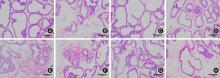
Fig.1
Histopathological analysis of prostate tissues was conducted for rats(HE,×100)"

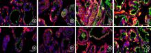
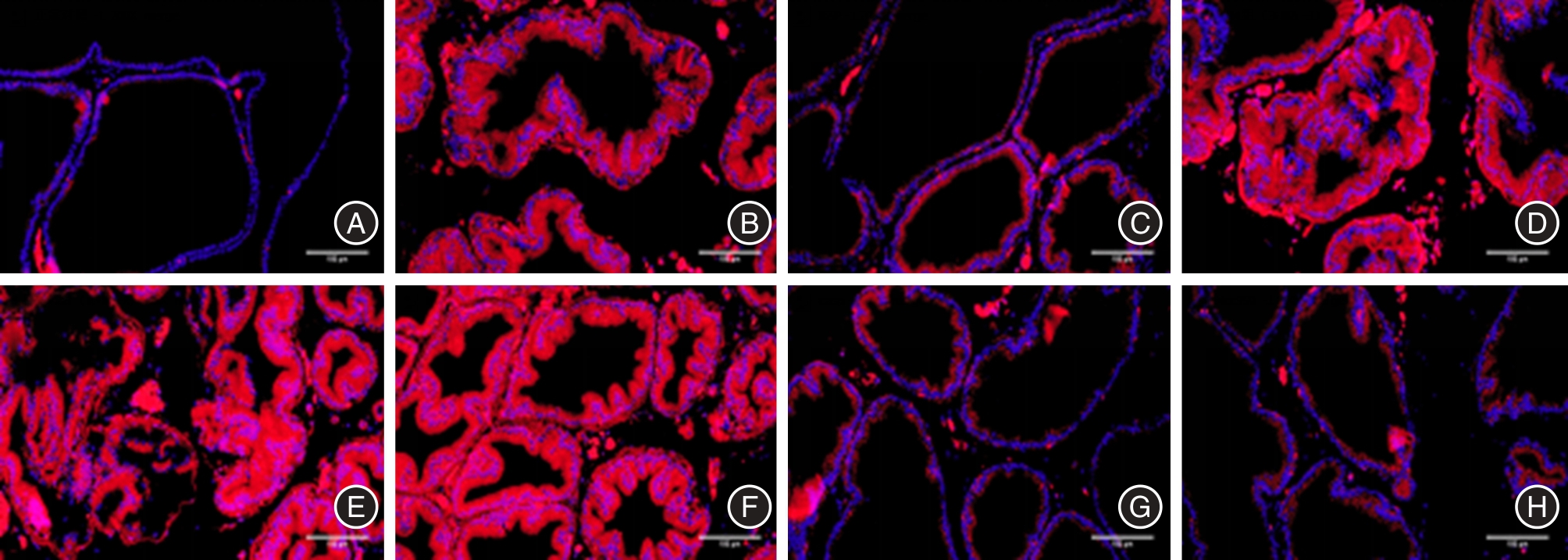
Fig.2
Fluorescence images of autophagic lysosomal assembly (LC3-Ⅱ and LAMP-1 co-expression) in prostate tissues of each group (scale 100 μm)"
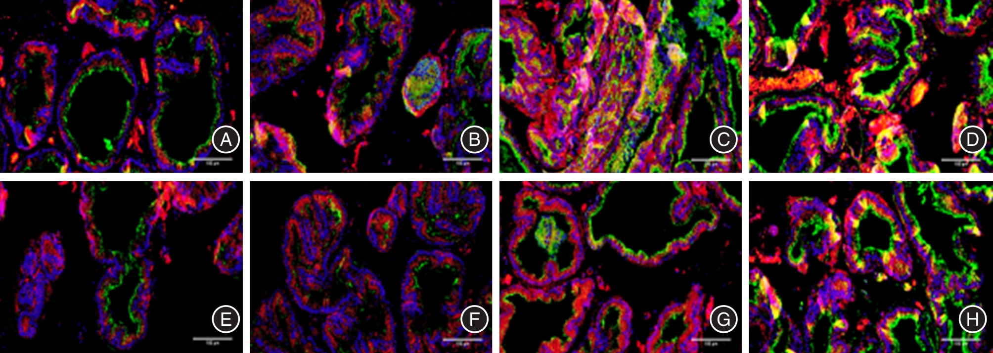
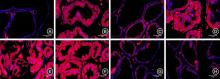
Fig.3
Fluorescence images of mtROS release level in prostate tissues of each group (scale 100 μm)"
Tab.1
Comparison of LC3-II/LAMP-1 co-expression, mtROS and mitochondrial membrane potential levels in prostate tissue(n = 3)"
| 组别 | LC3-II/LAMP-1共表达水平 | mtROS水平 | 线粒体膜电位 |
|---|---|---|---|
| N组 | 3.941 ± 0.255 | 2.686 ± 1.191 | 5.895 ± 0.189 |
| M组 | 7.006 ± 1.003△△ | 32.611 ± 3.192△△ | 0.575 ± 0.197△△ |
| RAP组 | 20.208 ± 1.248▲▲ | 10.916 ± 1.342▲▲ | 1.985 ± 0.271▲▲ |
| RAP+Mdivi-1组 | 13.771 ± 1.402▲▲ | 35.126 ± 1.271 | 0.738 ± 0.101 |
| 3-MA组 | 3.225 ± 0.633▲▲ | 42.046 ± 1.006▲▲ | 0.122 ± 0.021▲▲ |
| Mdivi-1组 | 3.583 ± 0.404▲▲ | 44.245 ± 3.362▲▲ | 0.217 ± 0.125▲▲ |
| Caspase1组 | 7.593 ± 0.703 | 7.871 ± 1.385▲▲ | 3.227 ± 0.162▲▲ |
| NLRP3组 | 13.735 ± 0.891▲▲ | 4.366 ± 0.864▲▲ | 4.779 ± 0.161▲▲ |
| F值 | 141.001 | 249.709 | 511.302 |
| P值 | < 0.001 | < 0.001 | < 0.001 |
Fig.4
Fluorescence images of mitochondrial membrane potential level in prostate tissues of each group (scale 100 μm)"
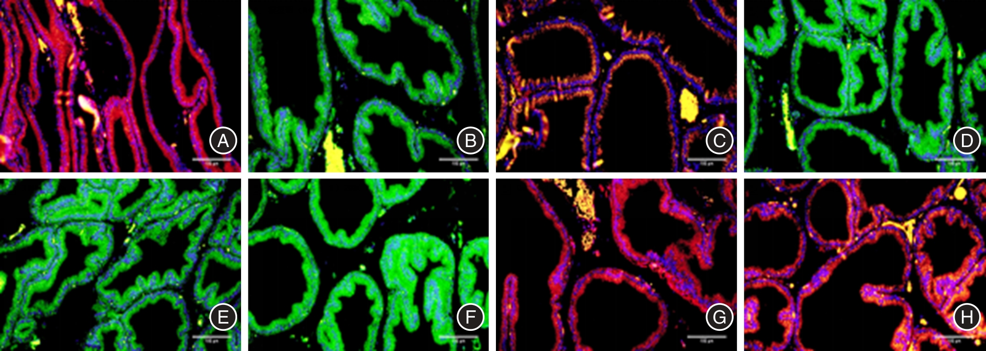

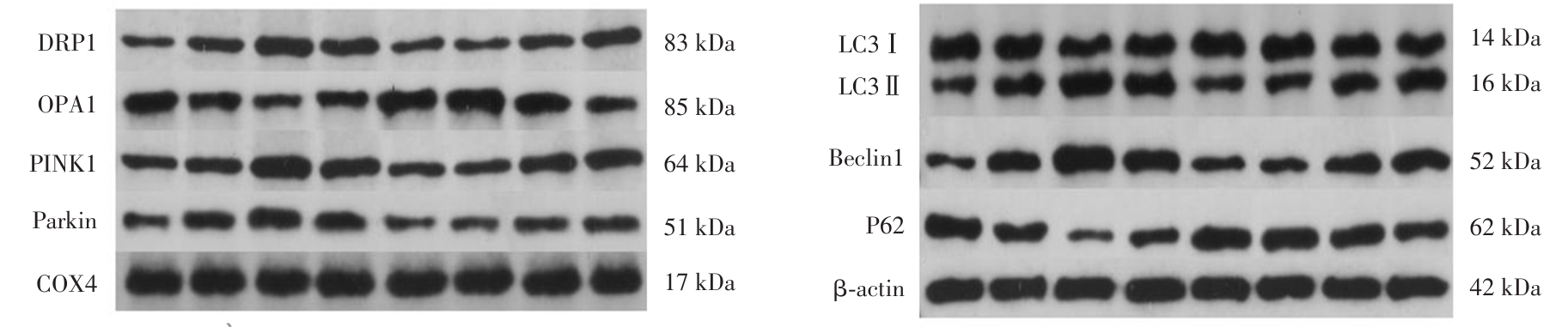
Fig.5
Proteins band diagrams of autophagy?related in prostate tissue of each group"
Tab.2
Comparison of protein expression of mitochondrial DRP1, OPA1, Parkin and PINK1 in prostate tissue(n = 5)"
| 组别 | DRP1 | OPA1 | Parkin | PINK1 |
|---|---|---|---|---|
| N组 | 0.206 ± 0.048 | 1.471 ± 0.157 | 0.304 ± 0.092 | 0.319 ± 0.116 |
| M组 | 0.441 ± 0.063△△ | 0.756 ± 0.121△△ | 0.571 ± 0.089△△ | 0.768 ± 0.101△△ |
| RAP组 | 1.114 ± 0.127▲▲ | 0.282 ± 0.075▲▲ | 1.287 ± 0.146▲▲ | 1.431 ± 0.157▲▲ |
| RAP+Mdivi-1组 | 0.705 ± 0.092▲▲ | 0.556 ± 0.090▲ | 0.651 ± 0.138 | 0.986 ± 0.099▲▲ |
| 3-MA组 | 0.222 ± 0.064▲▲ | 1.231 ± 0.155▲▲ | 0.229 ± 0.079▲▲ | 0.266 ± 0.076▲▲ |
| Mdivi-1组 | 0.184 ± 0.043▲▲ | 1.388 ± 0.18▲▲ | 0.232 ± 0.082▲▲ | 0.205 ± 0.054▲▲ |
| Caspase1组 | 0.349 ± 0.085 | 0.770 ± 0.10 | 0.397 ± 0.103▲ | 0.715 ± 0.088 |
| NLRP3组 | 0.595 ± 0.095▲▲ | 0.731 ± 0.126 | 0.746 ± 0.123▲ | 0.966 ± 0.137▲▲ |
| F值 | 76.512 | 51.333 | 52.693 | 77.849 |
| P值 | < 0.001 | < 0.001 | < 0.001 | < 0.001 |
Tab.3
Comparison of protein expression of LC3Ⅱ/LC3Ⅰ, Beclin1 and P62 in prostate tissue(n = 5)"
| 组别 | LC3Ⅱ/LC3Ⅰ | Beclin1 | P62 |
|---|---|---|---|
| N组 | 0.287 ± 0.102 | 0.161 ± 0.058 | 0.928 ± 0.085 |
| M组 | 0.851 ± 0.207△△ | 0.424 ± 0.092△△ | 0.585 ± 0.089△△ |
| RAP组 | 3.704 ± 0.336▲▲ | 1.227 ± 0.131▲▲ | 0.142 ± 0.037▲▲ |
| RAP+Mdivi-1组 | 2.003 ± 0.313▲▲ | 0.728 ± 0.102▲▲ | 0.313 ± 0.062▲▲ |
| 3-MA组 | 0.414 ± 0.163▲▲ | 0.188 ± 0.072▲▲ | 0.957 ± 0.082▲▲ |
| Mdivi-1组 | 0.393 ± 0.101▲▲ | 0.184 ± 0.041▲▲ | 0.961 ± 0.099▲▲ |
| Caspase1组 | 0.999 ± 0.233 | 0.446 ± 0.078 | 0.564 ± 0.051 |
| NLRP3组 | 1.783 ± 0.171▲▲ | 0.649 ± 0.094▲▲ | 0.410 ± 0.057▲▲ |
| F值 | 139.798 | 85.728 | 92.167 |
| P值 | < 0.001 | < 0.001 | < 0.001 |
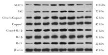
Fig.6
Proteins band diagrams of NLRP3 inflammasome related protein in prostate tissue of each group"

Tab.4
Comparison of NLRP3 inflammasome related protein expression in prostate tissue(n = 5)"
| 组别 | ASC | NLRP3 | Cleaved-Caspase1 | Cleaved-IL-1 | IL-18 |
|---|---|---|---|---|---|
| N组 | 0.052 ± 0.029 | 0.106 ± 0.069 | 0.220 ± 0.078 | 0.232 ± 0.071 | 0.163 ± 0.046 |
| M组 | 0.486 ± 0.088△△ | 0.887 ± 0.120△△ | 1.095 ± 0.089△△ | 1.305 ± 0.133△△ | 0.912 ± 0.108△△ |
| RAP组 | 0.213 ± 0.051▲▲ | 0.540 ± 0.114▲▲ | 0.612 ± 0.126▲▲ | 0.751 ± 0.122▲▲ | 0.493 ± 0.120▲▲ |
| RAP+Mdivi-1组 | 0.451 ± 0.093 | 0.891 ± 0.116 | 1.015 ± 0.062 | 1.219 ± 0.138 | 0.932 ± 0.084 |
| 3-MA组 | 0.621 ± 0.071▲▲ | 1.099 ± 0.100▲▲ | 1.371 ± 0.163▲▲ | 1.359 ± 0.235 | 1.212 ± 0.105▲▲ |
| Mdivi-1组 | 0.629 ± 0.083▲▲ | 1.185 ± 0.207▲▲ | 1.453 ± 0.162▲▲ | 1.382 ± 0.166 | 1.211 ± 0.096▲▲ |
| Caspase1组 | 0.124 ± 0.053▲▲ | 0.197 ± 0.088▲▲ | 0.410 ± 0.111▲▲ | 0.343 ± 0.122▲▲ | 0.291 ± 0.061▲▲ |
| NLRP3组 | 0.096 ± 0.043▲▲ | 0.198 ± 0.104▲▲ | 0.422 ± 0.064▲▲ | 0.256 ± 0.122▲▲ | 0.310 ± 0.071▲▲ |
| F值 | 62.268 | 64.154 | 84.951 | 63.448 | 112.284 |
| P值 | < 0.001 | < 0.001 | < 0.001 | < 0.001 | < 0.001 |
| 1 |
DEWITT-FOY M E, NICKEL J C, SHOSKES D A. Management of chronic prostatitis/chronic pelvic pain syndrome[J]. Eur Urol Focus,2019,5(1):2-4. doi:10.1016/j.euf.2018.08.027
doi: 10.1016/j.euf.2018.08.027 |
| 2 | 中华医学会男科学分会. 慢性前列腺炎/慢性盆腔疼痛综合征诊疗指南[J].中华男科学杂志,2022,28(6):544-559. |
| 3 |
FRANCO J V A, TURK T, JUNG J H, et al. Pharmacological interventions for treating chronic prostatitis/chronic pelvic pain syndrome: A Cochrane systematic review[J]. BJU Int,2020,125(4):490-496. doi:10.1111/bju.14988
doi: 10.1111/bju.14988 |
| 4 | 梁朝朝,慢性前列腺炎的研究进展——从基础到临床.中华腔镜泌尿外科杂志(电子版),2023,17(1): 96-96. |
| 5 |
KUZMENKO A V, GYAURGIEV T A, KUZMENKO V V, et al. The use of antioxidants in combination therapy of chronic prostatitis[J]. Urologiia,2024,1: 162-167. doi:10.18565/urology.2024.1.162-167
doi: 10.18565/urology.2024.1.162-167 |
| 6 |
LIU Y, ZHANG Y, ZHANG M, et al. Activated autophagy restored the impaired frequency and function of regulatory T cells in chronic prostatitis[J]. Prostate,2021,81(1):29-40. doi:10.1002/pros.24073
doi: 10.1002/pros.24073 |
| 7 |
SEOANE P I, LEE B, HOYLE C, et al. The NLRP3-inflammasome as a sensor of organelle dysfunction[J]. J Cell Biol,2020,219(12):e202006194. doi:10.1083/jcb.202006194
doi: 10.1083/jcb.202006194 |
| 8 |
陆佳伟,刘效谷,张文波. LncRNA及miRNA对NLRP3炎症小体信号的调控机制及其在相关疾病中的意义[J]. 实用医学杂志,2020,36(22):3149-3152. doi:10.3969/j.issn.1006-5725.2020.22.024
doi: 10.3969/j.issn.1006-5725.2020.22.024 |
| 9 |
何涛,黄华武,曾永龙,等. NLRP3炎症小体水平与老年慢性细菌性前列腺炎的相关性[J]. 中国老年学杂志,2021,41(9):1866-1869. doi:10.3969/j.issn.1005-9202.2021.09.024
doi: 10.3969/j.issn.1005-9202.2021.09.024 |
| 10 |
MA C G, LIU Y N, WANG H D. NLRP3 inflammasome in expressed prostatic secretions as a potential biomarker of chronic prostatitis/chronic pelvic pain syndrome[J]. Adv Clin Exp Med,2024,doi:10.17219/acem/192548. ahead of print
doi: 10.17219/acem/192548. ahead of print |
| 11 | 陆良喜,史宏,黄志敏,等. TLR4/NF-κB-NLRP3炎症小体信号通路在实验性自身免疫性前列腺炎大鼠中的作用机制[J]. 实用医学杂志,2025,41(6):800-805. |
| 12 | 中华中医药学会中药实验药理专业委员会. 慢性前列腺炎动物模型制备规范(草案)[J]. 中国实验方剂学杂志,2018,24(19):10-14. |
| 13 |
SANSON K V, DENG M, TING J P. The NLRP3 inflammasome: molecular activation and regulation to therapeutics[J]. Nat Rev Immunol,2019,19(8):477-489. doi:10.1038/s41577-019-0165-0
doi: 10.1038/s41577-019-0165-0 |
| 14 |
ZHANG L G, CHEN J, MENG J L, et al. Effect of alcohol on chronic pelvic pain and prostatic inflammation in a mouse model of experimental autoimmune prostatitis [J]. Prostate.2019,79(12):1439-1449. doi:10.1002/pros.23866
doi: 10.1002/pros.23866 |
| 15 |
LIU X, CHEN J, YUE S, et al. NLRP3-mediated IL-1β in regulating the imbalance between Th17 and Treg in experimental autoimmune prostatitis[J]. Sci Rep,2024,14(1):18829. doi:10.1038/s41598-024-69512-2
doi: 10.1038/s41598-024-69512-2 |
| 16 |
ZHANG F, MENG T, FENG R, et al. MIF aggravates experimental autoimmune prostatitis through activation of the NLRP3 inflammasome via the PI3K/AKT pathway[J]. Int Immunopharmacol,2024,141:112891. doi:10.1016/j.intimp.2024.112891
doi: 10.1016/j.intimp.2024.112891 |
| 17 |
ZHAO X, RUI FENG R, CHEN J, et al. 4-Octyl itaconate alleviates experimental autoimmune prostatitis by inhibiting the NLRP3 inflammasome- induced pyroptosis through activating Nrf2/HO-1 pathway[J]. Prostate,2024,84 (4):329-341. doi:10.1002/pros.24652
doi: 10.1002/pros.24652 |
| 18 |
CHEN L, WANG H, GE S, et al. IL-6/STAT3 pathway is involved in the regulation of autophagy in chronic non-bacterial prostatitis cells, and may be affected by the NLRP3 inflammasome[J].Ultrastruct Pathol,2021,45(4-5): 297-306. doi:10.1080/01913123.2021.1966149
doi: 10.1080/01913123.2021.1966149 |
| 19 |
LU J, SU Y, CHEN X, et al. Rapamycin-induced autophagy attenuates hormone-imbalance-induced chronic non-bacterial prostatitis in rats via the inhibition of NLRP3 inflamasome-mediated inflammation[J]. Mol Med Rep, 2019,19:221-230. doi:10.3892/mmr.2018.9683
doi: 10.3892/mmr.2018.9683 |
| 20 |
WU Z S, WANG H J, LEE W C, et al. Low-Energy Shock Wave Suppresses Prostatic Pain and Inflammation by Modulating Mitochondrial Dynamics Regulators on a Carrageenan-Induced Prostatitis Model in Rats[J]. Int J Mol Sci,2023,24(4): 3898. doi:10.3390/ijms24043898
doi: 10.3390/ijms24043898 |
| 21 |
NIU D, YUE S Y, WANG X, et al. High glucose intake exacerbates experimental autoimmune prostatitis through mitochondrial reactive oxygen species-dependent TGF-β activation-mediated Th17 differentiation[J]. Int Immunopharmacol,2024,130:111682. doi:10.1016/j.intimp.2024.111682
doi: 10.1016/j.intimp.2024.111682 |
| 22 |
LEE M J, CHO Y, HWANG Y, et al. Kaempferol Alleviates Mitochondrial Damage by Reducing Mitochondrial Reactive Oxygen Species Production in Lipopolysaccharide-Induced Prostate Organoids[J]. Foods,2023,12(20):3836. doi:10.3390/foods12203836
doi: 10.3390/foods12203836 |
| 23 |
ZHANG T, ZHAO J Y, LIU T M, et al. A novel mechanism for NLRP3 inflammasome activation[J]. Metabol Open,2022,13:100166. doi:10.1016/j.metop.2022.100166
doi: 10.1016/j.metop.2022.100166 |
| 24 |
ZHOU R, YAZDI A S, MENU P, et al. A role for mitochondria in NLRP3 inflammasome activation[J]. Nature,2011,469,221-225. doi:10.1038/nature09663
doi: 10.1038/nature09663 |
| 25 |
KIM M J, YOON J H, RYU J H. Mitophagy: A balance regulator of NLRP3 inflammasome activation[J]. BMB Rep,2016,49(10):529-535. doi:10.5483/bmbrep.2016.49.10.115
doi: 10.5483/bmbrep.2016.49.10.115 |
| 26 |
HSEU Y C, TSENG Y F, PANDEY S, et al. Coenzyme Q0 Inhibits NLRP3 Inflammasome Activation through Mitophagy Induction in LPS/ATP-Stimulated Macrophages[J]. Oxid Med Cell Longev,2022,2022:4266214. doi:10.1155/2022/4266214
doi: 10.1155/2022/4266214 |
| [1] | Lulin CHEN,Tingjie YANG,Meng SUN,Xin LI,Yiming GUO,Yuqing YANG,Yudong CAO,Wenzhe LI,Jiangshu YUAN,Honghui YANG. Association between coronary inflammation and malnutrition on prognosis in patients with coronary artery disease [J]. The Journal of Practical Medicine, 2025, 41(7): 1010-1017. |
| [2] | Liangxi LU,Hong SHI,Zhimin HUANG,Jie LU,Wenjie. WANG. Exploring mechanism of TLR4/NF⁃κB⁃NLRP3 inflammasome signaling pathway in experimental autoimmune prostatitis rats [J]. The Journal of Practical Medicine, 2025, 41(6): 800-805. |
| [3] | Chunxue KONG,Qiqi LIU,Liwei ZHANG,Chuansha WU,Longzhu XIONG,Guowei ZHANG,Minyue CAO,Ping LI,Ting ZHOU. Montelukast sodium inhibits airway inflammation through Phd2/Hif⁃1Α pathway in asthmatic mice [J]. The Journal of Practical Medicine, 2025, 41(5): 664-669. |
| [4] | Jiyue ZHU,Bo ZHANG,Yaru LI,Liuye. HUANG. The predictive value of the systemic inflammation response index for non⁃curative resection after endoscopic submucosal dissection for early colorectal cancer [J]. The Journal of Practical Medicine, 2025, 41(5): 716-723. |
| [5] | Rongxin LI,Li HUANG,Yueyang ZENG,Shuhui ZHANG,Yiran CHEN,Yuli LIU,Tieming. MA. Exploring the mechanism of electroacupuncture to improve cognitiveimpairment in alzheimer′s disease model rats based on NF⁃κB/NLRP3/Caspase⁃1 signaling pathway [J]. The Journal of Practical Medicine, 2025, 41(3): 322-329. |
| [6] | Xingwei WU,Jianying WANG,Chengxiao GUO,Ziyi LIU,Chao SUN,Fei. YU. The effect of remimazolam on modulating the ROS/RAGE/NF-κB signaling pathway in LPS-induced microglial inflammation [J]. The Journal of Practical Medicine, 2025, 41(2): 153-161. |
| [7] | Ying ZHOU,Dajun JIANG,Yong TIAN,Yongxiang GU,Guohui. YANG. Inhibition of TRAF6 ameliorates myocardial inflammatory injury and cardiac dysfunction via regulating cardiomyocyte inflammation in sepsis mice [J]. The Journal of Practical Medicine, 2024, 40(5): 608-614. |
| [8] | Xiaoyan WANG,Xiaoyi ZOU,Xiang ZHU,Ting WANG,Yetao QIANG,Siyuan ZHOU,Peng ZHANG,Ping ZHANG. Iron overload regulates atherosclerotic activity of foam cells induced by oxLDL [J]. The Journal of Practical Medicine, 2024, 40(3): 295-301. |
| [9] | Xiang JIA,Tianjie XU,Jiaxin FAN,Xiaoling GUO,Kainan LIU,Hui ZHANG,Yongsheng WANG,Qian. WANG. Metformin exerts a protective effect on articular cartilage in osteoarthritis rats by activating the SIRT1/p53 signaling pathway [J]. The Journal of Practical Medicine, 2024, 40(23): 3306-3316. |
| [10] | Zhe SHI,Xialin ZUO,Linhui PENG,Zhiwei LU,Kongping. LI. Effect of M1 microglial polarization on secondary damage in the thalamus after cerebral cortical infarction [J]. The Journal of Practical Medicine, 2024, 40(22): 3138-3145. |
| [11] | Shengnan JIANG,Wenbing ZHI,Jing CHEN,Tingting SUN,Zongren XU,Shuai LIU,Hong ZHANG,Ye LI,Yang LIU. Sophora davidii Hance leaves total alkaloids Attenuate Lipopolysaccharide-induced inflammatory response in RAW264.7 cell by Inhibiting the MAPK/NF-κB signaling pathway [J]. The Journal of Practical Medicine, 2024, 40(20): 2835-2840. |
| [12] | Guojiang YIN,Bixi LI,Pengxiao WEI,Yuqin YAN,Xiaoyang SONG,Kun LI. Effect of anterior quadratus lumborum block at the lateral supra⁃arcuate ligament on postoperative analgesia and inflammatory response in elderly patients undergoing robot⁃assisted radical prostatectomy [J]. The Journal of Practical Medicine, 2024, 40(2): 202-206. |
| [13] | Rui WANG,Duo LI,Zhao PENG,Lijun CUI,Xiang ZHANG,Kaili FAN,Wenyan. WU. Effect of endoscopic tumor resection by submucosal tunnel on recurrence in patients with submucosal tumors around cardia [J]. The Journal of Practical Medicine, 2024, 40(18): 2555-2560. |
| [14] | Xiaoqian WU,Xuexin LIU,Yulan GAO,Zhihua HAO,Leilei GUO,Qian. NIE. Application of immune inflammatory markers combined with magnetic controlled capsule internal examination in the diagnosis of gastric adenocarcinoma and precancerous lesions [J]. The Journal of Practical Medicine, 2024, 40(16): 2333-2339. |
| [15] | Qizhu FENG,Manman LU,Jie SUN,Jiaquan ZHANG,Sheng DING,Jian ZHANG,Qi. WANG. Novel markers of systemic inflammation in prediction of the early severity of acute pancreatitis [J]. The Journal of Practical Medicine, 2024, 40(14): 1963-1968. |
| Viewed | ||||||
|
Full text |
|
|||||
|
Abstract |
|
|||||