

The Journal of Practical Medicine ›› 2026, Vol. 42 ›› Issue (13): 2300-2308.doi: 10.3969/j.issn.1006-5725.2026.13.005
• Feature Reports:Cardiovascular diseases • Previous Articles
Qunxiang XIE( ),Xiaoling JIANG,Fang PAN
Received:2026-03-18
Online:2026-07-10
Published:2026-07-14
Contact:
Qunxiang XIE
E-mail:18933214330@163.com
CLC Number:
Qunxiang XIE,Xiaoling JIANG,Fang PAN. Application of SonoVCADheart for assessing fetal coronary artery morphology[J]. The Journal of Practical Medicine, 2026, 42(13): 2300-2308.
Fig.1
Two-dimensional ultrasound measurement of coronary artery inner diameter"
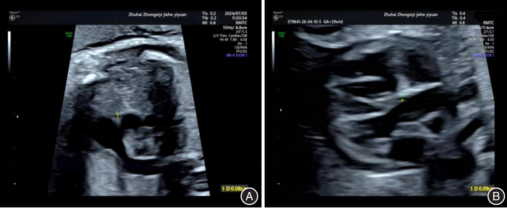
Fig.2
Basic imaging diagram of SonoVCADheart technology"
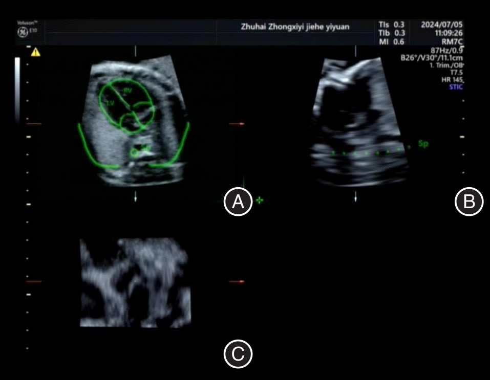
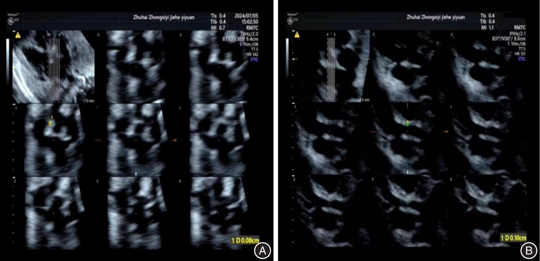
Fig.3
Measurement of coronary artery inner diameter using SonoVCADheart technology"
Tab.1
Comparison of diagnosis of coronary artery abnormalities between two examination methods"
| 产后超声心动图 | 胎儿超声心动图 | SonoVCADheart | ||
|---|---|---|---|---|
| 冠状动脉正常 | 冠状动脉异常 | 冠状动脉正常 | 冠状动脉异常 | |
| 合计 | 277 | 23 | 278 | 22 |
| 冠状动脉正常(n = 278) | 271(97.48) | 7(2.52) | 276(99.28) | 2(0.72) |
| 冠状动脉异常(n = 22) | 6(27.27) | 16(72.73) | 2(9.09) | 20(90.91) |
Tab.2
Diagnostic efficacy of two examination methods for coronary artery abnormalities"
| 检查方法 | 敏感度(95%CI) | 特异度(95%CI) | 准确率 | 阳性预测值 | 阴性预测值 |
|---|---|---|---|---|---|
| 胎儿超声心动图 | 72.73%(50.57% ~ 87.92%) | 97.48%(94.93% ~ 98.81%) | 287/300(95.67%) | 16/23(69.57%) | 271/277(97.83%) |
| SonoVCADheart | 90.91%(72.18% ~ 97.47%) | 99.28%(97.37% ~ 99.84%) | 296/300(98.67%) | 20/22(90.91%) | 276/278(99.28%) |
| χ2值 | - | - | 4.904 | - | 2.527 |
| P值 | 0.263 | 0.179 | 0.027 | 0.383 | 0.112 |
Tab.3
Comparison of correlation and consistency of coronary artery inner diameter measurements between two examination methods"
| 冠状动脉分支 | 胎儿超声心动图 | SonoVCADheart | r值 | ICC |
|---|---|---|---|---|
| 左冠状动脉主干 | 0.13 ± 0.03 | 0.12 ± 0.02 | 0.865 | 0.848 |
| 右冠状动脉 | 0.12 ± 0.03 | 0.13 ± 0.01 | 0.859 | 0.853 |
| [1] |
GÓMEZ-ARRIAGA P I, ESCRIBANO D, GÓMEZ-MONTES E, et al. Prenatal diagnosis of isolated coronary artery fistula: Systematic review, analysis of perinatal prognostic factors and case report[J]. J Matern Fetal Neonatal Med, 2023, 36(1): 2206938. doi:10.1080/14767058.2023.2206938 .
doi: 10.1080/14767058.2023.2206938 |
| [2] |
PISESKY A, NIELD L E, ROSENTHAL J, et al. Comparison of pre- and postnatally diagnosed coronary artery fistulae: Echocardiographic features and clinical outcomes[J]. J Am Soc Echocardiogr, 2022, 35(12): 1322-1335. doi:10.1016/j.echo.2022. 07.015 .
doi: 10.1016/j.echo.2022. 07.015 |
| [3] |
邹鹏, 孙善权, 刘琴, 等. 超声心动图对室间隔完整型肺动脉瓣闭锁和极重度肺动脉瓣狭窄胎儿的分型、评估及其出生后随访[J]. 中国循证儿科杂志, 2021, 16(3): 192-196. doi:10.3969/j.issn.1673-5501.2021.03.004 .
doi: 10.3969/j.issn.1673-5501.2021.03.004 |
| [4] |
KANAGALA S G, GUPTA V, DUNN G V, et al. Narrative review of anomalous origin of coronary arteries: Pathophysiology, management, and treatment[J]. Curr Cardiol Rev, 2023, 19(6): e300523217447. doi:10.2174/1573403x19666230530095341 .
doi: 10.2174/1573403x19666230530095341 |
| [5] |
谷孝艳, 王静怡, 范佳祺, 等. 胎儿超声心动图指征在先天性心脏病产前诊断中的应用价值[J]. 中国实用妇科与产科杂志, 2022, 38(10): 993-995.doi:10.19538/j.fk2022100109 .
doi: 10.19538/j.fk2022100109 |
| [6] |
沈珍珍, 沈双双, 汪晓敏, 等. 胎儿超声心动图联合母体血清标志物对胎儿先天性心脏病的诊断价值[J]. 中国妇幼保健, 2024, 39(1): 165-168.doi:10.19829/j.zgfybj.issn.1001-4411. 2024.01.043 .
doi: 10.19829/j.zgfybj.issn.1001-4411. 2024.01.043 |
| [7] |
励娜, 赵博文, 陈志鑫, 等. 超声心动图定量正常中晚孕期胎儿动脉导管Z-评分的研究[J]. 现代实用医学, 2023, 35(7): 906-908, 封3. doi:10.3969/j.issn.1671-0800.2023.07.020 .
doi: 10.3969/j.issn.1671-0800.2023.07.020 |
| [8] |
HU W Y, YU Y C, DAI L Y, et al. Reliability of sonography-based volume computer aided diagnosis in the normal fetal heart[J]. J Ultrasound Med, 2021, 40(5): 953-962. doi:10.1002/jum.15469 .
doi: 10.1002/jum.15469 |
| [9] |
UITTENBOGAARD L B, HAAK M C, VAN VUGT J M G. Feasibility of automated 3-dimensional fetal cardiac screening in routine ultrasound practice[J]. J Ultrasound Med, 2009, 28(7): 881-888. doi:10.7863/jum.2009.28.7.881 .
doi: 10.7863/jum.2009.28.7.881 |
| [10] |
ZENG Q H, CHEN S X, TIAN J. Application of fetal left and right ventricular outflow tract sections obtained by SonoVCADheart technology in the diagnosis of fetal cardiac malformations at 11-17 weeks of gestation [J]. Chin J Birth Health Hered,2023,31 (1):153-158. doi:10.13404/j.cnki.cjbhh.2023.01.028 .
doi: 10.13404/j.cnki.cjbhh.2023.01.028 |
| [11] |
戴丽雅, 赵博文, 余燕成, 等. 基于容积超声的胎儿心脏计算机辅助诊断技术在定量评估主动脉和肺动脉内径中的价值[J]. 中华超声影像学杂志, 2020, 29(1): 31-36. doi:10.3760/cma.j.issn.1004-4477.2020.01.006 .
doi: 10.3760/cma.j.issn.1004-4477.2020.01.006 |
| [12] |
WANG Y, WANG L, ZANG H, et al. Comparison of different ultrasonic screening methods and analysis of high risk factors for fetal cardiac malformation in second trimester of pregnancy[J]. Pediatr Cardiol, 2025, 46(4): 769-777. doi:10.1007/s00246-024-03525-6 .
doi: 10.1007/s00246-024-03525-6 |
| [13] |
MOON-GRADY A J, DONOFRIO M T, GELEHRTER S, et al. Guidelines and recommendations for performance of the fetal echocardiogram: An update from the American society of echocardiography[J]. J Am Soc Echocardiogr, 2023, 36(7): 679-723. doi:10.1016/j.echo.2023.04.014 .
doi: 10.1016/j.echo.2023.04.014 |
| [14] |
WANG A, DOAN T T, REDDY C, et al. Artificial intelligence in fetal and pediatric echocardiography[J]. Children, 2025, 12(1): 14. doi:10.3390/children12010014 .
doi: 10.3390/children12010014 |
| [15] |
GEMBICKI M, SCHARF J L, DRACOPOULOS C, et al. Maximal reduction of STIC acquisition time for volumetric assessment of the fetal heart-benefits and limitations of semiautomatic fetal intelligent navigation echocardiography (FINE) static mode[J]. J Clin Med, 2022, 11(14): 4062. doi:10.3390/jcm11144062 .
doi: 10.3390/jcm11144062 |
| [16] |
ZHAO L, WU Y, CHEN S, et al. Feasibility study on prenatal cardiac screening using four-dimensional ultrasound with spatiotemporal image correlation: A multicenter study[J]. PLoS One, 2016, 11(6): e0157477. doi:10.1371/journal.pone.0157477 .
doi: 10.1371/journal.pone.0157477 |
| [17] |
郭又问. 时空关联成像技术在胎儿心脏检查中的应用研究[D]. 长沙: 中南大学, 2010. .doi:10.7666/d.y1719460 .
doi: 10.7666/d.y1719460 |
| [18] |
陈晔, 唐颖, 朱善良, 等. 超声心动图诊断儿童冠状动脉异常起源于肺动脉[J]. 中国医学影像学杂志, 2023, 31(11): 1140-1144. doi:10.3969/j.issn.1005-5185.2023.11.003 .
doi: 10.3969/j.issn.1005-5185.2023.11.003 |
| [19] |
GARCIA-CANADILLA P, DE VRIES T, GONZALEZ-TENDE-RO A, et al. Structural coronary artery remodelling in the rabbit fetus as a result of intrauterine growth restriction[J]. PLoS One, 2019, 14(6): e0218192. doi:10.1371/journal.pone.0218192 .
doi: 10.1371/journal.pone.0218192 |
| [20] |
NIU K X, LI T G, YE J J, et al. Detection of fetal right coronary artery–right ventricle fistula with pulmonary valve atresia using HDlive Flow with spatiotemporal image correlation[J]. Quant Imaging Med Surg, 2025, 15(8): 7690-7696. doi:10.21037/qims-24-2214 .
doi: 10.21037/qims-24-2214 |
| [21] |
D′ALBERTI E, MAPPA I, RIZZO G. Accuracy of 3D ultrasound in the prenatal diagnosis of congenital heart diseases[M]//3D Ultrasound of Fetal Heart. Cham: Springer Nature Switzerland, 2025: 35-48. doi:10.1007/978-3-031-87175-7_3 .
doi: 10.1007/978-3-031-87175-7_3 |
| [22] |
HU X, SHEN J, HE C, et al. A retrospective analysis: CCTA vs. TTE in diagnosing coronary artery fistula[J]. Curr Med Imaging, 2025, 21: e15734056333289. doi:10.2174/0115734056333289 250311184106 .
doi: 10.2174/0115734056333289 250311184106 |
| [23] |
GONÇALVES L F. Four-dimensional ultrasound examination of the fetal heart using spatiotemporal image correlation (STIC)[M]//Fetal Cardiology. Boca Raton: CRC Press,2018:192-212. doi:10.1201/9781315117769-14 .
doi: 10.1201/9781315117769-14 |
| [24] |
CALVES L, LEE W, ESPINOZA J, et al. Examination of the fetal heart by four-dimensional (4D) ultrasound with spatio-temporal image correlation (STIC)[J]. Ultrasound Obstet Gynecol,2006,27 (3):336-348. doi:10.1002/uog.2707 .
doi: 10.1002/uog.2707 |
| [25] |
GINDES L, HEGESH J, WEISZ B, et al. Three and four dimensional ultrasound: a novel method for evaluating fetal cardiac anomalies [J]. Prenat Diagn,2009,29(7):645-653. doi:10.1002/pd.2279 .
doi: 10.1002/pd.2279 |
| [1] | Shouquan CHENG,Naifeng LIU,Ruoshui LI. Analysis of risk factors for aortic valve calcification in patients with coronary artery calcification and construction of a predictive model [J]. The Journal of Practical Medicine, 2026, 42(4): 668-676. |
| [2] | Yingxuan LI,Jun SHAO,Qian FENG,Tao CHEN,Jia LIU. Association of PON1-Q192R gene polymorphism with severity of coronary artery lesions in patients with coronary heart disease [J]. The Journal of Practical Medicine, 2026, 42(10): 1745-1751. |
| [3] | Guixian PAN,Sitao LI,Hu HAO,Wei LIU,Qiuping YANG,Xin XIAO,Yao. CAI. Pedigree analysis and prenatal diagnosis in a family with congenital ectopia lentis [J]. The Journal of Practical Medicine, 2025, 41(6): 824-828. |
| [4] | Yuqi ZHANG,Kesong ZHOU,Shiquan ZHANG,Lei TANG,Enxiu XIE,Hongquan LIAO,Tao. YANG. Correlation of fetal rectal ampulla abdominal diameter with gestational age and establishment of reference values in low⁃risk fetuses at 18 ~ 40 weeks of pregnancy [J]. The Journal of Practical Medicine, 2025, 41(6): 882-888. |
| [5] | Xiaoyu ZHANG,Yuanyuan PAN,Minghuan FU,Yun LIN,Yiran CHEN,Yu. PENG. Research progress of hybrid coronary revascularization in treating coronary atherosclerotic heart disease [J]. The Journal of Practical Medicine, 2025, 41(6): 916-920. |
| [6] | Gaokai HU,Ya'nan NIU,Yukang GONG,Yang HU,Ruixuan XU,Wenshan. GAO. Research progress on the application of deep learning in lumbar spine disease [J]. The Journal of Practical Medicine, 2025, 41(6): 921-928. |
| [7] | Xue HE,Huan MA,Cong LI,Yan WANG,Yu KUANG,Lei LIU,Honghua YU,Xiaohong. YANG. Retinal neurovascular characteristics and their correlation with depression severity in patients with coronary artery disease and depression [J]. The Journal of Practical Medicine, 2025, 41(4): 515-521. |
| [8] | Bin DENG,Ailian WANG,Boli CHENG,Jiahao CHEN,Yun HE,Chonghai. LIU. Expression of NLRP3 inflammatory vesicles, Cav-1, and S1P1 in children with Kawasaki disease and their association with coronary artery injury [J]. The Journal of Practical Medicine, 2025, 41(13): 2094-2099. |
| [9] | Chao SHENG,Zhijian. WANG. Early identification, standardized diagnosis and treatment of placenta accreta spectrum disorders [J]. The Journal of Practical Medicine, 2024, 40(21): 2965-2970. |
| [10] | Li′na LIU,Heming WU,Zhiyuan ZHENG,Shuxian HUANG,Lingna. SHE. Application of ultrasound evaluation of NT thickening and nasal bone dyscalcification combined with CMA in prenatal diagnosis of fetuses [J]. The Journal of Practical Medicine, 2024, 40(19): 2755-2759. |
| [11] | Sumeng WANG,Fangfang ZHANG,Liantao NIE,Kexin LUO,Shifeng. LI. The value of electrocardiographic parameters in assessment of severe coronary artery lesions [J]. The Journal of Practical Medicine, 2024, 40(17): 2465-2470. |
| [12] | Yapeng LIANG,Chaopu ZHANG,Hao ZHANG,Zhongqun. WANG. The relationship between the expression of serum ANGPTL8 and KLF2 and the degree of coronary artery disease and the occurrence of major adverse cardiac events in patients with acute myocardial infarction [J]. The Journal of Practical Medicine, 2024, 40(13): 1827-1832. |
| [13] | Jing XIE,Dong GUO,Shiqiang FENG,Yi ZHAO,Hong LI,Zhenhua. QU. Effect of different administration doses of dexmedetomidine on the circulatory system and stress response in patients undergoing coronary artery bypass grafting with noncorporeal circulation [J]. The Journal of Practical Medicine, 2024, 40(11): 1574-1579. |
| [14] | ZHANG Miaomiao, FANG Yuqin, TANG Junxing, WANG Chaohong, SUN Yuxiu, ZHU Jiansheng.. The variation nature of 16p13.11 microduplicates encompassing NDE1 gene in prenatal diagnosis [J]. The Journal of Practical Medicine, 2023, 39(14): 1825-1829. |
| [15] | ZHANG Miao⁃ miao, FANG Yuqin, LI Jingran, WANG Chaohong, SUN Yuxiu, ZHU Jiansheng. . Analysis of 85,525 cases of rare autosomal trisomy detected by noninvasive prenatal testing [J]. The Journal of Practical Medicine, 2023, 39(12): 1506-1511. |
| Viewed | ||||||
|
Full text |
|
|||||
|
Abstract |
|
|||||
