

The Journal of Practical Medicine ›› 2025, Vol. 41 ›› Issue (6): 896-903.doi: 10.3969/j.issn.1006-5725.2025.06.019
• Medical Examination and Clinical Diagnosis • Previous Articles
Minsui CAI,Qi CUI,Sujun DING,Xuejun. NI( )
)
Received:2024-11-19
Online:2025-03-25
Published:2025-03-31
Contact:
Xuejun. NI
E-mail:XuejunNixuejunni@163.com
CLC Number:
Minsui CAI,Qi CUI,Sujun DING,Xuejun. NI. Application value study of cervical shear wave elasticity imaging combined with cervical length and anterior cervical angle in assessing cervical function and predicting spontaneous preterm birth[J]. The Journal of Practical Medicine, 2025, 41(6): 896-903.
Tab.1
General information of the two groups of"
| 项目 | 足月组(n = 723) | 早产组(n = 63) | Z/t/χ2值 | P值 |
|---|---|---|---|---|
| 孕妇年龄[M(P25,P75 )]/岁 | 32(21,43) | 32(22,40) | 1.83 | 0.41 |
| 孕妇年龄≤ 35岁 | 670(92.7) | 57(90.3) | 0.31 | 0.74 |
| 孕妇年龄> 35岁 | 53(7.3) | 6(9.7) | 1.88 | 0.36 |
| 接受超声检查的时间 | ||||
| 妊娠龄19 ~ 23+6周 | 99(13.7) | 9(14.2) | 0.32 | 0.14 |
| 妊娠龄24 ~ 27+6周 | 562(77.7) | 47(74.6) | 0.31 | 0.58 |
| 妊娠龄28 ~ 33+6周 | 48(6.7) | 5(7.9) | 0.58 | 0.17 |
| 妊娠龄34 ~ 36+6周 | 14(1.9) | 2(3.2) | 0.45 | 0.36 |
| 初产妇 | 616(85.2) | 56(88.9) | 0.12 | 0.74 |
| 经产妇 | 107(14.8) | 7(11.1) | 0.22 | 0.71 |
| 流产史 | 129(17.8) | 15(23.8) | 1.79 | 0.02 |
| 早产史 | 5(0.7) | 23(36.5) | 121.18 | < 0.001 |

Fig.1
Schematic diagram of transvaginal ultrasound measurement of cervical length"
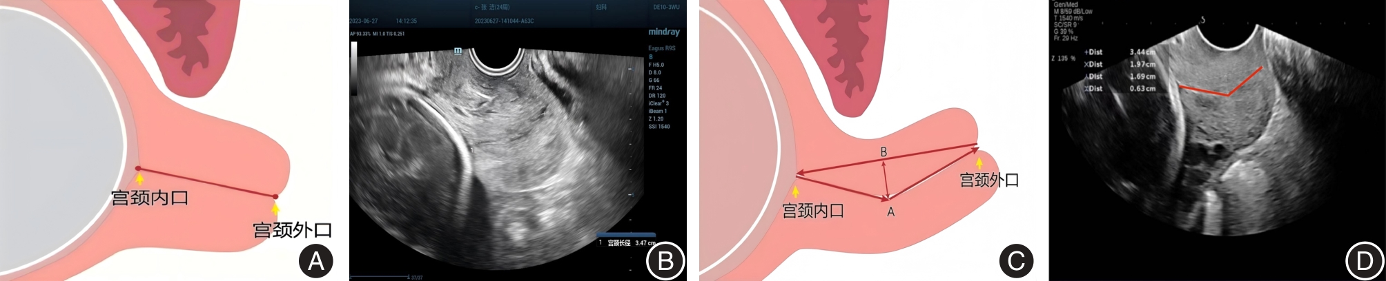
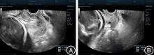
Fig.2
Schematic diagram of transvaginal ultrasoundmeasurement of the anterior cervical angle"
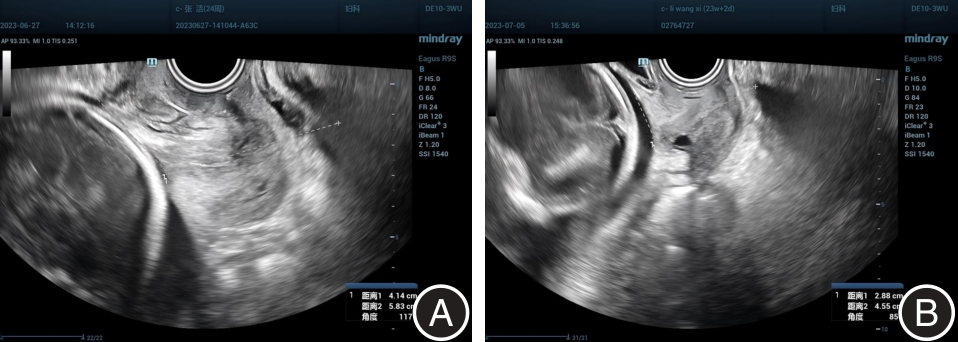
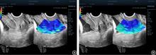
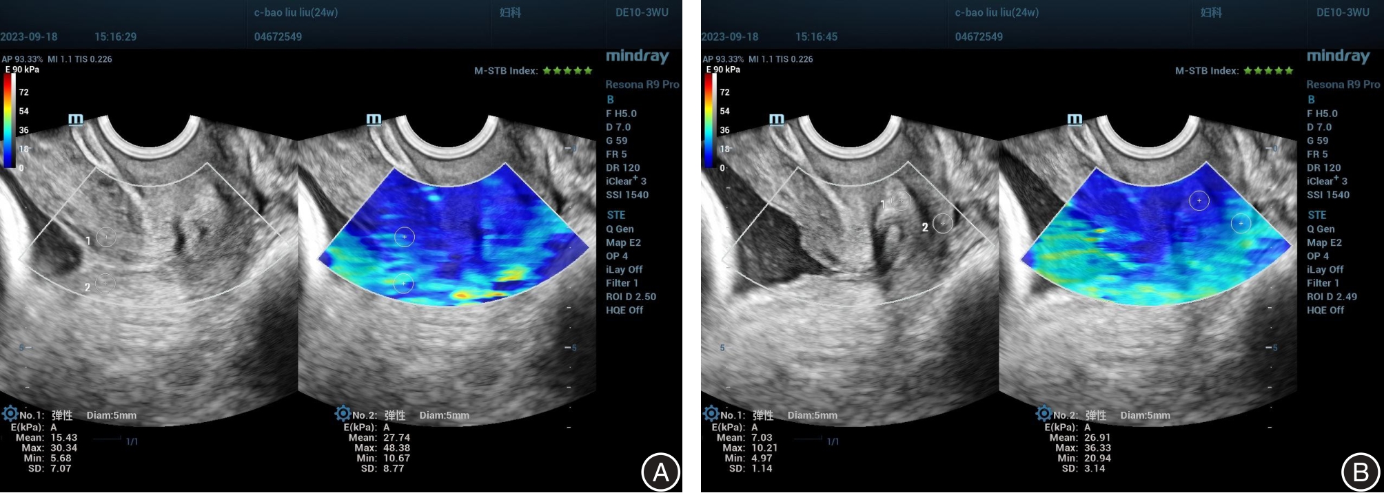
Fig.3
Schematic diagram of SWE of various parts of the cervix measured by transvaginal ultrasound"
Tab.2
Ultrasound examination results of the two groups of pregnant women"
| 项目 | 足月组(n = 723) | 早产组(n = 63) | t值 | P值 |
|---|---|---|---|---|
| CL/mm | 29.71 ± 10.85 | 25.42 ± 9.41 | 5.32 | < 0.001 |
| ACA/° | 99.42 ± 14 | 120.86 ± 10 | -13.87 | < 0.001 |
| SWE/kPa | ||||
| AE | 9.91 ± 2.14 | 6.47 ± 2.03 | 13.47 | < 0.001 |
| AI | 18.62 ± 6.97 | 10.98 ± 6.21 | 10.14 | < 0.001 |
| PI | 21.09 ± 5.04 | 11.32 ± 2.61 | 17.94 | < 0.001 |
| PE | 13.24 ± 5.04 | 8.16 ± 4.98 | 8.42 | < 0.001 |
Tab.3
Relationship between CL, ACA, SWE and gestational age"
| 项目 | 19 ~ 23+6周 | 24 ~ 27+6周 | 28 ~ 33+6周 | 34 ~ 36+6周 | t值 | P值 |
|---|---|---|---|---|---|---|
| 样本量 | 99 | 562 | 48 | 14 | ||
| CL/mm | 30.00 ± 10.22 | 29.91 ± 11.21 | 27.80 ± 9.42 | 27.20 ± 8.55 | 13.894 | 0.003 |
| ACA/° | 109.10 ± 5.23 | 100.16 ± 13.36 | 104.32 ± 10.17 | 93.16 ± 10.84 | 6.52 | 0.089 |
| SWE/kPa | ||||||
| AE | 10.08 ± 3.53 | 9.85 ± 2.12 | 5.36 ± 0.56 | 4.4 ± 0.12 | 17.29 | < 0.001 |
| AI | 16.94 ± 5.22 | 18.67 ± 6.86 | 10.51 ± 2.09 | 9.04 ± 0.09 | 14.96 | 0.002 |
| PI | 21.28 ± 6.67 | 20.99 ± 4.64 | 10.80 ± 3.02 | 10.59 ± 0.14 | 15.87 | 0.026 |
| PE | 13.13 ± 4.75 | 13.01 ± 5.22 | 7.39 ± 1.88 | 5.97 ± 0.11 | 13.69 | 0.001 |
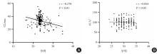
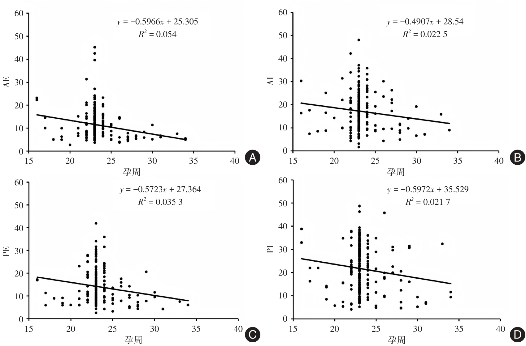
Fig.4
Correlation between CL, ACA and gestational age"
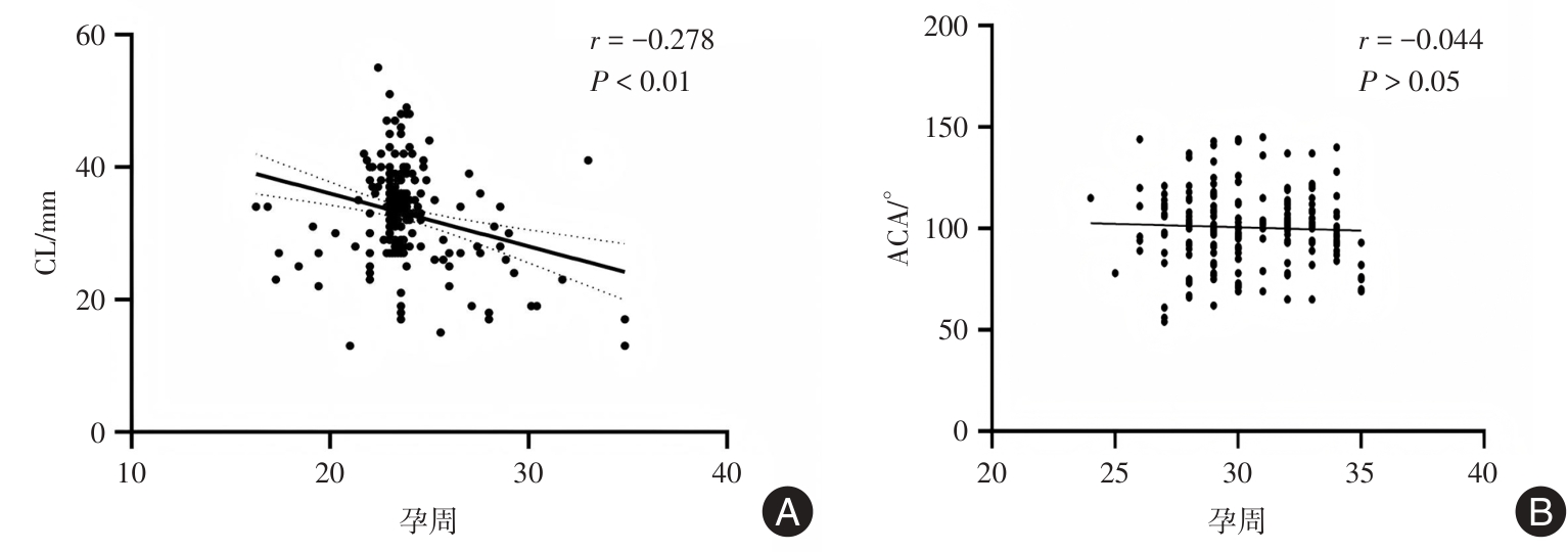
Fig.5
Relationship between SWE at different parts of the cervix and gestational age"
Tab.4
One-way ANOVA of ultrasound results"
| 危险因素 | 是否早产 | Z值 | P值 | |
|---|---|---|---|---|
| 否 | 是 | |||
| CL | 34.05(30.05,38.10) | 21.50(17.25,24.75) | -6.618 | < 0.001 |
| ACA | 95.21(76.13,118.46) | 132.36(118.97,142.02) | -4.445 | < 0.001 |
| AI SWE | 16.75(10.94,22.01) | 10.12(8.82,20.40) | -2.299 | 0.22 |
| PI SWE | 21.28(13.78,29.57) | 8.59(13.20,19.53) | -3.142 | 0.02 |
| AE SWE | 10.05(6.94,15.03) | 5.39(4.45,8.18) | -3.810 | < 0.001 |
| PE SWE | 12.71(8.37,19.47) | 7.01(5.92,12.84) | -3.358 | 0.01 |
Tab.5
Variable assignment table"
| 变量 | 分界值 | 赋值 |
|---|---|---|
| 既往早产史 | 0 = 无,1 = 有 | |
| 既往自然流产史 | 0 = 无,1 = 有 | |
| 宫颈SWE/kPa | 25th | 0 = 正常弹性区间 1 = 低弹性区间 2 = 高弹性区间 |
| CL/mm | 25 | 0 = <25 mm 1 = 25~35 mm 2 = >35 mm |
Tab.6
Multivariate logistic regression analysis of factors affecting premature birth"
| 自变量 | b | Sb | Wald χ2 | P值 | OR | 95%CI | |
|---|---|---|---|---|---|---|---|
| 下限 | 上限 | ||||||
| ACA ≥ 115.06° | 1.010 | 0.396 | 4.864 | 0.009 | 2.735 | 1.126 | 6.643 |
| PI ≤ 20.29 kPa | 1.066 | 0.462 | 5.339 | 0.010 | 2.904 | 1.177 | 7.165 |
| AE ≤ 5.78 kPa | 1.046 | 0.455 | 5.313 | 0.010 | 2.848 | 1.171 | 6.926 |
| PE ≤ 8.15 kPa | 1.058 | 0.461 | 5.283 | 0.011 | 2.881 | 1.170 | 7.095 |
| CL ≤ 25.0 mm | 1.038 | 0.488 | 4.537 | 0.022 | 2.823 | 1.087 | 7.331 |
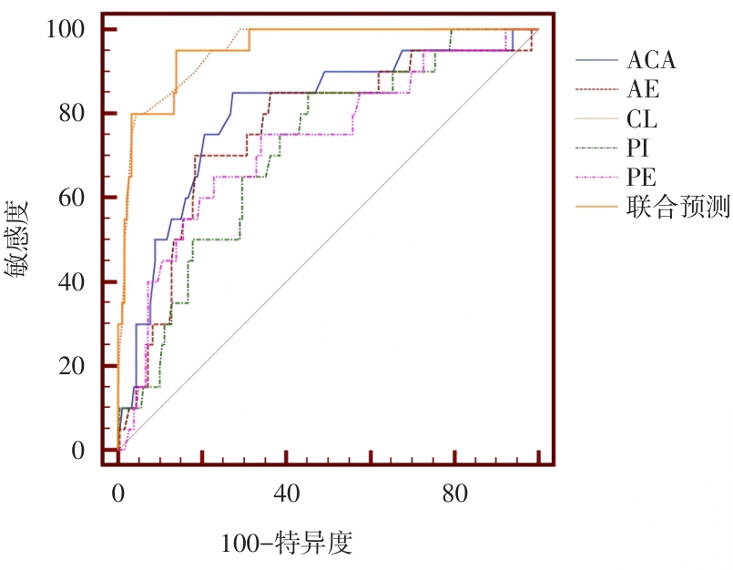
Tab.7
ROC curve analysis results"
| 参数 | AUC | 截断值 | 灵敏度/% | 特异度/% | P值 | 95%CI |
|---|---|---|---|---|---|---|
| ACA | 0.801 | 115.06° | 85.0 | 73.1 | 0.025 | 0.739 ~ 0.854 |
| PI | 0.714 | 20.29 kPa | 85.0 | 55.0 | < 0.01 | 0.646 ~ 0.776 |
| AE | 0.760 | 5.78 kPa | 70.0 | 81.7 | < 0.01 | 0.695 ~ 0.817 |
| PE | 0.729 | 8.15 kPa | 65.0 | 77.2 | < 0.01 | 0.662 ~ 0.789 |
| CL | 0.950 | 25.0 mm | 80.0 | 95.1 | < 0.01 | 0.911 ~ 0.976 |
| 联合预测 | 0.952 | / | 95.0 | 86.0 | < 0.01 | 0.913 ~ 0.977 |
Fig.6
ROC prediction curve"
| 1 | 中华医学会妇产科学分会产科学组. 早产的临床诊断与治疗指南(2014)[J]. 中国实用乡村医生杂志, 2015(12):3. |
| 2 |
TSIKOURAS P, BOTHOU A, GEREDE A, et al. Premature Birth, Management, Complication [M]. Global Women′s Health, 2021. doi:10.5772/intechopen.98324
doi: 10.5772/intechopen.98324 |
| 3 | 张文, 丁依玲. 子宫颈机能不全的可能病因 [J]. 中国实用妇科与产科杂志, 2023, 39(8): 780-783. |
| 4 | 孙丽洲, 杨娜娜. 子宫颈机能不全的热点问题与诊治策略 [J]. 中国实用妇科与产科杂志, 2023, 39(8): 769-771. |
| 5 | 孙笑, 时春艳. 子宫颈机能不全和短子宫颈的孕期超声检测和监管 [J]. 中国实用妇科与产科杂志, 2023, 39(8): 776-779. |
| 6 | 潘江皓, 孙健铭, 宋家明, 等. 急性下肢深静脉血栓形成患者行CDT联合ART治疗前后监测超声弹性成像参数变化对疗效的评估价值 [J]. 实用医学杂志, 2023, 39(18): 2362-2367. |
| 7 | 胡敏霞, 温德惠, 王立坤, 等. 高频超声与剪切波弹性成像在跟腱损伤术后评估中的应用研究 [J]. 实用医学杂志, 2022, 38(8): 1017-1021. |
| 8 |
SUTHASMALEE S, MOUNGMAITHONG S. Cervical shear wave elastography as a predictor of preterm delivery during 18⁃24 weeks of pregnancy [J]. J Obstet Gynaecol Res, 2019, 45(11): 2158-2168. doi:10.1111/jog.14094
doi: 10.1111/jog.14094 |
| 9 | 周汇恩, 陈婉明, 王梦蝶, 等. 基于临床-超声影像组学列线图模型预测孕晚期早产的价值 [J]. 实用医学杂志, 2023, 39(14): 1835-1841. |
| 10 |
QU Y, SUN H, CHEN J, et al. Systematic review and meta-analysis: Gray-scale ultrasound and shear wave elastography in the diagnosis of primipara pregnancy and delivery [J]. Ann Palliat Med, 2021, 10(11): 116641677-116611677. doi:10.21037/apm-21-2672
doi: 10.21037/apm-21-2672 |
| 11 |
FENG Q, CHAEMSAITHONG P, DUAN H, et al. Screening for spontaneous preterm birth by cervical length and shear-wave elastography in the first trimester of pregnancy [J]. Am J Obstet Gynecol, 2022, 227(3): 500.e1-500.e14. doi:10.1016/j.ajog.2022.04.014
doi: 10.1016/j.ajog.2022.04.014 |
| 12 |
SUN J, LI N, JIAN W, et al. Clinical application of cervical shear wave elastography in predicting the risk of preterm delivery in DCDA twin pregnancy [J]. BMC Pregnancy Childbirth, 2022, 22(1): 202. doi:10.1186/s12884-022-04526-0
doi: 10.1186/s12884-022-04526-0 |
| 13 | 中华医学会妇产科学分会产科学组. 早产临床防治指南(2024版) [J]. 中华妇产科杂志, 2024, 59(4): 257-269. |
| 14 | 唐修武, 倪观太. 宫颈机能不全的诊疗进展 [J]. 黑龙江医药, 2013, 26(4): 672-676. |
| 15 | 杨雅琴,李佳慧,赵莉萍,等.自发性早产预测的研究进展[J].中国生育健康杂志,2023,34(5):492-497. |
| 16 |
YANG X, DING Y, MEI J, et al. Second-trimester cervical shear wave elastography combined with cervical length for the prediction of spontaneous preterm birth [J]. Ultrasound Med Biol, 2022, 48(5): 820-829. doi:10.1016/j.ultrasmedbio.2022.01.003
doi: 10.1016/j.ultrasmedbio.2022.01.003 |
| 17 |
SHAO J, SHI G, QI Z, et al. Advancements in the application of ultrasound elastography in the cervix [J]. Ultrasound Med Biol, 2021, 47(8): 2048-2063. doi:10.1016/j.ultrasmedbio.2021.04.009
doi: 10.1016/j.ultrasmedbio.2021.04.009 |
| 18 |
AKBAS M, KOYUNCU F M. The Utility of Myometrial and Cervical Ultrasound Shear Wave Elastography in the Diagnosis of Ectopic Pregnancy [J]. Ultrasound Med Biol, 2020, 46(9): 2215-2221. doi:10.1016/j.ultrasmedbio.2020.04.035
doi: 10.1016/j.ultrasmedbio.2020.04.035 |
| 19 |
KUUSELA P, JACOBSSON B, HAGBERG H, et al. Second‐trimester transvaginal ultrasound measurement of cervical length for prediction of preterm birth: A blinded prospective multicentre diagnostic accuracy study [J]. BJOG, 2021, 128(2): 195-206. doi:10.1111/1471-0528.16519
doi: 10.1111/1471-0528.16519 |
| 20 | 丁苗苗, 栗河莉, 郭瑞霞. 经会阴超声测量宫颈长度及宫颈前角对孕期经阴道宫颈环扎术后早产的预测价值 [J]. 郑州大学学报(医学版), 2024, 59(1): 1-4. |
| 21 | 杜柳, 古裕均, 吴利红,等. E-Cervix弹性成像评估不同分娩史孕妇妊娠期宫颈组织差异的研究 [J]. 影像诊断与介入放射学, 2024, 33(1): 63-70. |
| 22 |
MYERS K M, FELTOVICH H, MAZZA E, et al. The mechanical role of the cervix in pregnancy [J]. J Biomech, 2015, 48(9): 1511-1523. doi:10.1016/j.jbiomech.2015.02.065
doi: 10.1016/j.jbiomech.2015.02.065 |
| 23 | 左喜芳, 杜雨峰, 杨静, 等. 双胎孕妇妊娠中晚期的子宫颈长度对早产的预测价值 [J]. 中华妇产科杂志, 2019, 54(5): 318-323. |
| 24 |
PACHULIIA O V, KHALENKO V V, SHENGELIIA M O, et al. Biomechanisms of cervical remodeling and current approaches to maturity assessment[J]. J Obstet Gynaecol, 2023, 72(1): 81-95. doi:10.17816/jowd114934
doi: 10.17816/jowd114934 |
| 25 | 李雪, 张弘. 宫颈机能不全环扎与早产 [J]. 中国实用妇科与产科杂志, 2018, 34(2): 146-150. |
| 26 | 柯宇莉, 魏玉梅. 超声监测宫颈长度预测单胎妊娠早产的研究进展 [J]. 现代妇产科进展, 2023, 32(8): 630-632. |
| [1] | Zhuolin YAO,Zhen LI,Caihua ZHANG,Mingmei ZHANG,Ruolin JIA,Yuan CAO,Yiping WANG,Yichun. GUAN. Analysis of clinical outcome of IVF/ICSI with dual⁃stimulation protocol in POSEIDON patients with low expected prognosis [J]. The Journal of Practical Medicine, 2025, 41(4): 580-588. |
| [2] | Ke XU,Mingzhe ZHANG,Mingyang WANG,Song. LIU. Correlation between plasma ADAMTS13 level, disease severity and prognosis in patients with endometriosis [J]. The Journal of Practical Medicine, 2025, 41(2): 238-243. |
| [3] | Hui WANG,Zhiheng CHEN,Li YANG,Yunhao LIANG,Huijiao WU,Yu JIANG,Shuai. LIU. Effects of short⁃term insemination and early rescue ICSI on pregnancy and neonatal outcomes [J]. The Journal of Practical Medicine, 2025, 41(2): 202-207. |
| [4] | Shuanghao ZHANG,Jie TAO,Zehua CAI,Xuerong RAN,Sisi WEI,Jinfeng PAN,Jinguo ZHAI. A study of the current status of female pelvic floor dysfunction patients′ knowledge of minimally invasive laser treatment of the reproductive tract and their intention to make treatment decisions [J]. The Journal of Practical Medicine, 2025, 41(1): 126-133. |
| [5] | Min ZHOU,Xiaoxiao ZHANG,Zhao WU. The effect of aspirin on frozen thawed embryo transfer in endometriosis with thin endometrium [J]. The Journal of Practical Medicine, 2025, 41(1): 95-99. |
| [6] | Ping HUANG,Kangyun LAN,Yanchun LIANG,Qing CHEN,Ying JIN,Guangyuan CHEN,Gang. NIU. Microbiome study of deep invasive endometriosis lesions [J]. The Journal of Practical Medicine, 2024, 40(21): 3023-3030. |
| [7] | Hongyan WANG,Wenying WANG,Guojing HE,Guoping CHEN,Lixia LUAN,Fangfang. XUE. Fractional CO2 laser combined with triamcinolone acetonide and econazole nitrate cream in the treatment of vulvar lichen sclerosus [J]. The Journal of Practical Medicine, 2024, 40(21): 3047-3053. |
| [8] | Jia ZHEN,Ziyuan ZHAO,Wei SHI,Zilu WANG,Zhiyong LIU,Li XU. The mechanism and treatment progress of endometrial receptivity change in adenomyosis [J]. The Journal of Practical Medicine, 2024, 40(20): 2941-2948. |
| [9] | Cheng LIU,Meiying SANG,Qunying FANG,Shun BAI,Meihong HU,Shengxia. ZHENG. Strategy for frozen⁃thawed cycle blastocyst transfer after hysteroscopic adhesiolysis [J]. The Journal of Practical Medicine, 2024, 40(17): 2390-2394. |
| [10] | Yuan LI,Huiling LIU,Dan WU,Baobao. LI. Research advances of virulence factors in recurrent vulvovaginal candidiasis [J]. The Journal of Practical Medicine, 2024, 40(16): 2347-2351. |
| [11] | Yanzhi WU,Yang. LIU. Research progress of m6A modification in benign ovarian related diseases [J]. The Journal of Practical Medicine, 2024, 40(16): 2352-2356. |
| [12] | Jie YANG,Chunhong LI,Jingfei HUANG,Zhiwei CHEN,Lin. LIU. The expression and clinical significance of hsa_circ_0003922 in endometrioid carcinoma [J]. The Journal of Practical Medicine, 2024, 40(14): 1975-1980. |
| [13] | Wenxin LI,Minjun LU,Li LIN,Yueqin LIU,Xiaolan. ZHU. circRAF1 regulates the proliferation and apoptosis of human ovarian granulosa cells [J]. The Journal of Practical Medicine, 2024, 40(7): 910-917. |
| [14] | Zhixia TANG,Shuangying MA,Ying ZHAGN,Jiajia SHENG,Juan LI,Jingjing HE,Henghua XUAN,Mingyun. HONG. Role of complement C3 in early predicting pregnancy outcomes of frozen⁃thawed embryo transfer [J]. The Journal of Practical Medicine, 2024, 40(7): 924-929. |
| [15] | Huaming ZHANG,Wanshan HE,Yun HAN,Guanqiao CHEN,Bin CHEN,Zhifu WEI,Hengying WU,Bin. WEN. Screening of gene differential expression of adenosine deaminase RNA specific 1 in cervical cancer cells based on transcriptome sequencing technology [J]. The Journal of Practical Medicine, 2023, 39(24): 3169-3174. |
| Viewed | ||||||
|
Full text |
|
|||||
|
Abstract |
|
|||||