

The Journal of Practical Medicine ›› 2025, Vol. 41 ›› Issue (4): 471-477.doi: 10.3969/j.issn.1006-5725.2025.04.003
• Basic Research • Previous Articles
Hui FANG1,Yiting YUAN1,Yongchun ZHANG1,Shanshan REN1,Lulu CHEN1,Wei LIAO1,Ai. TIAN1,2( )
)
Received:2024-10-11
Online:2025-02-25
Published:2025-02-28
Contact:
Ai. TIAN
E-mail:tianaident@foxmail.com
CLC Number:
Hui FANG,Yiting YUAN,Yongchun ZHANG,Shanshan REN,Lulu CHEN,Wei LIAO,Ai. TIAN. Regulation of PU.1 on apoptosis resistance of aging macrophages stimulated by Porphyromonas gingivalis lipopolysaccharide[J]. The Journal of Practical Medicine, 2025, 41(4): 471-477.
Tab.1
primer sequence"
| 基因 | 引物序列(5’-3’) |
|---|---|
| GAPDH | F:GGTTGTCTCCTGCGACTTCA R:TGGTCCAGGGTTTCTTACTCC |
| IL?6 | F:CTTCTTGGGACTGATGCTGGT R:AGGTCTGTTGGGAGTGGTATCC |
| IL?1β | F:AGCTTCAGGCAGGCAGTATC R:AAGGTCCACGGGAAAGACAC |
| TNF?α | F:ATGAGCACAGAAAGCATGATC R:TACAGGCTTGTCACTCGAATT |
| PU.1 | F:TACCTTCCAGTTCTCGTCCAAG R: GACTTTCTTCACCTCGCCTGT |
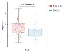
Fig.1
In GEO database, the expression of PU.1 increased in periodontitis gingival tissue"
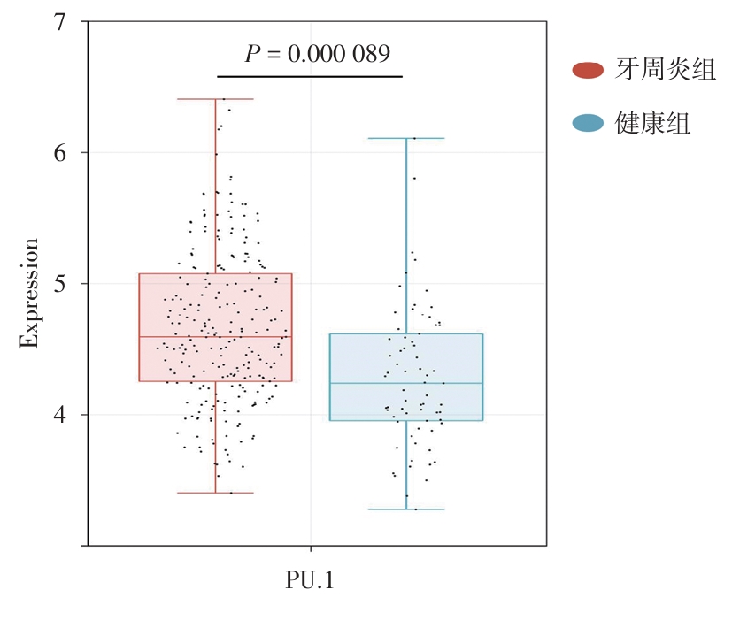
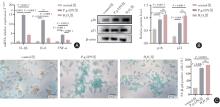
Fig.2
Macrophage senescence was induced by H2O2 and P.g-LPS"
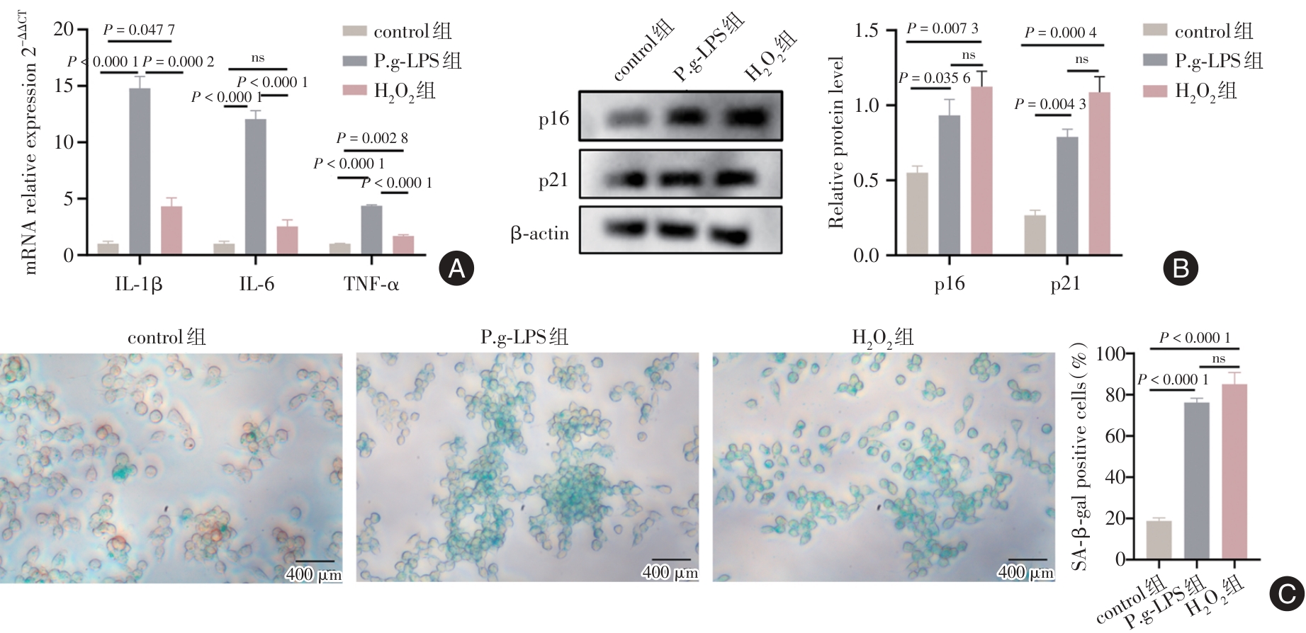
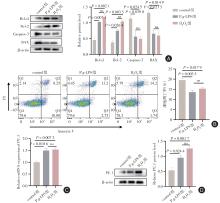
Fig.3
Apoptotic resistance was observed in senescent macrophages along with increased expression of PU.1"
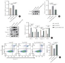
Fig.4
Inhibition of PU.1 promoted macrophage apoptosis"
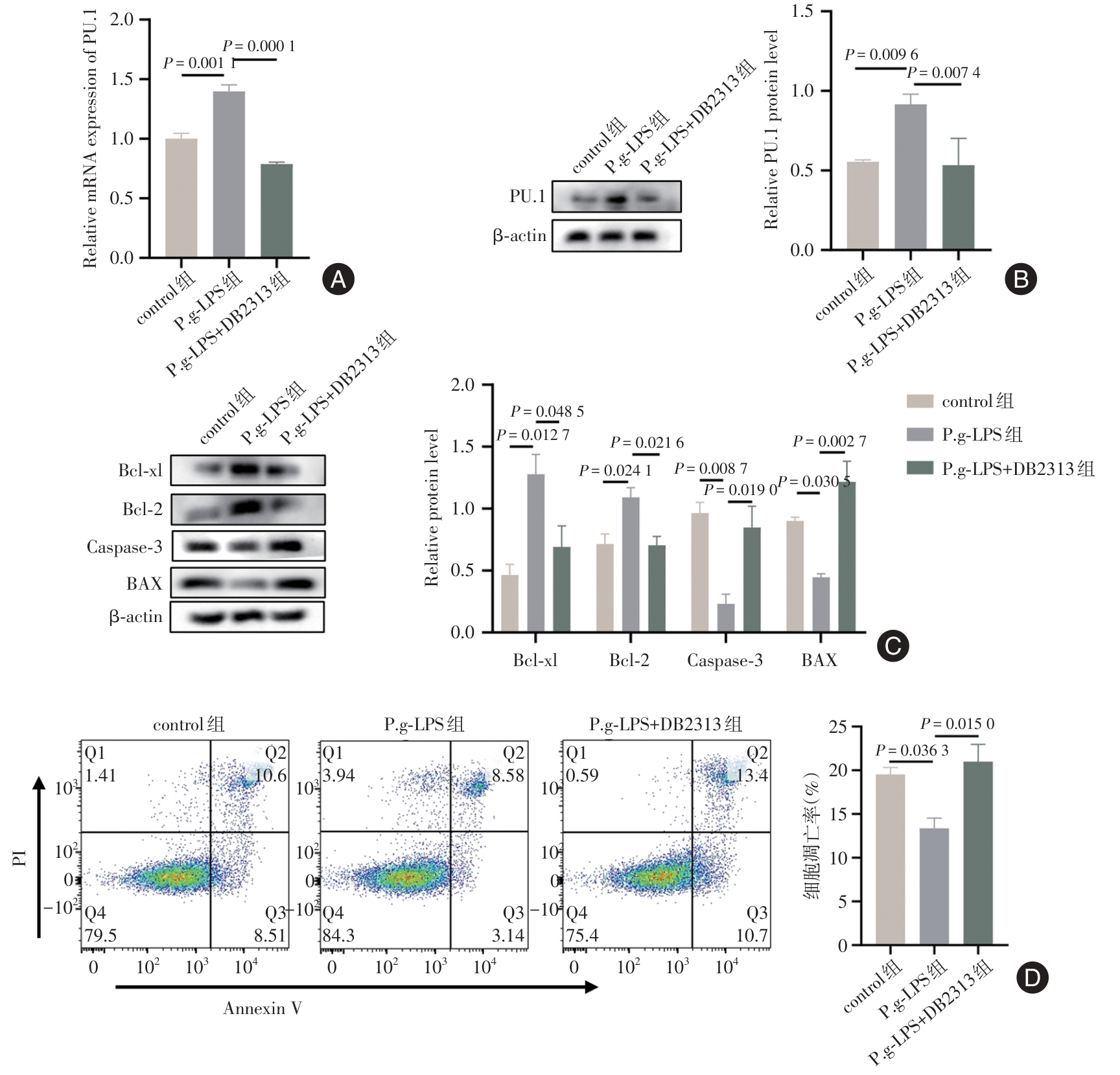
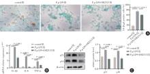
Fig.5
Inhibition of PU.1 can improve the senescence of macrophages"

| 1 |
HUANG X, XIE M, XIE Y, et al. The roles of osteocytes in alveolar bone destruction in periodontitis [J]. J Transl Med, 2020, 18(1): 479. doi:10.1186/s12967-020-02664-7
doi: 10.1186/s12967-020-02664-7 |
| 2 | 王兴. 第四次全国口腔流行病学调查报告 [M]. 北京: 人民卫生出版社, 2018. |
| 3 |
MARTÍNEZ-GARCÍA M, HERNÁNDEZ-LEMUS E. Periodontal Inflammation and Systemic Diseases: An Overview [J]. Frontiers in Physiology, 2021, 12: 709438. doi:10.3389/fphys.2021.709438
doi: 10.3389/fphys.2021.709438 |
| 4 |
YU N, VAN DYKE T E. Periodontitis: A host mediated disruption of microbial homeostasis [J]. Curr Oral Health Rep, 2020, 7(1): 3-11. doi:10.1007/s40496-020-00256-4
doi: 10.1007/s40496-020-00256-4 |
| 5 | 周征,齐霞,杨冬茹. 牙周炎的宿主反应调节治疗[J]. 口腔疾病防治, 2019, 27(11): 681-688. |
| 6 |
ZHANG P, WANG Q, NIE L, et al. Hyperglycemia-induced inflamm-aging accelerates gingival senescence via NLRC4 phosphorylation [J]. J Biol Chem, 2019, 294(49): 18807-18819. doi:10.1074/jbc.ra119.010648
doi: 10.1074/jbc.ra119.010648 |
| 7 |
WANG Q, NIE L, ZHAO P, et al. Diabetes fuels periodontal lesions via GLUT1-driven macrophage inflammaging [J]. Int J Oral Sci, 2021, 13(1): 11. doi:10.1038/s41368-021-00116-6
doi: 10.1038/s41368-021-00116-6 |
| 8 |
LUCAS V, CAVADAS C, ALEXANDRA AVELEIRA C. Cellular Senescence: From Mechanisms to Current Biomarkers and Senotherapies [J]. Pharmacol Rev, 2023, 75(4): 675-713. doi:10.1124/pharmrev.122.000622
doi: 10.1124/pharmrev.122.000622 |
| 9 | 安靖雯,冯俊云,饶磊,等. 细胞衰老与瘢痕纤维化的关系研究进展[J]. 实用医学杂志,2024,40(12):1749-1754. |
| 10 |
HU L, LI H, ZI M, et al. Why Senescent Cells Are Resistant to Apoptosis: An Insight for Senolytic Development [J]. Front Cell Dev Biol, 2022, 10: 822816. doi:10.3389/fcell.2022.822816
doi: 10.3389/fcell.2022.822816 |
| 11 |
SHEN M, FU J, ZHANG Y, et al. A novel senolytic drug for pulmonary fibrosis: BTSA1 targets apoptosis of senescent myofibroblasts by activating BAX [J]. Aging Cell, 2024,23(9): e14229. doi:10.1111/acel.14229
doi: 10.1111/acel.14229 |
| 12 |
CHEN J, CHEN K H, WANG L M, et al. Decoy receptor 2 mediates the apoptosis-resistant phenotype of senescent renal tubular cells and accelerates renal fibrosis in diabetic nephropathy [J]. Cell Death Dis, 2022, 13(6): 522. doi:10.1038/s41419-022-04972-w
doi: 10.1038/s41419-022-04972-w |
| 13 |
PRATTICHIZZO F, DE NIGRIS V, MANCUSO E,et al. Short-term sustained hyperglycaemia fosters an archetypal senescence-associated secretory phenotype in endothelial cells and macrophages [J]. Redox Biol, 2018, 15: 170-181. doi:10.1016/j.redox.2017.12.001
doi: 10.1016/j.redox.2017.12.001 |
| 14 |
YUE Z, NIE L, JI N, et al. Hyperglycaemia aggravates periodontal inflamm-aging by promoting SETDB1-mediated LINE-1 de-repression in macrophages [J]. J Clin Periodontol, 2023, 50(12): 1685-1696. doi:10.1111/jcpe.13871
doi: 10.1111/jcpe.13871 |
| 15 | 谭茗,李雯, 孙婴宁. 转录因子PU.1的最新研究进展 [J]. 中国细胞生物学学报,2021,43(1): 249-262. |
| 16 |
WANG T, WANG J, SUN T, et al. PU.1 regulates osteoarthritis progression via CSF1R in synovial cells [J]. Biochim Biophys Acta Mol Basis Dis, 2024, 1871(1): 167525. doi:10.1016/j.bbadis.2024.167525
doi: 10.1016/j.bbadis.2024.167525 |
| 17 |
LAUDANSKI K, ZAWADKA M, POLOSAK J, et al. Acquired immunological imbalance after surgery with cardiopulmonary bypass due to epigenetic over-activation of PU.1/M-CSF [J]. J Transl Med,2018,16(1):143. doi:10.1186/s12967-018-1518-3
doi: 10.1186/s12967-018-1518-3 |
| 18 |
ZHANG K, WANG S, WANG Z, et al. Critical roles of PU.1/cathepsin S activation in regulating inflammatory responses of macrophages in periodontitis [J]. J Periodontal Res, 2023, 58(5): 939-947. doi:10.1111/jre.13153
doi: 10.1111/jre.13153 |
| 19 |
TU J, CHEN W, FANG Y, et al. PU.1 promotes development of rheumatoid arthritis via repressing FLT3 in macrophages and fibroblast-like synoviocytes [J]. Ann Rheum Dis, 2023, 82(2): 198-211. doi:10.1136/ard-2022-222708
doi: 10.1136/ard-2022-222708 |
| 20 |
JENAL M, BATLINER J, REDDY V A, et al. The anti-apoptotic gene BCL2A1 is a novel transcriptional target of PU.1 [J]. Leukemia, 2010, 24(5): 1073-1076. doi:10.1038/leu.2010.26
doi: 10.1038/leu.2010.26 |
| 21 |
RIDINGER-SAISON M, EVANNO E, GALLAIS I, et al. Epigenetic silencing of Bim transcription by Spi-1/PU.1 promotes apoptosis resistance in leukaemia [J]. Cell Death Differ, 2013, 20(9): 1268-1278. doi:10.1038/cdd.2013.88
doi: 10.1038/cdd.2013.88 |
| 22 |
PIMENOVA A A, HERBINET M, GUPTA I, et al. Alzheimer's-associated PU.1 expression levels regulate microglial inflammatory response [J]. Neurobiol Dis, 2021, 148:105217. doi:10.1016/j.nbd.2020.105217
doi: 10.1016/j.nbd.2020.105217 |
| 23 |
DELESTRÉ L, CUI H, ESPOSITO M, et al. Senescence is a Spi1-induced anti-proliferative mechanism in primary hematopoietic cells [J]. Haematologica, 2017, 102(11): 1850-1860. doi:10.3324/haematol.2016.157636
doi: 10.3324/haematol.2016.157636 |
| 24 |
AQUINO-MARTINEZ R, KHOSLA S, FARR J N,et al. Periodontal Disease and Senescent Cells: New Players for an Old Oral Health Problem?[J]. Int J Mol Sci,2020, 21(20):7441. doi:10.3390/ijms21207441
doi: 10.3390/ijms21207441 |
| 25 |
BUDAMAGUNTA V, MANOHAR-SINDHU S, YANG Y, et al. Senescence-associated hyper-activation to inflammatory stimuli in vitro [J]. Aging (Albany NY), 2021, 13(15): 19088-19107. doi:10.18632/aging.203396
doi: 10.18632/aging.203396 |
| 26 |
ANTONY-DEBRÉ I, PAUL A,LEITE, J et al. Pharmacological inhibition of the transcription factor PU.1 in leukemia [J]. J Clin Invest, 2017, 127(12): 4297. doi:10.1172/jci92504
doi: 10.1172/jci92504 |
| 27 |
BAAR M P, BRANDT R M C, PUTAVET D A, et al. Targeted Apoptosis of Senescent Cells Restores Tissue Homeostasis in Response to Chemotoxicity and Aging [J]. Cell,2017, 169(1): 132-147. doi:10.1016/j.cell.2017.02.031
doi: 10.1016/j.cell.2017.02.031 |
| 28 |
YOSEF R, PILPEL N, TOKARSKY-AMIEL R, et al. Directed elimination of senescent cells by inhibition of BCL-W and BCL-XL [J]. Nature Communications, 2016, 7: 11190. doi:10.1038/ncomms11190
doi: 10.1038/ncomms11190 |
| 29 |
HAIMOVICI A, RUPP V, AMER T,et al. The caspase-activated DNase promotes cellular senescence [J]. EMBO J, 2024, 43(16): 3523-3544. doi:10.1038/s44318-024-00163-9
doi: 10.1038/s44318-024-00163-9 |
| 30 |
HE Y, ZHANG X, CHANG J, et al. Using proteolysis-targeting chimera technology to reduce navitoclax platelet toxicity and improve its senolytic activity [J]. Nat Commun, 2020, 11(1): 1996. doi:10.1038/s41467-020-15838-0
doi: 10.1038/s41467-020-15838-0 |
| [1] | Junjie ZHAI,Shaoying WEN,Xinru LI,Rui SUN,Ning QI,Qifan ZHANG,Li YANG,Hui HUANG,Lingju MA,Yinju HAO,Yideng JIANG,Guizhong LI,Shengchao. MA. Role of Toll⁃like receptor 4 in regulation of homocysteine⁃induced ferroptosis in macrophages [J]. The Journal of Practical Medicine, 2025, 41(3): 313-321. |
| [2] | Yijia LIU,Bin LIU,Kui HU,Yanfang CHEN,De. CAI. Regulatory effects and mechanisms of exosomes derived from stem cells and macrophages on colitis [J]. The Journal of Practical Medicine, 2025, 41(3): 447-453. |
| [3] | Chunyan LI,Ting XIAO,Bangcui WU,Yong CHEN,Mei TIAN. PKCβ inhibitor modulates macrophage phenotype and affects kidney ischemia-reperfusion injury during transplantation [J]. The Journal of Practical Medicine, 2025, 41(1): 23-29. |
| [4] | Yong LIU,XiaoLei CHENG,Xiangli CUI,Hao TANG,Huanzhen CHEN. ROBO3 deficiency promotes chemotherapy⁃induced transition of macrophage to foam cell [J]. The Journal of Practical Medicine, 2024, 40(6): 787-795. |
| [5] | Aiqin LI,Zhen ZHANG,Ya′nan XU,Jinyuan ZHU,Xu. ZHANG. Research progress on the interaction between macro⁃phages and fibroblasts in ARDS pulmonary fibrosis [J]. The Journal of Practical Medicine, 2024, 40(4): 571-574. |
| [6] | Xing CHEN,Yan DENG. The role of glutamine metabolic reprogramming in macrophages in cardiovascular disease [J]. The Journal of Practical Medicine, 2024, 40(22): 3262-3267. |
| [7] | Ji JIN,Hong SUN,Yong ZHUANG,Xu NING,Miao LIU. Research progress on mechanism and treatment of intervertebral disc aging [J]. The Journal of Practical Medicine, 2024, 40(22): 3268-3274. |
| [8] | Linlin GAI,Weice SUN,Jinjin CHU,Donghua XU. Changes of M1/M2 macrophages polarization associated with active pulmonary tuberculosis and the effect of ESAT6 on macrophage polarization [J]. The Journal of Practical Medicine, 2024, 40(20): 2867-2873. |
| [9] | Xing CAI,Xinglong MA,Changjian ZHOU,Peng XIE,Songxuan SHEN,Yanmei MIAO,Jiamei SONG,Leiyu. XIE. The research progress of macrophage glycolysis in sepsis [J]. The Journal of Practical Medicine, 2024, 40(19): 2783-2788. |
| [10] | Jianbo YANG,Jichun SHAO,Zhijun ZENG,Tao ZHAO,Xing. WANG. Effect of lncRNA MIF⁃AS1 on the malignant biological behavior of prostate cancer cells by regulating the miR⁃423⁃5p/PYCR1 axis [J]. The Journal of Practical Medicine, 2024, 40(18): 2544-2549. |
| [11] | Yushan CHEN,Tingting WANG,Xinyi HAN,Chengjun HUA,Boyuan JIN,Shasha SHANG,Yonghua ZONG,Yazhou. LIANG. Effect of M1 macrophage polarization regulated by berberine combined with curcumin on atherosclerosis [J]. The Journal of Practical Medicine, 2024, 40(14): 1915-1921. |
| [12] | Guirong CHEN,Minggang WANG,Huaming LIN,Xinxin CHEN,Juan LUO,Fengqin YE,Xiufeng. WANG. Potential value of liver macrophages and their plasticity in the treatment of ACLF [J]. The Journal of Practical Medicine, 2024, 40(14): 2035-2040. |
| [13] | Chun LONG,Hongying BI,Changzhen YANG,Jiakai WANG,Yan TANG,Xu. LIU. Emodin upregulates the Sirt2 to attenuate LPS-induced oxidative stress response in RAW264.7 cells [J]. The Journal of Practical Medicine, 2024, 40(13): 1785-1790. |
| [14] | Jingwen AN,Junyun FENG,Lei RAO,Dewu LIU. Research progress on relationship between cellular senescence and scar fibrosis [J]. The Journal of Practical Medicine, 2024, 40(12): 1749-1754. |
| [15] |
WANG Jing , GAO Yuru, CAI Qianwei, ZHUA Weiwei, HUANG Xiao, SUN Dakang, WANG Xiaozhi, WANG Tao..
Remimazolam alleviates LPS ⁃ induced acute lung injury by regulating macrophage polarization [J]. The Journal of Practical Medicine, 2023, 39(9): 1092-1097. |
| Viewed | ||||||
|
Full text |
|
|||||
|
Abstract |
|
|||||