

The Journal of Practical Medicine ›› 2025, Vol. 41 ›› Issue (18): 2920-2927.doi: 10.3969/j.issn.1006-5725.2025.18.020
• Medical Examination and Clinical Diagnosis • Previous Articles
Yangchun DU1,Hongyu ZHENG1,Haining CHEN1,Wenwen GUO2,Jinxiu YAO1,Tongliu LAN1,Yanju XIAO1( )
)
Received:2025-06-06
Online:2025-09-20
Published:2025-09-25
Contact:
Yanju XIAO
E-mail:13877196198@139.com
CLC Number:
Yangchun DU,Hongyu ZHENG,Haining CHEN,Wenwen GUO,Jinxiu YAO,Tongliu LAN,Yanju XIAO. Ultrasound⁃based deep learning radiomics nomogram to differentiate type Ⅰ and type Ⅱ epithelial ovarian cancer[J]. The Journal of Practical Medicine, 2025, 41(18): 2920-2927.
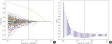
Fig.1
Selection of traditional radiomics features by the LASSO regression algorithm"

Tab.1
Clinical parameters and sonographic semantic features of the training and testing sets"
| 项目 | 训练集(n = 156) | 测试集(n = 39) | |||||||
|---|---|---|---|---|---|---|---|---|---|
| Ⅰ型EOC(n = 64) | Ⅱ型EOC(n = 92) | t/χ2 值 | P值 | Ⅰ型EOC (n = 16) | Ⅱ型EOC (n = 23) | t/χ2 值 | P值 | ||
| 年龄(x ± s)/岁 | 47.31 ± 10.34 | 54.04 ± 9.66 | -4.158 | < 0.001 | 50.06 ± 10.32 | 56.17 ± 8.10 | -2.07 | 0.045 | |
| 单核细胞计数(x ± s)/(109·L-1) | 0.60 ± 0.27 | 0.59 ± 0.27 | 0.228 | 0.747 | 0.45 ± 0.19 | 0.73 ± 0.24 | -3.89 | < 0.001 | |
| 血小板计数(x ± s)/(109·L-1) | 316.03 ± 88.64 | 365.91 ± 110.79 | -2.995 | 0.004 | 298.19 ± 75.92 | 368.17 ± 101.86 | -2.331 | 0.025 | |
| PLR(x ± s) | 200.27 ± 128.317 | 242.95 ± 130.71 | -2.021 | 0.045 | 199.09 ± 143.20 | 252.62 ± 118.34 | -1.275 | 0.21 | |
| LMR(x ± s) | 3.49 ± 1.63 | 3.66 ± 2.88 | -0.427 | 0.662 | 4.37 ± 1.93 | 2.46 ± 1.08 | 3.952 | < 0.001 | |
| FIGO分期 | 30.628 | < 0.001 | 11.493 | < 0.001 | |||||
| Ⅰ+Ⅱ | 49(76.56) | 29(31.52) | 13(81.25) | 6(26.09) | |||||
| Ⅲ+Ⅳ | 15(23.44) | 63(68.48) | 3(18.75) | 17(73.91) | |||||
| 月经状态 | 9.748 | 0.002 | 0.321 | 0.583 | |||||
| 绝经前 | 42(65.62) | 37(40.22) | 7(43.75) | 8(34.78) | |||||
| 绝经后 | 22(34.38) | 55(59.78) | 9(56.25) | 15(65.22) | |||||
| 肿块声像图特征 | 21.361 | < 0.001 | 5.252 | 0.020 | |||||
| 囊性 | 10(15.62) | 1(1.09) | 2(12.50) | 0(0.00) | |||||
| 囊实性 | 38(59.38) | 40(43.48) | 10(62.50) | 10(43.48) | |||||
| 实性 | 16(25.00) | 51(55.43) | 4(25.00) | 13(56.52) | |||||
| 彩色血流评分 | 17.311 | < 0.001 | 4.503 | 0.032 | |||||
| 1分 | 6(9.38) | 3(3.26) | 1(6.25) | 0(0.00) | |||||
| 2分 | 38(59.38) | 30(32.61) | 8(50.00) | 6(26.09) | |||||
| 3分 | 19(29.69) | 52(56.52) | 7(43.75) | 16(69.57) | |||||
| 4分 | 1(1.56) | 7(7.61) | 0(0.00) | 1(4.35) | |||||
| 肿瘤侧别 | 17.11 | < 0.001 | 5.564 | 0.018 | |||||
| 单侧 | 51(79.69) | 43(46.74) | 13(81.25) | 10(43.48) | |||||
| 双侧 | 13(20.31) | 49(53.26) | 3(18.75) | 13(56.52) | |||||
| CA125 | 18.686 | < 0.001 | 8.474 | 0.002 | |||||
| ≤ 35 U/mL | 19(29.69) | 7(7.61) | 8(50.00) | 2(8.70) | |||||
| > 35 U/mL且≤ 200 U/mL | 21(32.81) | 22(23.91) | 3(18.75) | 4(17.39) | |||||
| > 200 U/mL且> 500 U/mL | 8(12.50) | 19(20.65) | 2(12.50) | 5(21.74) | |||||
| ≥ 500 U/mL | 16(25.00) | 44(47.83) | 3(18.75) | 12(52.17) | |||||
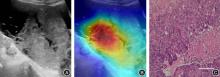
Fig.2
Visualization of Grad-CAM(× 400)"
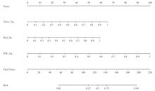
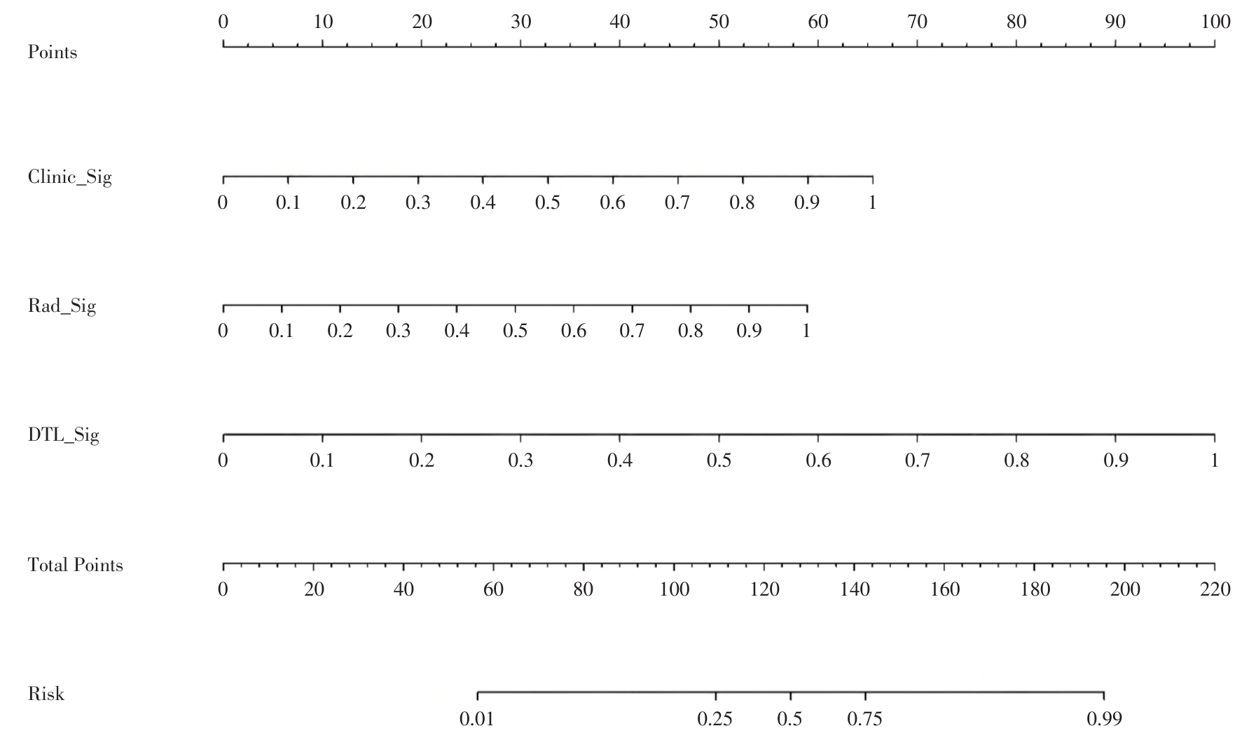
Fig.3
Nomogram of the combined model"
Tab.2
Prediction performance of each model"
| 项目 | 临床参数标签 | 影像组学标签 | 深度迁移 学习标签 | 深度学习影像组学列线图 |
|---|---|---|---|---|
| 训练集 | ||||
| AUC | 0.850* | 0.878* | 0.920* | 0.958 |
| 2.5%CI | 0.787 | 0.824 | 0.876 | 0.931 |
| 97.5%CI | 0.912 | 0.932 | 0.963 | 0.986 |
| 准确性 | 0.808 | 0.827 | 0.827 | 0.904 |
| 查准率 | 0.823 | 0.849 | 0.828 | 0.914 |
| 查全率 | 0.859 | 0.859 | 0.891 | 0.924 |
| F1 Score | 0.840 | 0.854 | 0.859 | 0.919 |
| 测试集 | ||||
| AUC | 0.916 | 0.709* | 0.842* | 0.951 |
| 2.5%CI | 0.827 | 0.539 | 0.712 | 0.876 |
| 97.5%CI | 1.000 | 0.880 | 0.972 | 1.000 |
| 准确性 | 0.872 | 0.641 | 0.744 | 0.923 |
| 查准率 | 0.821 | 0.680 | 0.710 | 0.885 |
| 查全率 | 1.000 | 0.739 | 0.957 | 1.000 |
| F1 Score | 0.902 | 0.708 | 0.815 | 0.939 |
Fig.4
ROC curves of each model"
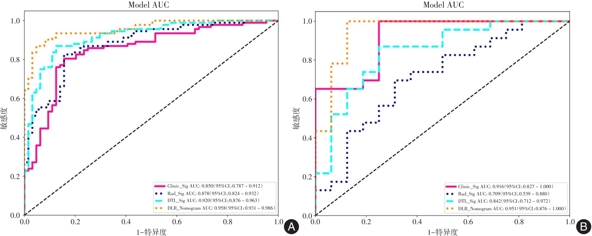
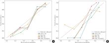
Fig.5
Calibration curves of each model"
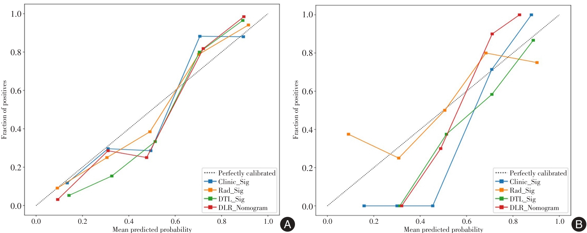
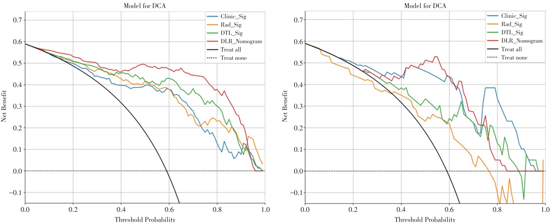
Fig.6
Decision curve analysis of each model"
| [1] | 张丹, 李燕东. 卵巢与附件区肿瘤的研究进展[J]. 中华医学超声杂志 ( 电子版 ), 2020, 17(3): 274-278. |
| [2] |
QIAN L, REN J, LIU A, et al. MR imaging of epithelial ovarian cancer: A combined model to predict histologic subtypes[J]. Eur Radiol, 2020, 30(11): 5815-5825. doi:10.1007/s00330-020-06993-5
doi: 10.1007/s00330-020-06993-5 |
| [3] |
BERA K, BRAMAN N, GUPTA A, et al. Predicting cancer outcomes with radiomics and artificial intelligence in radiology[J]. Nat Rev Clin Oncol, 2022, 19(2): 132-146. doi:10.1038/s41571-021-00560-7
doi: 10.1038/s41571-021-00560-7 |
| [4] |
LI X, YANG L, JIAO X. Comparison of Traditional Radiomics, Deep Learning Radiomics and Fusion Methods for Axillary Lymph Node Metastasis Prediction in Breast Cancer[J]. Acad Radiol, 2023, 30(7): 1281-1287. doi:10.1016/j.acra.2022.10.015
doi: 10.1016/j.acra.2022.10.015 |
| [5] |
PARK H J, PARK B, LEE S S. Radiomics and Deep Learning: Hepatic Applications[J]. Korean J Radiol, 2020, 21(4): 387-401. doi:10.3348/kjr.2019.0752
doi: 10.3348/kjr.2019.0752 |
| [6] |
罗刚, 泮思林, 乔思波, 等. 深度学习技术在胎儿超声心动图图像自动识别中的应用[J]. 实用医学杂志, 2022, 38(14): 1830-1833. doi:10.3969/j.issn.1006⁃5725.2022.14.022
doi: 10.3969/j.issn.1006?5725.2022.14.022 |
| [7] |
TONG T, GU J, XU D, et al. Deep learning radiomics based on contrast-enhanced ultrasound images for assisted diagnosis of pancreatic ductal adenocarcinoma and chronic pancreatitis[J]. BMC Med, 2022, 20(1): 74. doi:10.1186/s12916-022-02258-8
doi: 10.1186/s12916-022-02258-8 |
| [8] |
SOHN J H, FIELDS B K K. Radiomics and Deep Learning to Predict Pulmonary Nodule Metastasis at CT[J]. Radiology, 2024, 311(1): e233356. doi:10.1148/radiol.233356
doi: 10.1148/radiol.233356 |
| [9] |
DU Y, XIAO Y, GUO W, et al. Development and validation of an ultrasound-based deep learning radiomics nomogram for predicting the malignant risk of ovarian tumours[J]. Biomed Eng Online, 2024, 23(1): 41. doi:10.1186/s12938-024-01234-y
doi: 10.1186/s12938-024-01234-y |
| [10] |
DU Y, GUO W, XIAO Y, et al. Ultrasound-based deep learning radiomics model for differentiating benign, borderline, and malignant ovarian tumours: A multi-class classification exploratory study[J]. BMC Med Imaging, 2024, 24(1): 89. doi:10.1186/s12880-024-01251-2
doi: 10.1186/s12880-024-01251-2 |
| [11] |
CHEN H, YANG B W, QIAN L, et al. Deep Learning Prediction of Ovarian Malignancy at US Compared with O-RADS and Expert Assessment[J]. Radiology, 2022, 304(1): 106-113. doi:10.1148/radiol.211367
doi: 10.1148/radiol.211367 |
| [12] |
JIAN J, LI Y, PICKHARDT P J, et al. MR image-based radiomics to differentiate type Ⅰ and type Ⅱ epithelial ovarian cancers[J]. Eur Radiol, 2021, 31(1): 403-410. doi:10.1007/s00330-020-07091-2
doi: 10.1007/s00330-020-07091-2 |
| [13] |
ZHANG H, MAO Y, CHEN X, et al. Magnetic resonance imaging radiomics in categorizing ovarian masses and predicting clinical outcome: A preliminary study[J]. Eur Radiol, 2019, 29(7): 3358-3371. doi:10.1007/s00330-019-06124-9
doi: 10.1007/s00330-019-06124-9 |
| [14] |
CHEN X, WANG X, ZHANG K, et al. Recent advances and clinical applications of deep learning in medical image analysis[J]. Med Image Anal, 2022, 79: 102444. doi:10.1016/j.media.2022.102444
doi: 10.1016/j.media.2022.102444 |
| [15] |
郭润财, 王蕾, 黄振国, 等. 基于从头训练模式深度学习卷积神经网络模型评估急性肺栓塞的价值[J]. 实用医学杂志, 2023, 39(22): 2979-2983. doi:10.3969/j.issn.1006-5725.2023.22.021
doi: 10.3969/j.issn.1006-5725.2023.22.021 |
| [16] |
TOSEEF M, OLAYEMI PETINRIN O, WANG F, et al. Deep transfer learning for clinical decision-making based on high-throughput data: Comprehensive survey with benchmark results[J]. Brief Bioinform, 2023, 24(4): 1-16. doi:10.1093/bib/bbad254
doi: 10.1093/bib/bbad254 |
| [17] |
WANG R, CAI Y, LEE I K, et al. Evaluation of a convolutional neural network for ovarian tumor differentiation based on magnetic resonance imaging[J]. Eur Radiol, 2021, 31(7): 4960-4971. doi:10.1007/s00330-020-07266-x
doi: 10.1007/s00330-020-07266-x |
| [18] |
CHRISTIANSEN F, EPSTEIN E L, SMEDBERG E, et al. Ultrasound image analysis using deep neural networks for discriminating between benign and malignant ovarian tumors: Comparison with expert subjective assessment[J]. Ultrasound Obstet Gynecol, 2021, 57(1): 155-163. doi:10.1002/uog.23530
doi: 10.1002/uog.23530 |
| [19] |
GAO Y, ZENG S, XU X, et al. Deep learning-enabled pelvic ultrasound images for accurate diagnosis of ovarian cancer in China: A retrospective, multicentre, diagnostic study[J]. Lancet Digit Health, 2022, 4(3): e179-e187. doi:10.1016/s2589-7500(21)00278-8
doi: 10.1016/s2589-7500(21)00278-8 |
| [20] |
HSU S T, SU Y J, HUNG C H, et al. Automatic ovarian tumors recognition system based on ensemble convolutional neural network with ultrasound imaging[J]. BMC Med Inform Decis Mak, 2022, 22(1): 298. doi:10.1186/s12911-022-02047-6
doi: 10.1186/s12911-022-02047-6 |
| [21] |
WANG T, WANG H, WANG Y, et al. MR-based radiomics-clinical nomogram in epithelial ovarian tumor prognosis prediction: Tumor body texture analysis across various acquisition protocols[J]. J Ovarian Res, 2022, 15(1): 1-10. doi:10.1186/s13048-021-00941-7
doi: 10.1186/s13048-021-00941-7 |
| [22] |
ZHU H, AI Y, ZHANG J, et al. Preoperative Nomogram for Differentiation of Histological Subtypes in Ovarian Cancer Based on Computer Tomography Radiomics[J]. Front Oncol, 2021, 11: 642892. doi:10.3389/fonc.2021.642892
doi: 10.3389/fonc.2021.642892 |
| [23] |
YAN B C, MA X L, LI Y, et al. MRI-Based Radiomics Nomogram for Selecting Ovarian Preservation Treatment in Patients With Early-Stage Endometrial Cancer[J]. Front Oncol, 2021, 11: 1-11. doi:10.3389/fonc.2021.730281
doi: 10.3389/fonc.2021.730281 |
| [24] |
ZENG Q, LI H, ZHU Y, et al. Development and validation of a predictive model combining clinical, radiomics, and deep transfer learning features for lymph node metastasis in early gastric cancer[J]. Front Med (Lausanne), 2022, 9: 986437. doi:10.3389/fmed.2022.986437
doi: 10.3389/fmed.2022.986437 |
| [25] |
GAO W, WANG W, SONG D, et al. A predictive model integrating deep and radiomics features based on gadobenate dimeglumine-enhanced MRI for postoperative early recurrence of hepatocellular carcinoma[J]. Radiol Med, 2022, 127(3): 259-271. doi:10.1007/s11547-021-01445-6
doi: 10.1007/s11547-021-01445-6 |
| [26] |
GONG J, ZHANG W, HUANG W, et al. CT-based radiomics nomogram may predict local recurrence-free survival in esophageal cancer patients receiving definitive chemoradiation or radiotherapy: A multicenter study[J]. Radiother Oncol, 2022, 174: 8-15. doi:10.1016/j.radonc.2022.06.010
doi: 10.1016/j.radonc.2022.06.010 |
| [27] |
AFSHAR P, MOHAMMADI A, PLATANIOTIS K N, et al. From Handcrafted to Deep-Learning-Based Cancer Radiomics: Challenges and opportunities[J]. Ieee Signal Proc Mag, 2019, 36(4): 132-160. doi:10.1109/msp.2019.2900993
doi: 10.1109/msp.2019.2900993 |
| Viewed | ||||||
|
Full text |
|
|||||
|
Abstract |
|
|||||