| 1 |
PRIETO J, BANALES J M, MEDINA J F. Primary biliary cholangitis: pathogenic mechanisms [J]. Curr Opin Gastroenterol, 2021, 37(2): 91-98.
|
| 2 |
TANAKA A. Current understanding of primary biliary cholangitis [J]. Clin Mol Hepatol, 2021, 27(1): 1-21.
|
| 3 |
LEUNG K K, DEEB M, HIRSCHFIELD G M. Review article: pathophysiology and management of primary biliary cholangitis [J]. Aliment Pharmacol Ther, 2020, 52(7): 1150-1164.
|
| 4 |
TANAKA A, LEUNG P S C, GERSHWIN M E. Evolution of our understanding of PBC [J]. Best Pract Res Clin Gastroenterol, 2018, 34-35: 3-9.
|
| 5 |
CHASCSA D M H, LINDOR K D. Emerging therapies for PBC [J]. J Gastroenterol, 2020, 55(3): 261-272.
|
| 6 |
PHAW N A, DYSON J K, JONES D. Emerging drugs for the treatment of primary biliary cholangitis [J]. Expert Opin Emerg Drugs, 2020, 25(2): 101-112.
|
| 7 |
党富涛, 唐谭绪,徐加敏. 原发性胆汁性胆管炎患者中人工肝支持系统的应用 [J]. 实用医学杂志, 2020, 36(22): 3153-3156.
|
| 8 |
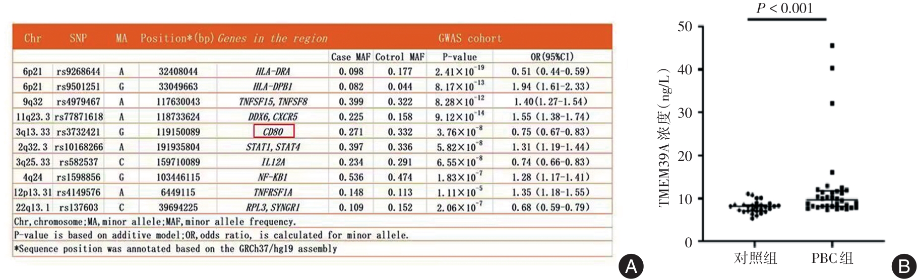
TRAN Q, PARK J, LEE H, et al. TMEM39A and Human Diseases: A Brief Review [J]. Toxicol Res, 2017, 33(3): 205-209.
|
| 9 |
MIAO G, ZHANG Y, CHEN D, et al. The ER-Localized Transmembrane Protein TMEM39A/SUSR2 Regulates Autophagy by Controlling the Trafficking of the PtdIns(4)P Phosphatase SAC1 [J]. Mol Cell, 2020, 77(3): 618-632.e615.
|
| 10 |
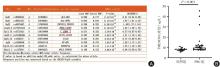
QIU F, TANG R, ZUO X,et al. A genome-wide association study identifies six novel risk loci for primary biliary cholangitis [J]. Nat Commun, 2017, 8: 14828.
|
| 11 |
SASAKI M, MIYAKOSHI M, SATO Y, et al. Autophagy may precede cellular senescence of bile ductular cells in ductular reaction in primary biliary cirrhosis [J]. Dig Dis Sci, 2012, 57(3): 660-666.
|
| 12 |
LI H, GUAN Y, HAN C,et al. The pathogenesis, models and therapeutic advances of primary biliary cholangitis [J]. Biomed Pharmacother, 2021, 140: 111754.
|
| 13 |
TANAKA A, LEUNG P S C, GERSHWIN M E. The genetics of primary biliary cholangitis [J]. Curr Opin Gastroenterol, 2019, 35(2): 93-98.
|
| 14 |
ASEEM S O, HYLEMON P B, ZHOU H. Bile Acids and Biliary Fibrosis [J]. Cells, 2023, 12(5):792.
|
| 15 |
HOFMANN A F, HAGEY L R, KRASOWSKI M D. Bile salts of vertebrates: structural variation and possible evolutionary significance [J]. J Lipid Res, 2010, 51(2): 226-246.
|
| 16 |
CHIANG J Y L, FERRELL J M. Bile Acids as Metabolic Regulators and Nutrient Sensors [J]. Annu Rev Nutr, 2019, 39: 175-200.
|
| 17 |
WOOLBRIGHT B L, DORKO K, ANTOINE D J, et al. Bile acid-induced necrosis in primary human hepatocytes and in patients with obstructive cholestasis [J]. Toxicol Appl Pharmacol, 2015, 283(3): 168-177.
|
| 18 |
KLIONSKY D J, PETRONI G, AMARAVADI R K,et al. Autophagy in major human diseases [J]. Embo J, 2021, 40(19): e108863.
|
| 19 |
PANZITT K, FICKERT P, WAGNER M. Regulation of autophagy by bile acids and in cholestasis - CholestoPHAGY or CholeSTOPagy [J]. Biochim Biophys Acta Mol Basis Dis, 2021, 1867(2): 166017.
|
| 20 |
SASAKI M, MIYAKOSHI M, SATO Y, et al. Autophagy mediates the process of cellular senescence characterizing bile duct damages in primary biliary cirrhosis [J]. Lab Invest, 2010, 90(6): 835-843.
|
| 21 |
SASAKI M, NAKANUMA Y. Bile Acids and Deregulated Cholangiocyte Autophagy in Primary Biliary Cholangitis [J]. Dig Dis, 2017, 35(3): 210-216.
|
| 22 |
LUO S, WANG X, BAI M, et al. The conserved autoimmune-disease risk gene TMEM39A regulates lysosome dynamics [J]. Proc Natl Acad Sci U S A, 2021, 118(6):e2011379118.
|


 ),Chan WANG1,Chuanli REN2,Mingming. ZHANG3