

The Journal of Practical Medicine ›› 2026, Vol. 42 ›› Issue (6): 1070-1077.doi: 10.3969/j.issn.1006-5725.2026.06.021
• Treatise: Clinical Practice • Previous Articles
Kai LI1,Xing WANG2,Zhijun ZENG2,Xu CHENG1,Bo SHI1( )
)
Received:2025-11-11
Revised:2025-12-26
Accepted:2025-12-31
Online:2026-03-25
Published:2026-03-26
Contact:
Bo SHI
E-mail:878017236@qq.com
CLC Number:
Kai LI,Xing WANG,Zhijun ZENG,Xu CHENG,Bo SHI. Application and imaging characteristics of transrectal real-time tissue elastography combined with MRI in the diagnosis of benign and malignant prostate lesions[J]. The Journal of Practical Medicine, 2026, 42(6): 1070-1077.
Tab.1
TRTE and MRI imaging characteristics"
| 病理诊断 | TRTE影像学特点 | 例数 | MRI影像学特点 | 例数 |
|---|---|---|---|---|
| 恶性(n = 79) | 病灶区全蓝色或大部分为蓝色 | 57 | T2WI病灶区低信号 | 67 |
| 病灶区见局部蓝色区域 | 7 | T1WI病灶区周边脂肪不对称或者消失 | 24 | |
| 病灶区夹杂少许蓝色 | 13 | DWI高信号、ADC低信号 | 67 | |
| 病灶区无蓝色 | 2 | 增强早期强化明显 | 62 | |
| T2WI等信号、DWI低信号 | 12 | |||
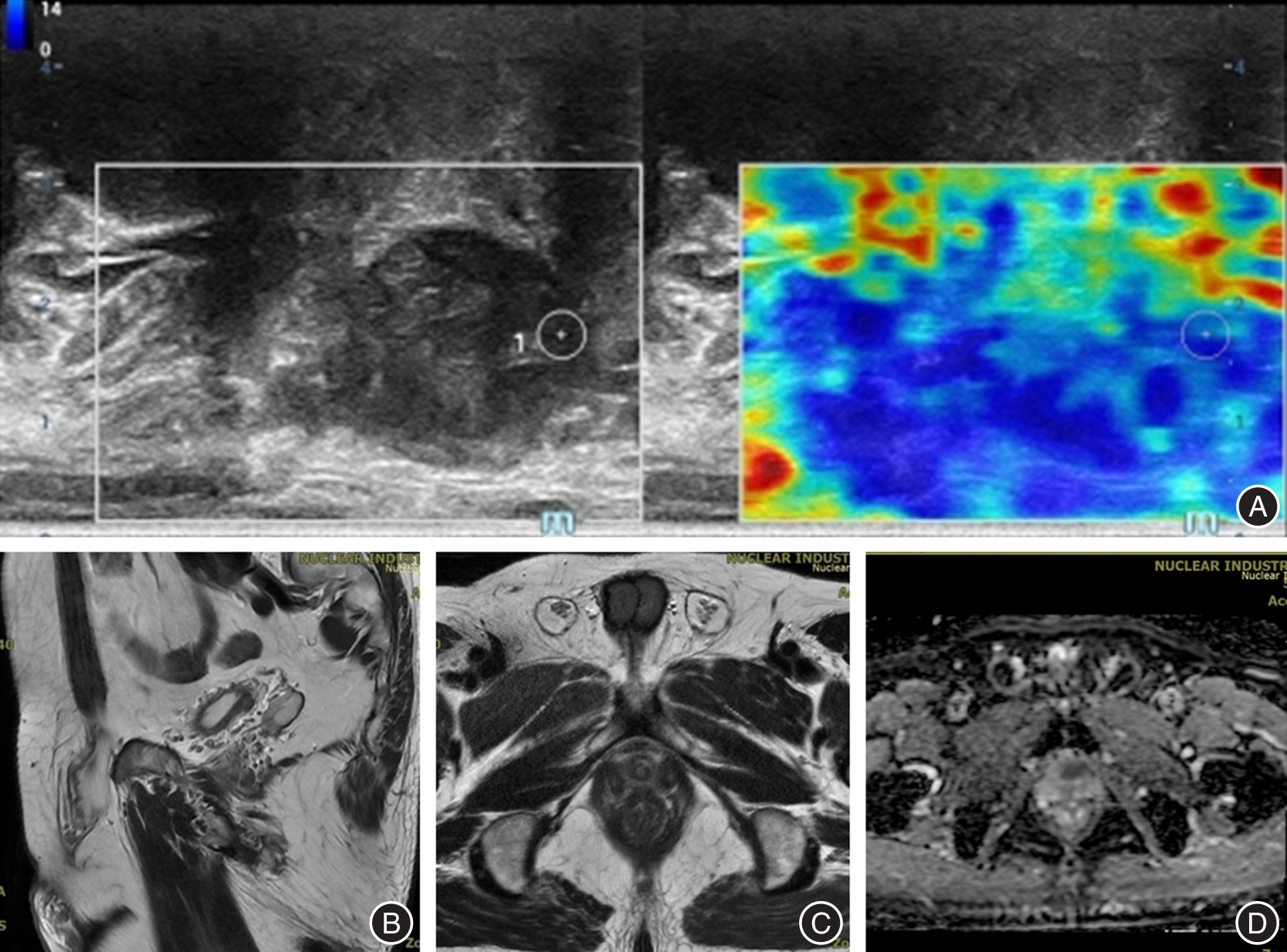
| 良性(n = 75) | 病灶区大部分为绿色 | 60 | T2WI呈现均匀高信号 | 62 |
| 病灶区夹杂少许蓝色 | 6 | T2WI低信号 | 13 | |
| 病灶区见局部蓝色区域 | 9 | 增强扫描未见明显强化 | 75 |

Fig.1
TRTE and MRI images of Case 1"
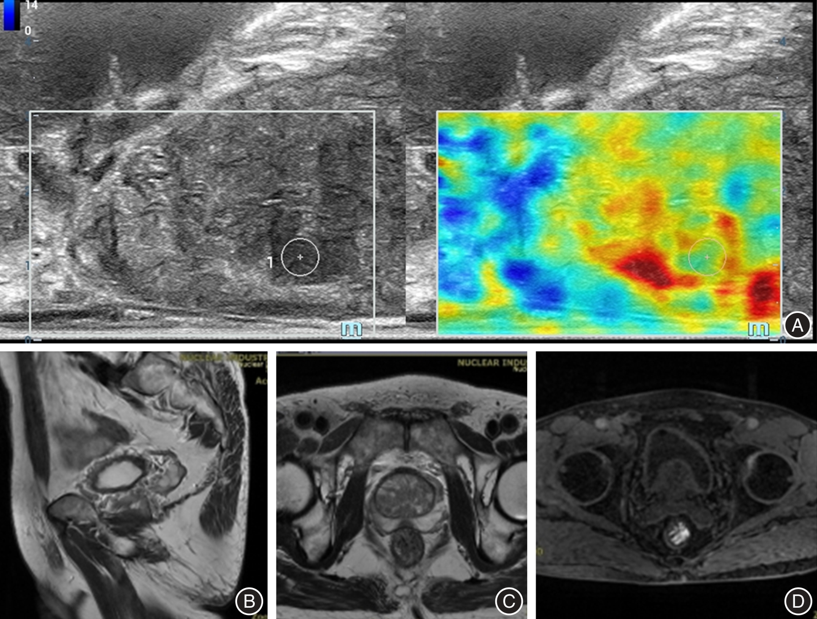

Fig.2
TRTE and MRI images of Case 2"
Tab.2
Comparison of baseline data and imaging parameters between patients with benign and malignant lesions"
| 项目 | 例数 | 年龄(x ± s)/岁 | 平均穿刺针数(x ± s)/针 | tPSA/(ng/mL) | PSAD | 弹性评分/分 | PI-RADS V2.1/分 |
|---|---|---|---|---|---|---|---|
| 恶性 | 79 | 71.76 ± 6.49 | 13.40 ± 0.60 | 41.58(8.36,231.88) | 0.62(0.13,3.68) | 4(2,4) | 4(2,4) |
| 良性 | 75 | 68.47 ± 5.83 | 12.50 ± 0.80 | 12.42(4.62,32.14) | 0.17(0.06,0.40) | 1(1,2) | 2(1,2) |
| t/Z值 | 3.303 | 7.924 | 7.628 | 5.494 | 5.832 | 6.927 | |
| P值 | 0.001 | < 0.001 | < 0.001 | < 0.001 | < 0.001 | < 0.001 |
Tab.3
Comparison of consistency of TRTE, MRI and clinical pathology in the diagnosis of benign and malignant prostate lesions 例"
| 检查方式 | 临床病理 | 合计 | ||
|---|---|---|---|---|
| 恶性 | 良性 | |||
| 合计 | 79 | 75 | 154 | |
| TRTE | 恶性 | 64 | 9 | 73 |
| 良性 | 15 | 66 | 81 | |
| MRI | 恶性 | 67 | 13 | 80 |
| 良性 | 12 | 62 | 74 | |
Tab.4
Diagnostic value of TRTE and MRI based on binary classification in distinguishing benign from malignant prostate lesions"
| 方法 | 敏感度 | 特异度 | 准确率 | 阳性预测值 | 阴性预测值 |
|---|---|---|---|---|---|
| TRTE | 81.01(64/79) | 88.00(66/75) | 84.42(130/154) | 87.67(64/73) | 81.48(66/81) |
| MRI | 84.81(67/79) | 82.67(62/75) | 83.77(129/154) | 83.75(67/80) | 83.78(62/74) |
Tab.5
Multivariate logistic regression analysis of influencing factors of malignant prostate lesions"
| 变量 | β | SE | Wald | P值 | OR | 95%CI |
|---|---|---|---|---|---|---|
| 年龄 | 0.072 | 0.038 | 3.590 | 0.059 | 1.078 | 0.998 ~ 1.158 |
| PSAD | 0.087 | 0.029 | 9.000 | 0.003 | 1.091 | 1.031 ~ 1.155 |
| 弹性评分 | 0.181 | 0.065 | 7.754 | 0.006 | 1.198 | 1.055 ~ 1.361 |
| PI-RADS V2.1 | 0.358 | 0.126 | 8.073 | 0.005 | 1.430 | 1.117 ~ 0.831 |
| 常数项 | -0.421 | 0.107 | 15.481 | < 0.001 |
Tab.6
ROC curve analysis of three diagnostic regimens for the diagnosis of malignant prostate lesions"
| 方法 | AUC | 标准误 | 95%CI |
|---|---|---|---|
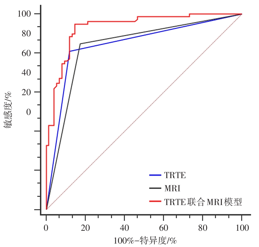
| TRTE | 0.845 | 0.0292 | 0.778 ~ 0.898 |
| MRI | 0.837 | 0.0299 | 0.769 ~ 0.892 |
| TRTE联合MRI模型 | 0.933 | 0.0200 | 0.881 ~ 0.967 |
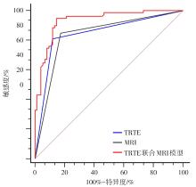
Fig.3
ROC curve analysis of three diagnostic regimens for the diagnosis of malignant prostate lesions"
| [1] |
YU X D, YAN S S, LIU R J, et al. Apparent differences in prostate zones: Susceptibility to prostate cancer, benign prostatic hyperplasia and prostatitis[J]. Int Urol Nephrol, 2024, 56(8): 2451-2458. doi:10.1007/s11255-024-04012-w .
doi: 10.1007/s11255-024-04012-w |
| [2] |
王芸, 李萍, 陈璐, 等. 以IKAP理论为指导的延续性护理对老年前列腺癌术后患者尿失禁及生活质量的影响[J]. 护理实践与研究, 2024, 21(1): 125-131. doi:10.3969/j.issn.1672-9676.2024.01.019 .
doi: 10.3969/j.issn.1672-9676.2024.01.019 |
| [3] |
DESTOUNI M, LAZARIS A C, TZELEPI V. Cribriform patterned lesions in the prostate gland with emphasis on differential diagnosis and clinical significance[J]. Cancers, 2022, 14(13): 3041. doi:10.3390/cancers14133041 .
doi: 10.3390/cancers14133041 |
| [4] |
胡博文, 胡亚兰, 梁辉. 前列腺癌早期筛查的常见方法及最新研究进展[J/OL]. 中华腔镜泌尿外科杂志(电子版), 2025, 19(6): 800-808. doi:10.3877/cma.j.issn.1674-3253.2025.06.019 .
doi: 10.3877/cma.j.issn.1674-3253.2025.06.019 |
| [5] |
WANG Y, FENG Y, YANG X, et al. Enhanced transrectal ultrasound, real-time sonoelastography, and contrast-enhanced transrectal ultrasound in heavily prescreened Chinese men with naive and repetitive biopsy: A comparison of detection rate of prostate cancer per man and per lesion[J]. Ultrasound Q, 2022, 38(3): 237-245. doi:10.1097/RUQ.0000000000000589 .
doi: 10.1097/RUQ.0000000000000589 |
| [6] |
FERNANDES M C, YILDIRIM O, WOO S, et al. The role of MRI in prostate cancer: Current and future directions[J]. MAGMA, 2022, 35(4): 503-521. doi:10.1007/s10334-022-01006-6 .
doi: 10.1007/s10334-022-01006-6 |
| [7] |
KAMOI K, OKIHARA K, OCHIAI A, et al. The utility of transrectal real-time elastography in the diagnosis of prostate cancer[J]. Ultrasound Med Biol, 2008, 34(7): 1025-1032. doi:10.1016/j.ultrasmedbio.2007.12.002 .
doi: 10.1016/j.ultrasmedbio.2007.12.002 |
| [8] |
TURKBEY B, ROSENKRANTZ A B, HAIDER M A, et al. Prostate imaging reporting and data system version 2.1: 2019 update of prostate imaging reporting and data system version 2[J]. Eur Urol, 2019, 76(3): 340-351. doi:10.1016/j.eururo.2019.02.033 .
doi: 10.1016/j.eururo.2019.02.033 |
| [9] |
WILLIAMS I S, MCVEY A, PERERA S, et al. Modern paradigms for prostate cancer detection and management[J]. Med J Aust, 2022, 217(8): 424-433. doi:10.5694/mja2.51722 .
doi: 10.5694/mja2.51722 |
| [10] |
GOURDIN T, VELAYATI A. Treatments and challenges in advanced prostate cancer[J]. Curr Opin Oncol, 2023, 35(3): 200-205. doi:10.1097/CCO.0000000000000938 .
doi: 10.1097/CCO.0000000000000938 |
| [11] |
IPPOLITI S, FLETCHER P, ORECCHIA L, et al. Optimal biopsy approach for detection of clinically significant prostate cancer[J]. Br J Radiol, 2022, 95(1131): 20210413. doi:10.1259/bjr.20210413 .
doi: 10.1259/bjr.20210413 |
| [12] |
张继燊, 谢玉洁, 杨婷, 等. 前列腺特异性膜抗原PET/CT对减少前列腺癌过度穿刺活检的应用价值[J]. 中山大学学报(医学科学版), 2025, 46(2): 311-317. doi:10.13471/j.cnki.j.sun.yat-sen.univ(med.sci).2025.0215 .
doi: 10.13471/j.cnki.j.sun.yat-sen.univ(med.sci).2025.0215 |
| [13] |
沈波, 李安域, 朱正, 等. 磁共振/超声成像融合引导的经会阴前列腺穿刺活检对前列腺癌的诊断价值[J]. 转化医学杂志, 2024, 13(10): 1712-1717. doi:10.3639/i.issn.2095-3097. 2024.10.034 .
doi: 10.3639/i.issn.2095-3097. 2024.10.034 |
| [14] |
INOUE T, SHIN T. Current magnetic resonance imaging-based diagnostic strategies for prostate cancer[J]. Int J Urol, 2023, 30(12): 1078-1086. doi:10.1111/iju.15281 .
doi: 10.1111/iju.15281 |
| [15] |
LIU Y, ZENG S, ZHOU D, et al. Diagnostic performance of multiple ultrasonic modalities for prostate cancer[J]. Clinics, 2025, 80: 100680. doi:10.1016/j.clinsp.2025.100680 .
doi: 10.1016/j.clinsp.2025.100680 |
| [16] |
ALMALKI Y E, MANSOUR M G E, ALI S A, et al. Advanced strain elastography is a reliable approach for prostate cancer detection in patients with elevated PSA levels[J]. Sci Rep, 2024, 14: 2917. doi:10.1038/s41598-024-53440-2 .
doi: 10.1038/s41598-024-53440-2 |
| [17] |
LI J, ZHU C, YANG S, et al. Non-invasive diagnosis of prostate cancer and high-grade prostate cancer using multiparametric ultrasonography and serological examination[J]. Ultrasound Med Biol, 2024, 50(4): 600-609. doi:10.1016/j.ultrasmedbio. 2024. 01.003 .
doi: 10.1016/j.ultrasmedbio. 2024. 01.003 |
| [18] | 杨秋子, 毛星刚, 孙季冬, 等. 头颅磁共振成像在轻型颅脑损伤诊断与预后评估中的应用进展[J]. 中华神经外科疾病研究杂志, 2024, 18(6): 90-94. |
| [19] |
FIARD G, GIGANTI F. How MRI is changing prostate cancer management: A focus on early detection and active surveillance: Comment l’IRM est en train de révolutionner la prise en charge du cancer de la prostate: Focus sur la détection précoce et la surveillance active[J]. Prog Urol, 2022, 32(6S1): 6S19-6S25. doi:10.1016/S1166-7087(22)00171-3 .
doi: 10.1016/S1166-7087(22)00171-3 |
| [20] |
刘玉姗, 徐冉, 曾施, 等. 多种超声模式在前列腺癌诊断中的应用价值比较[J]. 中国临床医学影像杂志, 2023, 34(4): 250-254. doi:10.12117/jccmi.2023.04.006 .
doi: 10.12117/jccmi.2023.04.006 |
| [21] |
NG A B C D, ASIF A, AGARWAL R, et al. Biparametric vs multiparametric MRI for prostate cancer diagnosis: The PRIME diagnostic clinical trial[J]. JAMA, 2025, 334(13): 1170-1179. doi:10.1001/jama.2025.13722 .
doi: 10.1001/jama.2025.13722 |
| [22] |
GREY A D R, SCOTT R, SHAH B, et al. Multiparametric ultrasound versus multiparametric MRI to diagnose prostate cancer (CADMUS): A prospective, multicentre, paired-cohort, confirmatory study[J]. Lancet Oncol, 2022, 23(3): 428-438. doi:10.1016/S1470-2045(22)00016-X .
doi: 10.1016/S1470-2045(22)00016-X |
| [23] |
张同莉, 王长春, 祖拜热·依斯坎代尔, 等. 经直肠超声造影联合MRI在PSA灰区前列腺癌诊断中的价值[J]. 中国超声医学杂志, 2024, 40(8): 924-927. doi:10.3969/j.issn.1002-0101. 2024.08.027 .
doi: 10.3969/j.issn.1002-0101. 2024.08.027 |
| [24] |
DARYANANI A, TURKBEY B. Recent advancements in CT and MR imaging of prostate cancer[J]. Semin Nucl Med, 2022, 52(3): 365-373. doi:10.1053/j.semnuclmed.2021.11.013 .
doi: 10.1053/j.semnuclmed.2021.11.013 |
| [25] |
DITONNO F, FRANCO A, MANFREDI C, et al. Novel non-MRI imaging techniques for primary diagnosis of prostate cancer: Micro-ultrasound, contrast-enhanced ultrasound, elastography, multiparametric ultrasound, and PSMA PET/CT[J]. Prostate Cancer Prostatic Dis, 2024, 27(1): 29-36. doi:10.1038/s41391-023-00708-9 .
doi: 10.1038/s41391-023-00708-9 |
| [26] |
郭艳娜, 朱益麟, 张萌迪. 经直肠实时组织弹性成像、MRI联合检查参数预测前列腺恶性病变列线图模型的构建及验证[J]. 癌症进展, 2025, 23(17): 2096-2101. doi:10.11877/j.issn.1672-1535.2025.23.17.26 .
doi: 10.11877/j.issn.1672-1535.2025.23.17.26 |
| [27] |
SINGH D, CHANDRAN A, PANEBIANCO V, et al. MRI capacity assessment for prostate cancer screening in five sites of Europe[J]. Eur J Radiol, 2025, 190: 112235. doi:10.1016/j.ejrad. 2025.112235 .
doi: 10.1016/j.ejrad. 2025.112235 |
| [1] | Yingmei HAN,Yijie LI,Heng ZHANG,Weiqing LI,Ze FENG,Feng WANG. The application value of deep learning in imaging studies for predicting the conversion of Alzheimer′s disease [J]. The Journal of Practical Medicine, 2025, 41(9): 1413-1424. |
| [2] | Li TANG,Yurong GONG,Liye ZENG,Yanfang GAO,Chengzhe. DENG. Application value of 3.0T magnetic resonance imaging T2 mapping sequence combined with serum nesfatin⁃1 level detection in the diagnosis of elderly knee early osteoarthritis [J]. The Journal of Practical Medicine, 2025, 41(8): 1238-1242. |
| [3] | Huiling YE,Zhengchaoyi CHEN,Yihan HUANG,Yingjie ZHANG,Xiangbin ZHANG,Yuehu PU,Renming ZHONG. Radiotherapy treatment comparison of liver SBRT between 4D⁃CT and deep inspiration breath hold troughmagnetic resonance imaging [J]. The Journal of Practical Medicine, 2025, 41(7): 1044-1049. |
| [4] | Huiliang CAI,Qianying ZHANG,Ying HUANG,Weisheng PENG,Chengli WANG,Cuiting YANG,Na DENG,Sizhu ZHANG,Nina XU,Xiaobing HAN. Assessments of ki⁃67 expression in hepatocellular carcinoma using enhanced MRI intratumoral and peritumoral radiomics and clinical imaging features [J]. The Journal of Practical Medicine, 2025, 41(15): 2311-2319. |
| [5] | Yuling DUAN,Xuezhi ZHOU,Yongyi LI,Lixia MA,Desheng YANG,Jiao CHENG,Yan WU,Tao LIU,Guoyuan JIANG,Mei. WANG. Clinical value analysis of different MRI measurement methods in evaluating the efficacy of neoadjuvant therapy for breast cancer [J]. The Journal of Practical Medicine, 2025, 41(14): 2152-2159. |
| [6] | Yuexian LYU,Xiu BI,Ying LIU,Shujing CUI,Lixin ZHAO,Ge GAO,Jianxia WANG,Juan LI,Jun LI. The efficacy of plasma gasdermin D C⁃terminal fragment in the early diagnosis of sepsis [J]. The Journal of Practical Medicine, 2025, 41(12): 1899-1906. |
| [7] | Xiaoxiao QIN,Xiaozhuo LI,Hongli GUO,Lijing ZHANG. Combined value of multimodal fMRI and MRS in the differential diagnosis of postoperative recurrence and pseudoprogression of glioma [J]. The Journal of Practical Medicine, 2025, 41(11): 1736-1741. |
| [8] | Zhilong SI,Hao WANG,Fei XIAO. Feasibility of modified LIFT guided by magnetic resonance imaging in the treatment of deep anorectal abscess [J]. The Journal of Practical Medicine, 2025, 41(1): 65-70. |
| [9] | Yingmei HAN,Yijie LI,Heng ZHANG,Jing LV,Yi ZHANG,Yingbo QIAO,Nan LIN,Huiyong XU,Feng. WANG. Research progress of large-scale brain network of Alzheimer’s disease based on MRI analysis [J]. The Journal of Practical Medicine, 2024, 40(4): 575-579. |
| [10] | Yuke SONG,Jinfan XU,Xiaoming HE,Tianye LIN,Mincong HE,Qiushi. WEI. Correlation of high signal intensity of infrapatellar fat pad on symptoms and structure of knee osteoarthritis [J]. The Journal of Practical Medicine, 2024, 40(23): 3373-3378. |
| [11] | Huiling YE,Renming. ZHONG. Progress in assessing the treatment accuracy of liver stereotactic body radiotherapy through post-therapeutic magnetic resonance imaging morphologic alterations [J]. The Journal of Practical Medicine, 2024, 40(22): 3119-3123. |
| [12] | Cheng CHEN,Xinyang YU,Hua. ZHANG. Advances in imaging diagnosis of placenta accreta spectrum [J]. The Journal of Practical Medicine, 2024, 40(21): 2976-2981. |
| [13] | Jiarui ZHAO,Yulai GONG. Effectiveness of resting-state fMRI in the diagnosis of temporal lobe epilepsy-associated cognitive impairment: A review of literature [J]. The Journal of Practical Medicine, 2024, 40(20): 2954-2959. |
| [14] | Di WANG,Dan LIAO,Yuancheng LIU,Rui XU,Qinghong. DUAN. Analysis of global local consistency changes in first-episode depression with childhood maltreatment based on resting-state magnetic resonance [J]. The Journal of Practical Medicine, 2024, 40(16): 2311-2315. |
| [15] | Zhe ZENG,Lin LUO,Qiang CHEN,Siqi HOU,Shengzhe. JIANG. Default mode network analysis associated with memory impairment in acute mild traumatic brain injury [J]. The Journal of Practical Medicine, 2024, 40(10): 1412-1417. |
| Viewed | ||||||
|
Full text |
|
|||||
|
Abstract |
|
|||||