

The Journal of Practical Medicine ›› 2025, Vol. 41 ›› Issue (14): 2152-2159.doi: 10.3969/j.issn.1006-5725.2025.14.005
• Feature Reports:Breast carcinoma • Previous Articles
Yuling DUAN1,Xuezhi ZHOU1,Yongyi LI1,Lixia MA1,Desheng YANG2,Jiao CHENG3,Yan WU1,Tao LIU1,Guoyuan JIANG1,Mei. WANG4( )
)
Received:2025-03-20
Online:2025-07-25
Published:2025-07-29
Contact:
Mei. WANG
E-mail:15685295689@163.com
CLC Number:
Yuling DUAN,Xuezhi ZHOU,Yongyi LI,Lixia MA,Desheng YANG,Jiao CHENG,Yan WU,Tao LIU,Guoyuan JIANG,Mei. WANG. Clinical value analysis of different MRI measurement methods in evaluating the efficacy of neoadjuvant therapy for breast cancer[J]. The Journal of Practical Medicine, 2025, 41(14): 2152-2159.
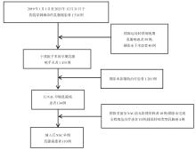
Fig.1
Patient inclusion and exclusion process diagram"
Tab.1
Comparison of the efficacy evaluation criteria for solid tumours"
| 疗效评估 | RISIST 1.1 | 二维面积评估标准 | 体积评估标准 |
|---|---|---|---|
| CR | 所有肿瘤病灶完全消失 | 所有肿瘤病灶完全消失 | 所有肿瘤病灶完全消失 |
| PR | 病灶最长直径缩小≥ 30% | 病灶最长直径及其垂直径乘积缩小≥50% | 病灶体积缩小≥ 65% |
| SD | 病灶变化介于PR与PD之间 | 病灶变化介于PR与PD之间 | 病灶变化介于PR与PD之间 |
| PD | 靶病灶长径和增加20%或出现新病灶 | 靶病灶最长直径及其垂直径乘积增加≥ 25%或出现新病灶 | 增加73%或出现新病灶 |
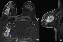
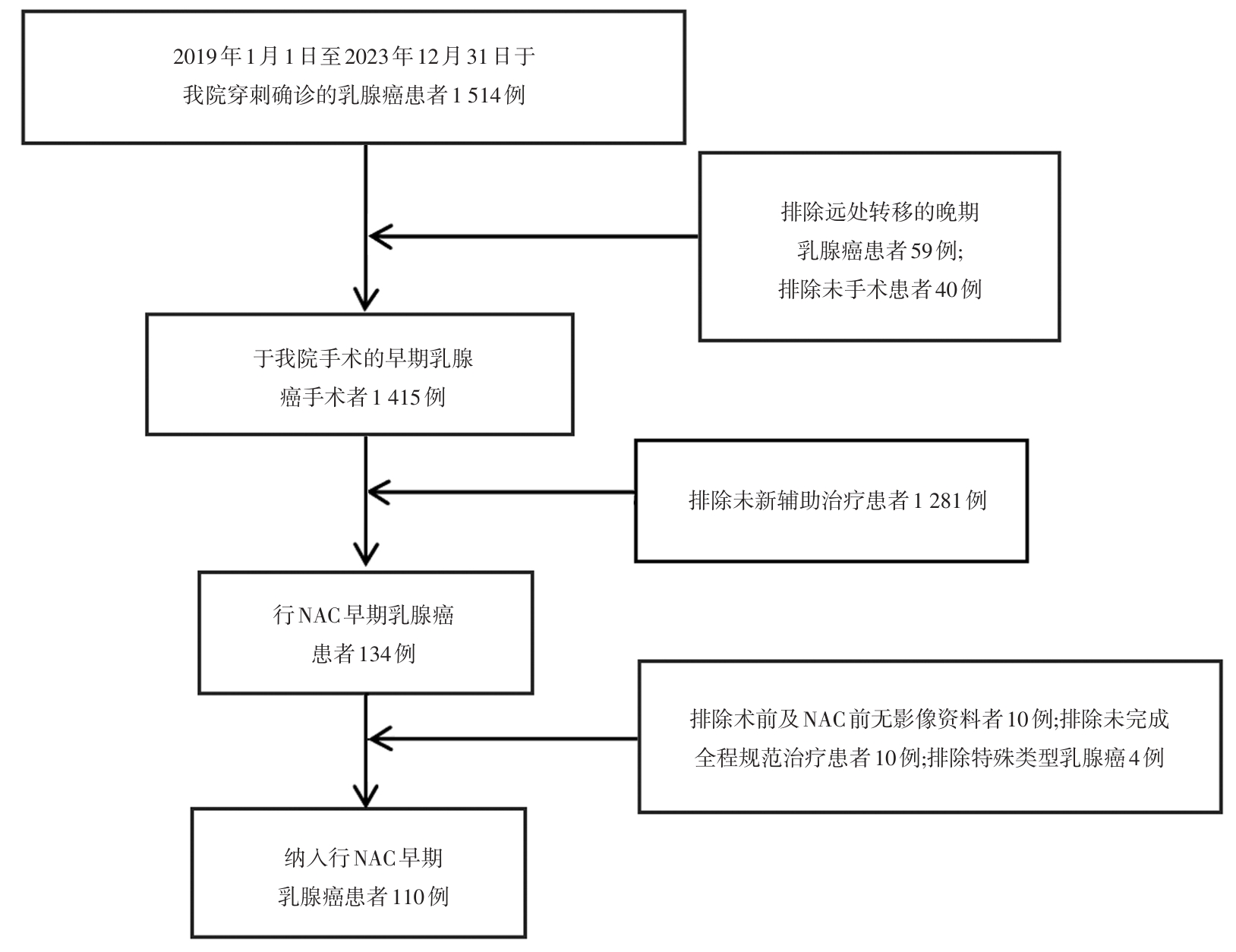
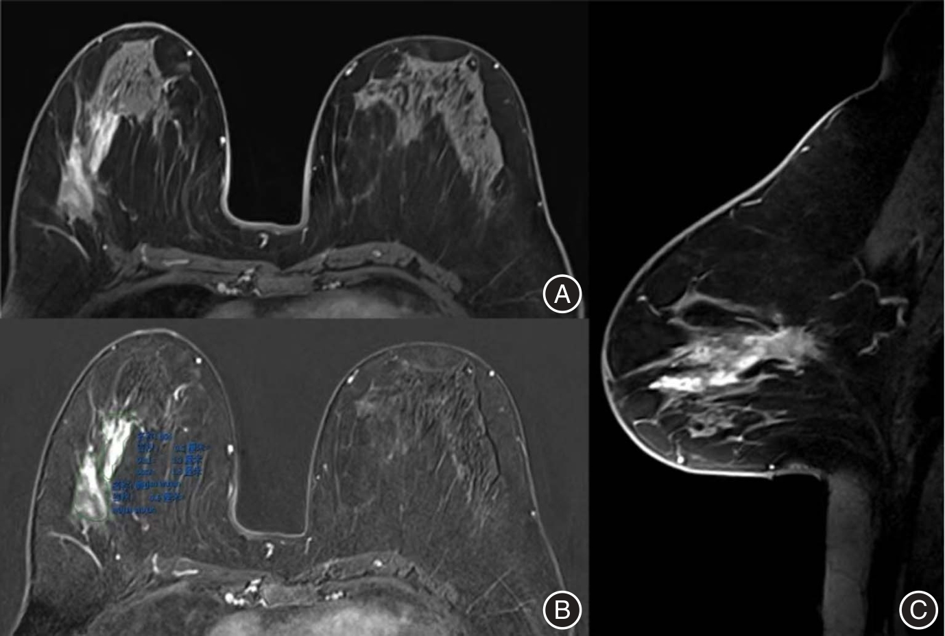
Fig.2
Imaging image before neoadjuvant chemotherapy"
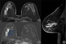
Fig.3
Imaging image after neoadjuvant chemotherapy"
Tab.2
Comparison of the diagnostic capabilities of the three measurement methods"
| 检查方法 | 敏感度 | 特异度 | PPV | NPV | 假阴性率 | 假阳性率 | 准确性 |
|---|---|---|---|---|---|---|---|
| RECIST 1.1 | 92.4 | 61.1 | 92.4 | 61.1 | 7.6 | 38.9 | 87.3 |
| 最优法 | 97.8 | 61.1 | 92.8 | 84.6 | 2.2 | 38.9 | 91.8 |
| 3D体积法 | 98.9 | 77.8 | 95.8 | 93.3 | 1.1 | 22.2 | 95.5 |
Tab.3
Comparison of diagnostic capabilities of 3D measurement standards and optimal measurement methods"
| 项目 | 3D体积测量法 | 最优法 | Z值 | P值 |
|---|---|---|---|---|
| 敏感度 | 98.9 | 97.8 | 0.636 | 0.52 |
| 特异度 | 77.8 | 61.1 | 2.683 | < 0.01 |
| 准确性 | 95.5 | 91.8 | 1.105 | 0.27 |
| PPV | 95.8 | 92.4 | 0.960 | 0.34 |
| NPV | 93.3 | 84.6 | 2.064 | < 0.05 |
Tab.5
Comparison of diagnostic capabilities of optimal measurement criteria and RECIST 1.1 measurement criteria"
| 项目 | 最优法 | RECIST 1.1 | Z值 | P值 |
|---|---|---|---|---|
| 敏感度 | 97.8 | 92.4 | 1.869 | 0.06 |
| 特异度 | 61.1 | 61.1 | 0.000 | 1.00 |
| 准确性 | 91.8 | 87.3 | 1.102 | 0.27 |
| PPV | 92.4 | 92.4 | 0.111 | 0.91 |
| NPV | 84.6 | 61.1 | 3.920 | < 0.01 |
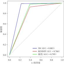
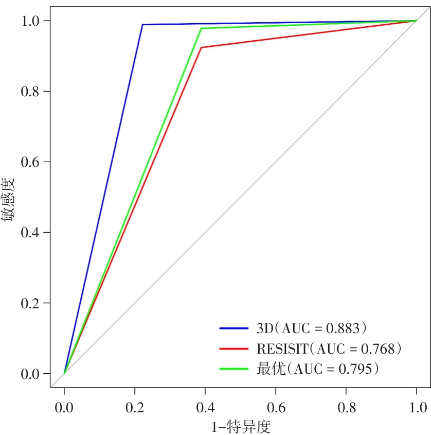
Fig.4
ROC curves of three measurement methods"
Tab.6
Area under the roc curve for three methods"
| 项目 | 区域 | 标准误 | 95%CI | |
|---|---|---|---|---|
| 下限 | 上限 | |||
| 3D | 0.883 | 0.059 | 0.767 | 1.000 |
| RESISIT | 0.768 | 0.072 | 0.626 | 0.909 |
| 最优 | 0.795 | 0.073 | 0.653 | 0.937 |
| [1] | 中国抗癌协会乳腺癌专业委员会,中华医学会肿瘤学分会乳腺肿瘤学组. 中国抗癌协会乳腺癌诊治指南与规范(2024年版)[J]. 中国癌症杂志, 2023,33(12):1092-1187. |
| [2] | 刘芬,赵辉,郭利敏. 多组学联合检测对乳腺癌临床病理特征、新辅助化疗效果的评估效能[J]. 实用医学杂志,2024,40(24):3539-3546. |
| [3] | 邵志敏,吴炅,江泽飞,等. 中国乳腺癌新辅助治疗专家共识(2022年版) [J]. 中国癌症杂志,2022, 32 (1): 80-89. |
| [4] | 肖晶晶,黄美玲,延常姣,等. Her-2阳性乳腺癌新辅助化疗联合靶向治疗获得病理完全缓解的影响因素[J]. 实用医学杂志,2022,38(5):542-546. |
| [5] | THERESE B B, BETHANY L N, JENNIFER L B, et al. NCCN Guidelines® Insights: Breast Cancer Screening and Diagnosis, Version 1.2023 [J]. J Natl Compr Canc Netw,2023,21(9):900-909. |
| [6] |
LU N, DONG J, FANG X, et al. Predicting pathologic response to neoadjuvant chemotherapy in patients with locally advanced breast cancer using multiparametric MRI [J]. BMC Med Imaging,2021,21(1):1-13. doi:10.1186/s12880-021-00688-z
doi: 10.1186/s12880-021-00688-z |
| [7] |
LIANG X, CHEN X, YANG Z, et al. Early prediction of pathological complete response to neoadjuvant chemotherapy combining DCE-MRI and apparent diffusion coefficient values in breast Cancer [J]. BMC Cancer, 2022, 22(1):1250. doi:10.1186/s12885-022-10315-x
doi: 10.1186/s12885-022-10315-x |
| [8] |
WANG X, HUA H, HAN J, et al. Evaluation of Multiparametric MRI Radiomics-Based Nomogram in Prediction of Response to Neoadjuvant Chemotherapy in Breast Cancer: A Two-Center study [J]. Clin Breast Cancer,2023,23(6):e331-e344. doi:10.1016/j.clbc.2023.05.010
doi: 10.1016/j.clbc.2023.05.010 |
| [9] |
SUDHIR R, KOPPULA V C, RAO T S, et al. Accuracy of digital mammography, ultrasound and MRI in predicting the pathological complete response and residual tumor size of breast cancer after completion of neoadjuvant chemotherapy [J]. Indian J Cancer,2022,59(3):345-353. doi:10.4103/ijc.ijc_795_19
doi: 10.4103/ijc.ijc_795_19 |
| [10] |
MIN K, TONG L, TANG L, et al. Head-to-head comparison of contrast-enhanced mammography and contrast-enhanced MRI for assessing pathological complete response to neoadjuvant therapy in patients with breast cancer: A meta-analysis [J]. Cancer Treat Res Commun,2023,202(1):1-9. doi:10.1007/s10549-023-07034-7
doi: 10.1007/s10549-023-07034-7 |
| [11] |
PENG Y, YUAN F, XIE F, et al. Comparison of automated breast volume scanning with conventional ultrasonography, mammography, and MRI to assess residual breast cancer after neoadjuvant therapy by molecular type [J]. Clin Radiol, 2023,78(5):e393-e400. doi:10.1016/j.crad.2022.12.002
doi: 10.1016/j.crad.2022.12.002 |
| [12] |
KO C C, YEH L R, KUO Y T, et al. Imaging biomarkers for evaluating tumor response: RECIST and beyond. [J]. Biomark Res,2021,9(1):52. doi:10.1186/s40364-021-00306-8
doi: 10.1186/s40364-021-00306-8 |
| [13] |
LITIÈRE S, BOGAERTS J. Imaging endpoints for clinical trial use: A RECIST perspective [J]. J Immunother Cancer,2022,10(11):e005092. doi:10.1136/jitc-2022-005092
doi: 10.1136/jitc-2022-005092 |
| [14] |
GAURAV JYOTI B, AMARTA J, GENIE K C W, et al. Diagnostic accuracy of magnetic resonance imaging to evaluate axillary lymph node status in breast cancer patients receiving neoadjuvant chemotherapy[J].Br J Radiol,2023,96(1143):20220904. doi:10.1259/bjr.20220904
doi: 10.1259/bjr.20220904 |
| [15] |
KWON M R, CHU J, KOOK S H, et al. Factors associated with radiologic-pathologic discordance in magnetic resonance imaging after neoadjuvant chemotherapy for breast cancer [J]. Clin Imaging,2022,89:1-9. doi:10.1016/j.clinimag.2022.05.002
doi: 10.1016/j.clinimag.2022.05.002 |
| [16] |
KIM Y, SIM S H, PARK B, et al. Criteria for identifying residual tumours after neoadjuvant chemotherapy of breast cancers: A magnetic resonance imaging study [J]. Sci Rep, 2021,11(1):634. doi:10.1038/s41598-020-79743-8
doi: 10.1038/s41598-020-79743-8 |
| [17] |
CHANG Y C, HUANG C S, LIU Y J, et al. Angiogenic response of locally advanced breast cancer to neoadjuvant chemotherapy evaluated with parametric histogram from dynamic contrast-enhanced MRI [J]. Phys Med Biol,2004,49(16):3593-3602. doi:10.1088/0031-9155/49/16/007
doi: 10.1088/0031-9155/49/16/007 |
| [18] |
SHARMA U, DANISHAD K K, SEENU V, et al. Longitudinal study of the assessment by MRI and diffusion-weighted imaging of tumor response in patients with locally advanced breast cancer undergoing neoadjuvant chemotherapy [J]. NMR Biomed,2009,22(1):104-113. doi:10.1002/nbm.1245
doi: 10.1002/nbm.1245 |
| [19] |
RAM S. One Size Fits All?-Not Anymore: Personalizing Breast Cancer Treatment with Use of a Semiautomated Functional Tumor Volume-based Predictive Model in the Assessment of Neoadjuvant Therapy Response [J]. Radiol Imaging Cancer,2023,5(4):e230089. doi:10.1148/rycan.230089
doi: 10.1148/rycan.230089 |
| [20] | ONISHI N, BARENG T J, GIBBS J, et al. Effect of Longitudinal Variation in Tumor Volume Estimation for MRI-guided Personalization of Breast Cancer Neoadjuvant Treatment [J]. Radiol Imaging Cancer,2023,5(4):e220126. |
| [21] |
PANTHI B, ADRADA B E, CANDELARIA R P, et al. Assessment of Response to Neoadjuvant Systemic Treatment in Triple-Negative Breast Cancer Using Functional Tumor Volumes from Longitudinal Dynamic Contrast-Enhanced MRI [J]. Cancers (Basel),2023,15(4):1025. doi:10.3390/cancers15041025
doi: 10.3390/cancers15041025 |
| [22] |
SCHWARTZ L H, LITIÈRE S, DE VRIES E, et al. RECIST 1.1-Update and clarification: From the RECIST committee [J]. Eur J Cancer,2016,62:132-137. doi:10.1016/j.ejca.2016.03.081
doi: 10.1016/j.ejca.2016.03.081 |
| [23] |
BRUIX J, SHERMAN M, LLOVET J M, et al. Clinical management of hepatocellular carcinoma. Conclusions of the Barcelona-2000 EASL conference. European Association for the Study of the Liver [J]. J Hepatol,2001,35(3):421-430. doi:10.1016/s0168-8278(01)00130-1
doi: 10.1016/s0168-8278(01)00130-1 |
| [24] |
BONEKAMP S, HALAPPA V G, GESCHWIND J F, et al. Unresectable hepatocellular carcinoma: MR imaging after intraarterial therapy. Part II. Response stratification using volumetric functional criteria after intraarterial therapy [J]. Radiology, 2013,268(2):431-439. doi:10.1148/radiol.13122307
doi: 10.1148/radiol.13122307 |
| [25] |
CHAPIRO J, LIN M, DURAN R, et al. Assessing tumor response after loco-regional liver cancer therapies: The role of 3D MRI [J]. Expert Rev Anticancer Ther,2015,15(2):199-205. doi:10.1586/14737140.2015.978861
doi: 10.1586/14737140.2015.978861 |
| [26] |
ZABOROWSKI A M, WONG S M. Neoadjuvant systemic therapy for breast cancer [J]. Br J Surg,2023,110(7):765-772. doi:10.1093/bjs/znad103
doi: 10.1093/bjs/znad103 |
| [27] |
TAMIRISA N, HUNT K K. Neoadjuvant Chemotherapy, Endocrine Therapy, and Targeted Therapy for Breast Cancer: ASCO Guideline [J]. Ann Surg Oncol,2022,29(3):1489-1492. doi:10.1245/s10434-021-11223-3
doi: 10.1245/s10434-021-11223-3 |
| [28] |
VAN LA PARRA R F D, CLOUGH K B, THYGESEN H H, et al. Oncological Safety of Oncoplastic Level Ⅱ Mammoplasties After Neoadjuvant Chemotherapy for Large Breast Cancers: A Matched-Cohort Analysis [J]. Ann Surg Oncol, 2021,28(11):5920-5928. doi:10.1245/s10434-021-09829-8
doi: 10.1245/s10434-021-09829-8 |
| [29] |
IANNESSI A, BEAUMONT H, LIU Y, et al. RECIST 1.1 and lesion selection: How to deal with ambiguity at baseline? [J]. Insights Imaging,2021,12(1):36. doi:10.1186/s13244-021-00976-w
doi: 10.1186/s13244-021-00976-w |
| [30] |
BEAUMONT H, IANNESSI A. Can we predict discordant RECIST 1.1 evaluations in double read clinical trials? [J]. Front Oncol, 2023,13:1239570. doi:10.3389/fonc.2023.1239570
doi: 10.3389/fonc.2023.1239570 |
| [31] |
YIN X, HADJILOUCAS S, ZHANG Y, et al. MRI radiogenomics for intelligent diagnosis of breast tumors and accurate prediction of neoadjuvant chemotherapy responses-a review [J]. Comput Methods Programs Biomed,2022,214: 106510. doi:10.1016/j.cmpb.2021.106510
doi: 10.1016/j.cmpb.2021.106510 |
| [32] |
LIANG X, CHEN X, YANG Z, et al. Early prediction of pathological complete response to neoadjuvant chemotherapy combining DCE-MRI and apparent diffusion coefficient values in breast Cancer [J]. BMC Cancer,2022,22(1):1250. doi:10.1186/s12885-022-10315-x
doi: 10.1186/s12885-022-10315-x |
| [33] |
CHANG Y, HUANG C, LIU Y, et al. Angiogenic response of locally advanced breast cancer to neoadjuvant chemotherapy evaluated with parametric histogram from dynamic contrast-enhanced MRI [J]. Phys Med Biol,2004,49(16):3593-3602. doi:10.1088/0031-9155/49/16/007
doi: 10.1088/0031-9155/49/16/007 |
| [34] |
SHARMA U, DANISHAD K, SEENU V, et al. Longitudinal study of the assessment by MRI and diffusion-weighted imaging of tumor response in patients with locally advanced breast cancer undergoing neoadjuvant chemotherapy [J]. NMR Biomed,2009,22(1):104-113. doi:10.1002/nbm.1245
doi: 10.1002/nbm.1245 |
| [35] |
DESMAISON C, NICCOLI P, OZIEL TAIEB S, et al. Transarterial chemoembolization (TACE) for neuroendocrine liver metastasis (NELM): Predictive value of volumetric arterial enhancement (VAE) on baseline MRI [J]. Bull Cancer, 2023,110(3):308-319. doi:10.1016/j.bulcan.2022.12.007
doi: 10.1016/j.bulcan.2022.12.007 |
| [36] |
RAHIMPOUR M, SAINT MARTIN M, FROUIN F, et al. Visual ensemble selection of deep convolutional neural networks for 3D segmentation of breast tumors on dynamic contrast enhanced MRI [J]. Eur Radiol,2023,33(2):959-969. doi:10.1007/s00330-022-09113-7
doi: 10.1007/s00330-022-09113-7 |
| [37] |
PARK G, KIM S, NAM Y, et al. 3D Breast Cancer Segmentation in DCE-MRI Using Deep Learning With Weak Annotation [J]. J Magn Reson Imaging,2024,59(6):2252-2262. doi:10.1002/jmri.28960
doi: 10.1002/jmri.28960 |
| [38] |
WANG L, LUO R, CHEN Y, et al. Breast Cancer Growth on Serial MRI: Volume Doubling Time Based on 3-Dimensional Tumor Volume Assessment [J]. J Magn Reson Imaging,2023,58(4):1303-1313. doi:10.1002/jmri.28670
doi: 10.1002/jmri.28670 |
| [39] |
MARTINCICH L, MONTEMURRO F, DE ROSA G, et al. Monitoring response to primary chemotherapy in breast cancer using dynamic contrast-enhanced magnetic resonance imaging [J]. Breast Cancer Res Treat,2004,83(1):67-76. doi:10.1023/b:brea.0000010700.11092.f4
doi: 10.1023/b:brea.0000010700.11092.f4 |
| [40] |
HYLTON N, GATSONIS C, ROSEN M, et al. Neoadjuvant Chemotherapy for Breast Cancer: Functional Tumor Volume by MR Imaging Predicts Recurrence-free Survival-Results from the ACRIN 6657/CALGB 150007 I-SPY 1 TRIAL [J]. Radiology,2016,279(1):44-55. doi:10.1148/radiol.2015150013
doi: 10.1148/radiol.2015150013 |
| [41] | GRADISHAR W, MORAN M, ABRAHAM J, et al. Breast Cancer, Version 3.2022, NCCN Clinical Practice Guidelines in Oncology [J]. J Natl Compr Canc Netw,2022,20(6): 691-722. |
| [42] |
LOIBL S, ANDRÉ F, BACHELOT T, et al. Early breast cancer: ESMO Clinical Practice Guideline for diagnosis, treatment and follow-up [J]. Ann Oncol,2024,35(2):159-182. doi:10.1016/j.annonc.2023.11.016
doi: 10.1016/j.annonc.2023.11.016 |
| Viewed | ||||||
|
Full text |
|
|||||
|
Abstract |
|
|||||