

The Journal of Practical Medicine ›› 2025, Vol. 41 ›› Issue (15): 2311-2319.doi: 10.3969/j.issn.1006-5725.2025.15.004
• Feature Reports:Hepatology • Previous Articles
Huiliang CAI1,Qianying ZHANG1,Ying HUANG1,Weisheng PENG1,Chengli WANG1,Cuiting YANG1,Na DENG1,Sizhu ZHANG1,Nina XU2,Xiaobing HAN1( )
)
Received:2025-05-15
Online:2025-08-10
Published:2025-08-11
Contact:
Xiaobing HAN
E-mail:542128723@qq.com
CLC Number:
Huiliang CAI,Qianying ZHANG,Ying HUANG,Weisheng PENG,Chengli WANG,Cuiting YANG,Na DENG,Sizhu ZHANG,Nina XU,Xiaobing HAN. Assessments of ki⁃67 expression in hepatocellular carcinoma using enhanced MRI intratumoral and peritumoral radiomics and clinical imaging features[J]. The Journal of Practical Medicine, 2025, 41(15): 2311-2319.
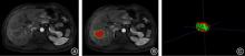
Fig.1
Delineation of the region of interest of the tumor"

Tab.1
Clinical and imaging characteristics of all HCC patients"
| 变量 | 训练集 (n = 84) | 阴性组 (n = 41) | 阳性组 (n = 43) | t/U/χ2值 | P值 | 验证集 (n = 36) | 阴性组 (n = 18) | 阳性组 (n = 18) | t/U/χ2值 | P值 |
|---|---|---|---|---|---|---|---|---|---|---|
| MTD/mm | 47.96 ± 37.02 | 53.78 ± 42.94 | 43.38 ± 31.32 | 959.5 | 0.42 | 42.97 ± 36.98 | 54.00 ± 44.05 | 31.94 ± 24.87 | 221.5 | 0.06 |
| 年龄/岁 | 57.87 ± 11.39 | 58.68 ± 12.77 | 57.23 ± 10.28 | 0.57 | 0.57 | 59.36 ± 10.14 | 60.56 ± 9.44 | 58.17 ± 10.92 | 0.702 | 0.49 |
| AST/(U/L) | 73.80 ± 110.35 | 56.30 ± 43.92 | 87.58 ± 141.50 | 887 | 0.88 | 109.76 ± 194.51 | 122.82 ± 246.61 | 96.69 ± 129.29 | 173.0 | 0.74 |
| ALT/(U/L) | 61.33 ± 105.25 | 41.79 ± 24.10 | 76.71 ± 137.79 | 829 | 0.72 | 90.98 ± 138.42 | 66.13 ± 59.38 | 115.82 ± 186.05 | 173.0 | 0.74 |
| PT/s | 14.15 ± 11.21 | 15.60 ± 16.75 | 13.01 ± 2.04 | 830.5 | 0.73 | 12.94 ± 1.98 | 12.56 ± 1.52 | 13.32 ± 2.33 | 138.5 | 0.47 |
| PLT/(×109/L) | 180.40 ± 81.72 | 176.81 ± 88.28 | 183.23 ± 77.01 | 805.5 | 0.57 | 170.17 ± 80.12 | 176.17 ± 84.45 | 164.17 ± 77.50 | 0.444 | 0.66 |
| 组织学分化程度/[例(%)] | 4.87 | 0.03 | 1.829 | 0.18 | ||||||
| 高分化 | 31(36.90) | 19(51.35) | 12(25.53) | 21(58.33) | 13(72.22) | 8(44.44) | ||||
| 非高分化 | 53(63.10) | 18(48.65) | 35(74.47) | 15(41.67) | 5(27.78) | 10(55.56) | ||||
| AFP/[例(%)] | 0 | 1 | 5.512 | 0.02 | ||||||
| < 20 μg/mL | 43(51.19) | 19(51.35) | 24(51.06) | 16(44.44) | 4(22.22) | 12(66.67) | ||||
| ≥ 20 μg/mL | 41(48.81) | 18(48.65) | 23(48.94) | 20(55.56) | 14(77.78) | 6(33.33) | ||||
| 性别/[例(%)] | 0.015 | 0.90 | 0.2 | 0.66 | ||||||
| 女 | 6(7.14) | 2(5.41) | 4(8.51) | 6(16.67) | 2(11.11) | 4(22.22) | ||||
| 男 | 78(92.86) | 35(94.59) | 43(91.49) | 30(83.33) | 16(88.89) | 14(77.78) | ||||
| 病史/[例(%)] | 0.015 | 0.90 | 0.93 | 0.34 | ||||||
| 非乙肝 | 6(7.14) | 2(5.41) | 4(8.51) | 5(13.89) | 4(22.22) | 1(5.56) | ||||
| 乙肝 | 78(92.86) | 35(94.59) | 43(91.49) | 31(86.11) | 14(77.78) | 17(94.44) | ||||
| 出血/[例(%)] | 3.247 | 0.07 | 0.2 | 0.66 | ||||||
| 无 | 75(89.29) | 30(81.08) | 45(95.74) | 30(83.33) | 14(77.78) | 16(88.89) | ||||
| 有 | 9(10.71) | 7(18.92) | 2(4.26) | 6(16.67) | 4(22.22) | 2(11.11) | ||||
| 坏死/[例(%)] | 1.884 | 0.17 | 4.012 | 0.05 | ||||||
| 无 | 40(47.62) | 14(37.84) | 26(55.32) | 19(52.78) | 6(33.33) | 13(72.22) | ||||
| 有 | 44(52.38) | 23(62.16) | 21(44.68) | 17(47.22) | 12(66.67) | 5(27.78) | ||||
| 包膜/[例(%)] | 0 | 1 | 2.922 | 0.09 | ||||||
| 无 | 42(50.00) | 19(51.35) | 23(48.94) | 14(38.89) | 4(22.22) | 10(55.56) | ||||
| 有 | 42(50.00) | 18(48.65) | 24(51.06) | 22(61.11) | 14(77.78) | 8(44.44) | ||||
| 脂肪/[例(%)] | 1.199 | 0.27 | 0 | 1 | ||||||
| 无 | 62(73.81) | 30(81.08) | 32(68.09) | 25(69.44) | 13(72.22) | 12(66.67) | ||||
| 有 | 22(26.19) | 7(18.92) | 15(31.91) | 11(30.56) | 5(27.78) | 6(33.33) | ||||
| 边界/[例(%)] | 0.618 | 0.43 | 0.138 | 0.71 | ||||||
| 清楚 | 54(64.29) | 26(70.27) | 28(59.57) | 26(72.22) | 12(66.67) | 14(77.78) | ||||
| 模糊 | 30(35.71) | 11(29.73) | 19(40.43) | 10(27.78) | 6(33.33) | 4(22.22) |
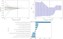
Fig.2
Construction of radiomics model"
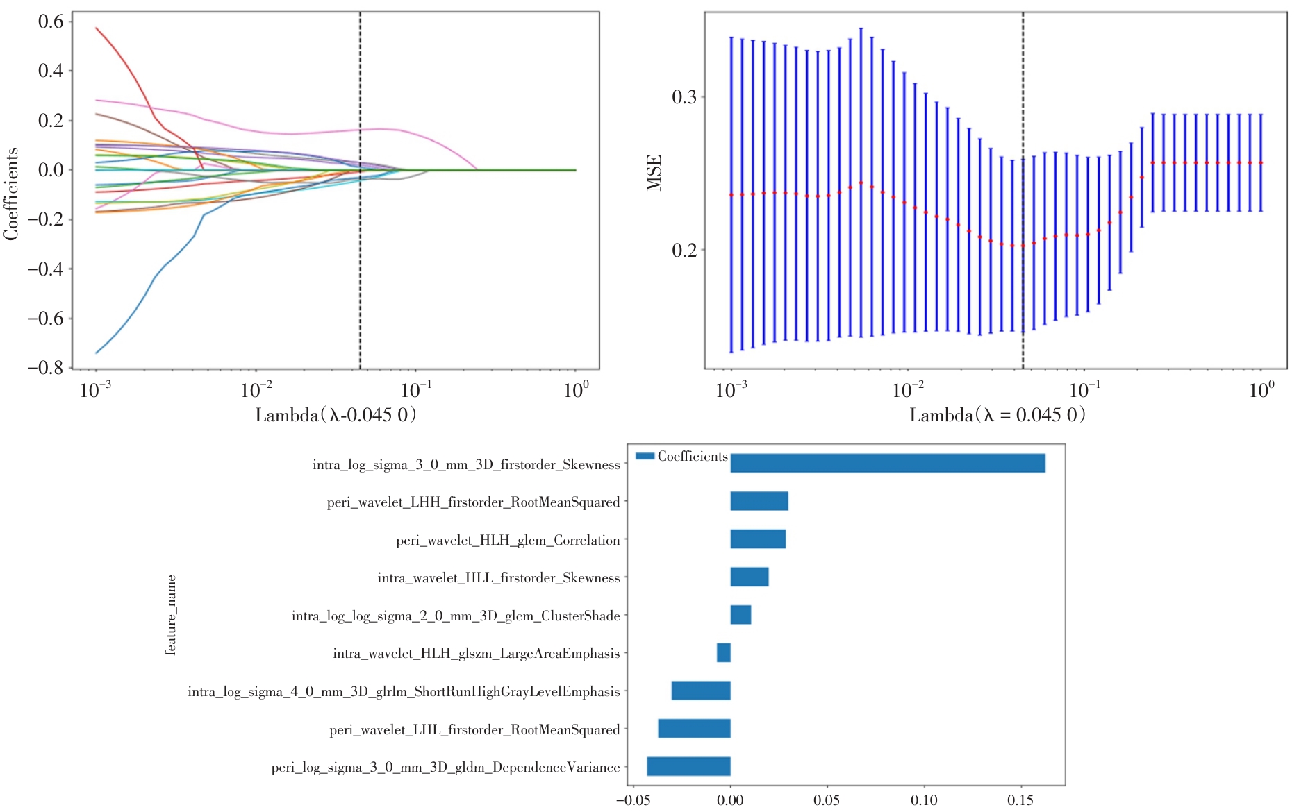
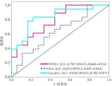
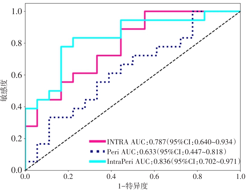
Fig.3
Receiver operating characteristic curve (ROC) and area under the curve (AUC) of the intratumoral (Intra), peritumoral (Peri), and combined intratumoral and peritumoral (IntraPeri) model performance in the validation set"
Tab.2
Univariate and multivariate analysis of clinical imaging characteristics using logistic regression"
| 变量 | OR(95%CI) | P值 | OR(95%CI) | P值 |
|---|---|---|---|---|
| 出血 | 0.286(0.076 ~ 1.068) | 0.118 | ||
| 坏死 | 0.913(0.056 ~ 1.499) | 0.763 | ||
| MTD | 1.000(0.094 ~ 1.006) | 0.930 | ||
| PLT | 1.001(0.999 ~ 1.023) | 0.256 | ||
| PT | 1.003(0.982 ~ 1.023) | 0.837 | ||
| 年龄 | 1.004(0.097 ~ 1.01) | 0.338 | ||
| AST | 1.004(1 ~ 1.007) | 0.127 | ||
| ALT | 1.006(1 ~ 1.012) | 0.092 | ||
| 性别 | 1.229(0.845 ~ 1.786) | 0.366 | ||
| 病史 | 1.229(0.845 ~ 1.786) | 0.366 | ||
| AFP | 1.278(0.761 ~ 2.145) | 0.436 | ||
| 包膜 | 1.333(0.799 ~ 2.228) | 0.356 | ||
| 边界 | 1.727(0.926 ~ 3.222) | 0.149 | ||
| 组织学分化程度 | 1.944(1.207 ~ 3.133) | 0.022 | 1.944(1.207 ~ 3.133) | 0.022 |
| 脂肪 | 2.143(1.009 ~ 4.549) | 0.096 |
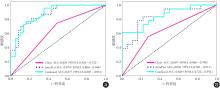
Fig.4
Receiver operating characteristic curve"
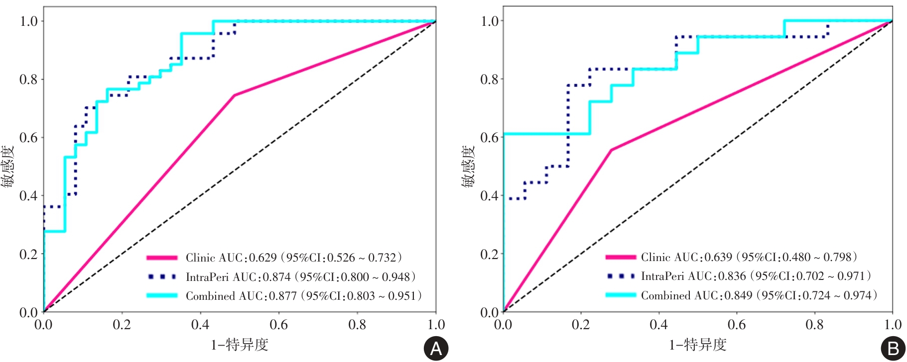

Fig.5
The Delong test and decision curve of the validation set"
Tab.3
Diagnostic performance of different models in predicting Ki-67 expression in liver cancer on the training set and validation set"
| 模型 | 准确性 | AUC | 95% CI | 敏感度 | 特异度 | 阳性预测值 | 阴性预测值 | |
|---|---|---|---|---|---|---|---|---|
| 训练集 | 临床模型 | 0.643 | 0.629 | 0.526 ~ 0.732 | 0.745 | 0.514 | 0.660 | 0.613 |
| 瘤内瘤周模型 | 0.786 | 0.874 | 0.801 ~ 0.948 | 0.702 | 0.892 | 0.892 | 0.702 | |
| 联合模型 | 0.821 | 0.877 | 0.803 ~ 0.951 | 0.957 | 0.649 | 0.776 | 0.923 | |
| 测试集 | 临床模型 | 0.639 | 0.639 | 0.480 ~ 0.798 | 0.556 | 0.722 | 0.667 | 0.619 |
| 瘤内瘤周模型 | 0.806 | 0.836 | 0.702 ~ 0.971 | 0.778 | 0.833 | 0.824 | 0.789 | |
| 联合模型 | 0.806 | 0.849 | 0.724 ~ 0.974 | 0.611 | 1.000 | 1.000 | 0.720 |
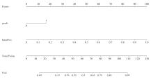
Fig.6
Nomograph of the combined model"
| [1] | 苏秋铭. CD97通过调控PI3K/AKT/mTOR通路促进肝细胞癌的增殖、侵袭和转移的机制研究[D]. 昆明:昆明医科大学, 2024. |
| [2] |
JIANG C, SUN X, QIU W, et al. Conversion therapy in liver transplantation for hepatocellular carcinoma: What's new in the era of molecular and immune therapy?[J]. Hepatobiliary Pancreat Dis Int, 2023, 22(1): 7-13. doi:10.1016/j.hbpd.2022.10.006
doi: 10.1016/j.hbpd.2022.10.006 |
| [3] | ZHIYING M, PEIYIN C, JIE L, et al. Pre-operative MRI features predict early post-operative recurrence of hepatocellular carcinoma with different degrees of pathological differentiation[J]. Radiol Med, 2023, 128(3):261-273. |
| [4] | 冯加其,刘雄青,黄鑫昱,等. m6A修饰在肝细胞癌药物治疗中作用研究进展[J]. 新医学,2024,55(6):480-488. |
| [5] | 段建平,杜镭,宋霆,等. 甲胎蛋白、AFP-L3%和DCP在HBV相关肝细胞癌早期诊断中的临床价值[J]. 新医学,2023,54(8):564-568. |
| [6] |
HU B, WANG R, ZHANG H, et al. Postnatal development of rat retina: A continuous observation and comparison between the organotypic retinal explant model and in vivo development[J]. Neural Regen Res, 2025, 20(3):900-912. doi:10.4103/nrr.nrr-d-23-01557
doi: 10.4103/nrr.nrr-d-23-01557 |
| [7] |
DENG J, TANG Q, WEI Q, et al. Preoperative assessment of Ki-67 expression in hepatocellular carcinoma using a multi-parametric spectral CT approach[J]. Quant Imaging Med Surg, 2025, 15(5):4262-4273. doi:10.21037/qims-24-2313
doi: 10.21037/qims-24-2313 |
| [8] | LIANG Y, SHENG G, GUO Y, et al. Prognostic significance of grade of malignancy based on histopathological differentiation and Ki-67 in pancreatic ductal adenocarcinoma[J]. Cancer BiolMed, 2024, 21(5): 416-432. |
| [9] |
ZHUO J, LU D, WANG J, et al. Molecular phenotypes reveal heterogeneous engraftments of patient-derived hepatocellular carcinoma xenografts[J]. Chin J Cancer Res, 2021, 33(4): 470-479. doi:10.21147/j.issn.1000-9604.2021.04.04
doi: 10.21147/j.issn.1000-9604.2021.04.04 |
| [10] |
SEUNGHYUP J, JIN O M, UNYONG K, et al. Glycosylation of serum haptoglobin as a marker of gastric cancer: an overview for clinicians[J]. Expert Rev Proteomics, 2020, 17(2):109-117. doi:10.1080/14789450.2020.1740091
doi: 10.1080/14789450.2020.1740091 |
| [11] | KONIARI I, ARTOPOULOU E, VELISSARIS D, et al. Biomarkers in the clinical management of patients with atrial fibrillation and heart failure[J]. J Geriatr Cardiol, 2021, 18(11): 908-951. |
| [12] | NOEL S W. Study on the Mechanisms of SAA/TLR4/NETs Inducing Angiogenesis in Rheumatoid Arthritis[D]. 天津:天津医科大学, 2024. |
| [13] |
JI J, BU Z, CHEN J. National guidelines for diagnosis and treatment of gastric cancer 2022 in China(English version)[J]. Chin J Cancer Res, 2022, 34(3): 207-242. doi:10.21147/j.issn.1000-9604.2022.03.04
doi: 10.21147/j.issn.1000-9604.2022.03.04 |
| [14] |
WANG J G. Chinese Guidelines for the Prevention and Treatment of Hypertension(2024 revision)[J]. J Geriatr Cardiol, 2025, 22(1): 1-149. doi:10.26599/1671-5411.2025.01.008
doi: 10.26599/1671-5411.2025.01.008 |
| [15] |
WU G, ZHU R, LU Y, et al. Optical scanning endoscope via a single multimode optical fiber[J]. Opto-Electron Sci, 2024, 3(3): 4-36. doi:10.29026/oes.2024.230041
doi: 10.29026/oes.2024.230041 |
| [16] |
REN X, LUO X, WANG F, et al. Recent advances in copper homeostasis-involved tumor theranostics[J]. Asian J Pharm Sci, 2024, 19(5): 56-89. doi:10.1016/j.ajps.2024.100948
doi: 10.1016/j.ajps.2024.100948 |
| [17] | 赖丹惠, 江燕辉, 叶思婷, 等. 颈动脉斑块超声影像组学特征对颈动脉支架置入术后再狭窄的预测能力[J]. 实用医学杂志, 2025, 41(5): 742-750. |
| [18] | WANG Q, ZHAO F, ZHANG H, et al. Deep learning-based multi-task prediction of response to neoadjuvant chemotherapy using multiscale whole slide images in breast cancer:A multicenter study[J]. Chin J Cancer Res, 2025, 37(1): 28-54. |
| [19] |
SHI Z X, LI C F, ZHAO L F, et al. Computed tomography radiomic features and clinical factors predicting the response to first transarterial chemoembolization in intermediate-stage hepatocellular carcinoma[J]. Hepatobiliary Pancreat Dis Int, 2024, 23(4): 361-369. doi:10.1016/j.hbpd.2023.06.011
doi: 10.1016/j.hbpd.2023.06.011 |
| [20] |
LI H, QIAO Y, DAI X, et al. 3D bioprinting of tumor models and potential applications[J]. Bio-Design and Manuf, 2024, 7(6): 857-888. doi:10.1007/s42242-024-00317-y
doi: 10.1007/s42242-024-00317-y |
| [21] | GHAZAL M. WDR76 Exerts Oncogene Function in Hepatocellular Carcinoma Via the G2/M Checkpoint Signaling Pathway[D]. 济南:山东大学, 2025. |
| [22] |
QI X, CHEN S, HE H, et al. The role and potential application of extracellular vesicles in liver cancer[J]. Sci ChinaLife Sci, 2021, 64(8): 1281-1294. doi:10.1007/s11427-020-1905-7
doi: 10.1007/s11427-020-1905-7 |
| [23] |
GANBIN Q, JINCAN C, WEIXIONGe L, et al. Gadoxetic acid-enhanced MRI combined with T1 mapping and clinical factors to predict Ki-67 expression of hepatocellular carcinoma[J]. Front Oncol, 2023, 13:1134646. doi:10.3389/fonc.2023.1134646
doi: 10.3389/fonc.2023.1134646 |
| [24] |
WANG X, CHEN X, NIU D, et al. National guidelines for diagnosis and treatment of breast cancer 2022 in China(English version)[J]. Chin J Cancer Res, 2022, 34(3): 151-175. doi:10.21147/j.issn.1000-9604.2022.03.02
doi: 10.21147/j.issn.1000-9604.2022.03.02 |
| [25] | 陈丽萍, 罗菊玉, 彭章艳, 等. 肿瘤病灶体积与子宫体积比值和组织中Ki-67、p16蛋白表达与子宫内膜癌病理特征及复发的关联[J]. 实用医学杂志, 2024, 40(23): 3367-3372. |
| [26] | 吕清清. 钆塞酸二钠增强MRI联合T1 mapping序列在预测肝细胞癌微血管侵犯及Ki-67表达中的价值[D]. 青岛:青岛大学, 2024. |
| [27] | 李思琪. 基于MRI影像组学对HBV相关性肝细胞癌及肝细胞癌GPC3表达水平预测价值的研究[D]. 保定:河北大学, 2025. |
| [1] | Yingmei HAN,Yijie LI,Heng ZHANG,Weiqing LI,Ze FENG,Feng WANG. The application value of deep learning in imaging studies for predicting the conversion of Alzheimer′s disease [J]. The Journal of Practical Medicine, 2025, 41(9): 1413-1424. |
| [2] | Li TANG,Yurong GONG,Liye ZENG,Yanfang GAO,Chengzhe. DENG. Application value of 3.0T magnetic resonance imaging T2 mapping sequence combined with serum nesfatin⁃1 level detection in the diagnosis of elderly knee early osteoarthritis [J]. The Journal of Practical Medicine, 2025, 41(8): 1238-1242. |
| [3] | Huiling YE,Zhengchaoyi CHEN,Yihan HUANG,Yingjie ZHANG,Xiangbin ZHANG,Yuehu PU,Renming ZHONG. Radiotherapy treatment comparison of liver SBRT between 4D⁃CT and deep inspiration breath hold troughmagnetic resonance imaging [J]. The Journal of Practical Medicine, 2025, 41(7): 1044-1049. |
| [4] | Danhui LAI,Yanhui JIANG,Siting YE,Shulian ZHUANG,Shuang YANG,Wen XUE,Jianxing ZHANG. Analysis of prediction of carotid in-stent restenosis based on ultrasonographic carotid plaque radiomics [J]. The Journal of Practical Medicine, 2025, 41(5): 742-750. |
| [5] | Yue LV,Yanna MENG,Panpan LI,Yinghong. CHEN. Value of SWD in preoperative assessment of liver fibrosis in patients with hepatocellular carcinoma [J]. The Journal of Practical Medicine, 2025, 41(5): 751-755. |
| [6] | Yuling DUAN,Xuezhi ZHOU,Yongyi LI,Lixia MA,Desheng YANG,Jiao CHENG,Yan WU,Tao LIU,Guoyuan JIANG,Mei. WANG. Clinical value analysis of different MRI measurement methods in evaluating the efficacy of neoadjuvant therapy for breast cancer [J]. The Journal of Practical Medicine, 2025, 41(14): 2152-2159. |
| [7] | Shangpeng HE,Weixian HUANG,Yanhui JIANG,Xiongqiang PENG,Lingcui MENG,Jianxing ZHANG. Value of ultrasound radiomics in re⁃evaluating the benign or malignant of Bethesda Ⅲ nodules [J]. The Journal of Practical Medicine, 2025, 41(12): 1892-1898. |
| [8] | Xiaoxiao QIN,Xiaozhuo LI,Hongli GUO,Lijing ZHANG. Combined value of multimodal fMRI and MRS in the differential diagnosis of postoperative recurrence and pseudoprogression of glioma [J]. The Journal of Practical Medicine, 2025, 41(11): 1736-1741. |
| [9] | Yanhao SUN,Yi ZHOU,Yilong. HU. The predictive value of rectus abdominis area and visceral fat distribution for the risk of surgical site infection after open radical surgery for hepatocellular carcinoma [J]. The Journal of Practical Medicine, 2025, 41(10): 1445-1452. |
| [10] | Yi WANG,Jie LIU,Mianjing LI,Li. TANG. Efficacy and safety of lobaplatin application on liver wound in hepatectomy of patients with primary hepatocellular carcinoma [J]. The Journal of Practical Medicine, 2025, 41(10): 1453-1459. |
| [11] | Zhilong SI,Hao WANG,Fei XIAO. Feasibility of modified LIFT guided by magnetic resonance imaging in the treatment of deep anorectal abscess [J]. The Journal of Practical Medicine, 2025, 41(1): 65-70. |
| [12] | Fazhu FEI,Jiajun LU,Shuai ZHANG,Hao LI,Bin REN. Clinical application progress of immunization and targeted therapy for Hepatocellular Carcinoma in special populations [J]. The Journal of Practical Medicine, 2024, 40(6): 738-742. |
| [13] | Zhipeng WU,Yuqin ZHANG,Minggang WANG,Rongzhen ZHANG,Dewen. MAO. Expression and biological function of TRP signaling pathway in hepatocellular carcinoma [J]. The Journal of Practical Medicine, 2024, 40(6): 743-747. |
| [14] | Yuxin CHENG,Liang LIU,Shiyu DONG,Shengchao LI,Meng ZHANG. Research advances in exosomal proteins, mRNA and non⁃coding RNA regulation of Hepatocellular Carcinoma [J]. The Journal of Practical Medicine, 2024, 40(6): 748-755. |
| [15] | Junhong XU,Hongbing YAO,Xueyao WANG,Wei GUO,Caijin LU,Jiaxing WU,Jianhui JIANG,Dongkang ZHAO. Clinical efficacy of FOLFOX⁃HAIC combined with lenvatinib and PD⁃1 inhibitor in the treatment of intermediate and advanced Hepatocellular Carcinoma [J]. The Journal of Practical Medicine, 2024, 40(6): 762-767. |
| Viewed | ||||||
|
Full text |
|
|||||
|
Abstract |
|
|||||