

The Journal of Practical Medicine ›› 2025, Vol. 41 ›› Issue (20): 3267-3275.doi: 10.3969/j.issn.1006-5725.2025.20.017
• Medical Examination and Clinical Diagnosis • Previous Articles
Yuying SONG,Saimaiti KHALIBINUR,Xu ZHOU,Xiangxin SONG,Yidai Abdigiti XIE,Aizezi DILRABA,Yibulayin. HASYATI( )
)
Received:2025-06-19
Online:2025-10-25
Published:2025-11-05
Contact:
Yibulayin. HASYATI
E-mail:3236321936@qq.com
CLC Number:
Yuying SONG,Saimaiti KHALIBINUR,Xu ZHOU,Xiangxin SONG,Yidai Abdigiti XIE,Aizezi DILRABA,Yibulayin. HASYATI. Quantitative susceptibility mapping analysis of brain iron deposition characteristics and their correlation with cognitive function in Alzheimer's disease patients[J]. The Journal of Practical Medicine, 2025, 41(20): 3267-3275.
Tab.1
General information comparison between AD group and control group"
| 变量 | 对照组 | AD组 | t/χ2值 | P值 |
|---|---|---|---|---|
| 性别 | 0.565 | 0.452 | ||
| 男 | 20(47.62) | 17(39.53) | ||
| 女 | 22(52.38) | 26(60.47) | ||
| 年龄( | 71.095 ± 8.384 | 69.977 ± 9.787 | -0.565 | 0.573 |
| 身高( | 162.286 ± 8.379 | 164.233 ± 7.656 | 1.119 | 0.266 |
| 体质量( | 64.000 ± 11.756 | 69.698 ± 11.135 | 2.295 | 0.024 |
| BMI( | 24.329 ± 3.332 | 25.794 ± 3.225 | 2.060 | 0.043 |
| 受教育水平 | 1.989 | 0.370 | ||
| 初中以下 | 20(47.62) | 27(62.79) | ||
| 高中、中专 | 12(28.57) | 9(20.93) | ||
| 大专、本科 | 10(23.81) | 7(16.28) | ||
| 高血压 | 27(64.29) | 29(67.44) | 0.094 | 0.759 |
| 冠心病 | 15(35.71) | 8(18.60) | 3.151 | 0.076 |
| 糖尿病 | 8(19.05) | 13(30.23) | 1.429 | 0.232 |
| 吸烟史 | 15(35.71) | 10(23.26) | 1.588 | 0.208 |
| 饮酒史 | 9(21.43) | 7(16.28) | 0.369 | 0.544 |
| MMSE[M(P25,P75)]/分 | 27.50(27.00,28.00) | 18.00(12.50,21.00) | -7.966 | <0.01 |
| MOCA [M(P25,P75)]/分 | 26.00(26.00,27.00) | 13.00(8.00,17.50) | -8.032 | <0.01 |
| ADL[M(P25,P75)]/分 | 100.00(100.00,100.00) | 85.000(55.00,100.00) | -6.352 | <0.01 |
Tab.2
Comparison of magnetic sensitivity values of frontal cortex, basal nucleus, and hippocampus between AD group and control group [M(P25,P75)]/(×10-4 ppb)"
| 变量 | 对照组(n = 42) | AD组(n = 43) | Z值 | P值 |
|---|---|---|---|---|
| 额叶皮质 | -15.827(-26.2,-4.6) | -11.529(-21.4,2.8) | -1.802 | 0.072 |
| 基底节区 | 389.802(314.1,481.9) | 479.586(408.8,577.4) | -2.980 | 0.003 |
| 海马 | 35.448(-10.8,90.7) | 102.686(48.6,141.7) | -2.792 | 0.005 |
Tab.3
Single factor analysis of factors affecting AD occurrence"
| 变量 | β | S.E | Z值 | P值 | OR(95%CI) |
|---|---|---|---|---|---|
| 性别 | |||||
| 男 | 1.000(Reference) | ||||
| 女 | 0.330 | 0.439 | 0.751 | 0.453 | 1.390(0.588 ~ 3.287) |
| 受教育水平 | |||||
| 初中以下 | 1.000 (Reference) | ||||
| 大专、本科 | -0.657 | 0.574 | -1.143 | 0.253 | 0.519(0.168 ~ 1.598) |
| 高中、中专 | -0.588 | 0.531 | -1.108 | 0.268 | 0.556(0.196 ~ 1.572) |
| 年龄/岁 | -0.014 | 0.024 | -0.570 | 0.569 | 0.986(0.941 ~ 1.034) |
| 身高/cm | 0.031 | 0.028 | 1.117 | 0.264 | 1.031(0.977 ~ 1.089) |
| 体质量/kg | 0.045 | 0.021 | 2.181 | 0.029 | 1.046(1.005 ~ 1.089) |
| BMI/(kg/m2) | 0.139 | 0.070 | 1.990 | 0.047 | 1.149(1.002 ~ 1.317) |
| 额叶皮质MSV | 0.028 | 0.016 | 1.812 | 0.070 | 1.029(0.998 ~ 1.061) |
| 基底节区MSV | 0.004 | 0.002 | 2.315 | 0.021 | 1.004(1.001 ~ 1.007) |
| 海马MSV | 0.005 | 0.003 | 2.009 | 0.045 | 1.005(1.001 ~ 1.011) |
Tab.4
Multivariate logistic regression analysis of factors affecting the occurrence of Alzheimer's disease"
| 变量 | β | S.E | Z值 | P值 | OR(95%CI) |
|---|---|---|---|---|---|
| 体质量/kg | 0.047 | 0.038 | 1.257 | 0.209 | 1.048 (0.974 ~ 1.129) |
| BMI/(kg/m2) | 0.025 | 0.124 | 0.197 | 0.844 | 1.025 (0.803 ~ 1.308) |
| 额叶皮质MSV | 0.035 | 0.019 | 1.823 | 0.068 | 1.036 (0.997 ~ 1.075) |
| 基底节区MSV | 0.004 | 0.002 | 2.251 | 0.024 | 1.004 (1.001 ~ 1.007) |
| 海马MSV | 0.008 | 0.003 | 2.565 | 0.010 | 1.008 (1.002 ~ 1.014) |
Tab.5
Comparison of magnetic sensitivity values in each subregion of ROI between AD group and control group [M(P25,P75)]/(×10-4 ppb)"
| 变量 | 对照组(n = 42) | AD组(n = 43) | Z值 | P值 |
|---|---|---|---|---|
| 左侧尾状核 | -4.614(-120.4,50.3) | 116.928(10.1,190.0) | -3.744 | < 0.01 |
| 右侧尾状核 | 22.949(-149.3,111.1) | 98.025(-33.0,201.2) | -2.215 | 0.027 |
| 左侧苍白球 | 788.759(546.4,957.3) | 845.302(666.4,1063.8) | -1.459 | 0.145 |
| 右侧苍白球 | 1 013.062(806.8,1 181.0) | 864.178(571.5,1150.6) | -1.863 | 0.062 |
| 左侧壳核 | 194.925(132.2,277.9) | 299.232(223.4,436.9) | -3.586 | < 0.01 |
| 右侧壳核 | 225.323(90.8,354.5) | 228.868(147.0,312.2) | -0.439 | 0.660 |
| 左侧海马头 | 28.978(-52.4,88.5) | 71.965(-4.5,202.3) | -2.151 | 0.031 |
| 右侧海马头 | 13.229(-110.1,121.9) | 99.851(16.5,231.5) | -2.816 | 0.005 |
| 左侧海马体 | -11.640(-56.3,52.1) | -10.206(-77.4,114.2) | -0.784 | 0.433 |
| 右侧海马体 | 38.372(-45.0,93.0) | 33.993(-17.1,129.2) | -0.186 | 0.852 |
| 左侧海马尾 | 52.521(-56.2,174.6) | 136.694(41.5,194.3) | -2.161 | 0.031 |
| 右侧海马尾 | 102.905(19.4,209.5) | 145.831(25.9,255.1) | -0.775 | 0.438 |
Tab.6
Correlation analysis between magnetic sensitivity values and MMSE"
| 部位 | 统计量 | MMSE |
|---|---|---|
| 左侧额叶皮质 | 相关系数 | -0.090 |
| P值 | 0.411 | |
| 右侧额叶皮质 | 相关系数 | 0.097 |
| P值 | 0.379 | |
| 左侧尾状核 | 相关系数 | -0.291 |
| P值 | 0.007 | |
| 右侧尾状核 | 相关系数 | -0.212 |
| P值 | 0.051 | |
| 左侧苍白球 | 相关系数 | -0.173 |
| P值 | 0.114 | |
| 右侧苍白球 | 相关系数 | 0.158 |
| P值 | 0.150 | |
| 左侧壳核 | 相关系数 | -0.150 |
| P值 | 0.170 | |
| 右侧壳核 | 相关系数 | 0.092 |
| P值 | 0.404 | |
| 左侧海马头 | 相关系数 | -0.17 |
| P值 | 0.132 | |
| 右侧海马头 | 相关系数 | -0.198 |
| P值 | 0.080 | |
| 左侧海马体 | 相关系数 | -0.164 |
| P值 | 0.145 | |
| 右侧海马体 | 相关系数 | -0.026 |
| P值 | 0.82 | |
| 左侧海马尾 | 相关系数 | -0.250 |
| P值 | 0.026 | |
| 右侧海马尾 | 相关系数 | -0.03 |
| P值 | 0.793 |
Tab.7
Correlation analysis between magnetic sensitivity values and MOCA"
| 部位 | 统计量 | MOCA |
|---|---|---|
| 左侧额叶皮质 | 相关系数 | -0.024 |
| P值 | 0.824 | |
| 右侧额叶皮质 | 相关系数 | 0.092 |
| P值 | 0.403 | |
| 左侧尾状核 | 相关系数 | -0.303 |
| P值 | 0.005 | |
| 右侧尾状核 | 相关系数 | -0.177 |
| P值 | 0.105 | |
| 左侧苍白球 | 相关系数 | -0.156 |
| P值 | 0.154 | |
| 右侧苍白球 | 相关系数 | 0.172 |
| P值 | 0.116 | |
| 左侧壳核 | 相关系数 | -0.258 |
| P值 | 0.017 | |
| 右侧壳核 | 相关系数 | 0.036 |
| P值 | 0.742 | |
| 左侧海马头 | 相关系数 | -0.136 |
| P值 | 0.228 | |
| 右侧海马头 | 相关系数 | -0.217 |
| P值 | 0.055 | |
| 左侧海马体 | 相关系数 | -0.240 |
| P值 | 0.032 | |
| 右侧海马体 | 相关系数 | 0.044 |
| P值 | 0.700 | |
| 左侧海马尾 | 相关系数 | -0.268 |
| P值 | 0.016 | |
| 右侧海马尾 | 相关系数 | -0.041 |
| P值 | 0.722 |
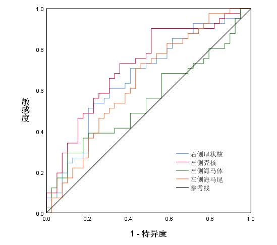
Tab.8
ROC model for diagnosing AD by iron deposition"
| 部位 | AUC | 最佳界值 | 敏感度 | 特异度 | Cut-off | P值 | 95%CI |
|---|---|---|---|---|---|---|---|
| 左侧尾状核 | 0.758 | 0.556 | 0.659 | 0.897 | 72.000 | < 0.01 | 0.649 ~ 0.867 |
| 左侧壳核 | 0.719 | 0.390 | 0.902 | 0.487 | 176.000 | 0.001 | 0.606 ~ 0.831 |
| 左侧海马体 | 0.551 | 0.190 | 0.293 | 0.897 | 91.667 | 0.433 | 0.424 ~ 0.678 |
| 左侧海马尾 | 0.640 | 0.271 | 0.707 | 0.564 | 63.685 | 0.031 | 0.518 ~ 0.763 |
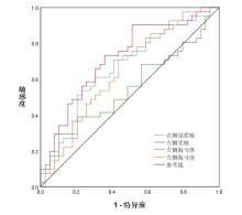
Fig.1
ROC curve of iron deposition diagnosis for AD"
| [1] |
SCHELTENS P, DE STROOPER B, KIVIPELTO M, et al. Alzheimer's disease[J]. Lancet, 2021, 397(10284): 1577-1590. doi:10.1016/s0140-6736(20)32205-4
doi: 10.1016/s0140-6736(20)32205-4 |
| [2] |
SPOTORNO N, ACOSTA-CABRONERO J, STOMRUD E, et al. Relationship between cortical iron and tau aggregation in Alzheimer's disease[J]. Brain, 2020, 143(5): 1341-1349. doi:10.1093/brain/awaa089
doi: 10.1093/brain/awaa089 |
| [3] | 郁金泰. 阿尔茨海默病精准诊疗现状和展望[J]. 中华医学信息导报, 2024, 39(17): 16. |
| [4] | 周显波, 裴中. 阿尔茨海默病早发现和早诊断与药物研发现状[J]. 阿尔茨海默病及相关病, 2024, 7(4): 243-246. |
| [5] |
PORSTEINSSON A P, ISAACSON R S, KNOX S, et al. Diagnosis of Early Alzheimer's Disease: Clinical Practice in 2021[J]. J Prev Alzheimers Dis, 2021, 8(3): 371-386. doi:10.14283/jpad.2021.23
doi: 10.14283/jpad.2021.23 |
| [6] |
FENG L, SUN J, XIA L, et al. Ferroptosis mechanism and Alzheimer's disease[J]. Neural Regen Res, 2024, 19(8): 1741-1750. doi:10.4103/1673-5374.389362
doi: 10.4103/1673-5374.389362 |
| [7] | 张其华, 谭艳. 磁共振定量磁敏感图在中枢神经系统的研究进展[J]. 磁共振成像, 2022, 13(1): 151-153,170. |
| [8] | 张琴, 侯勇哲, 王琳. 阿尔茨海默病脑铁沉积的QSM定量分析研究进展[J]. 医疗卫生装备, 2022, 43(9): 91-96. |
| [9] |
JACK C R Jr, ANDREWS S J, BEACH T G, et al. Revised criteria for the diagnosis and staging of Alzheimer's disease[J]. Nat Med, 2024, 30(8): 2121-2124. doi:10.1038/s41591-024-02988-7
doi: 10.1038/s41591-024-02988-7 |
| [10] |
QUINTAS-NEVES M, ALMEIDA F C, GAUTHREAUX K, et al. Fazekas scale magnetic resonance imaging assessment in Alzheimer's disease and primary age-related tauopathy[J]. Neuroradiology, 2024, 66(12): 2185-2193. doi:10.1007/s00234-024-03464-2
doi: 10.1007/s00234-024-03464-2 |
| [11] | 王金佩,蒋琦,李桂丽,等.新型冠状病毒肺炎对阿尔兹海默症影响的研究进展[J].中国热带医学,2023,23(3):304-309. |
| [12] |
MIELKE M M, EVANS J K, NEIBERG R H, et al. Alzheimer Disease Blood Biomarkers and Cognition Among Individuals with Diabetes and Overweight or Obesity[J]. JAMA Netw Open, 2025, 8(2): e2458149. doi:10.1001/jamanetworkopen.2024.58149
doi: 10.1001/jamanetworkopen.2024.58149 |
| [13] |
LU Q, SHAO N, FANG Z, et al. The anti-Alzheimer's disease effects of ganoderic acid A by inhibiting ferroptosis-lipid peroxidation via activation of the NRF2/SLC7A11/GPX4 signaling pathway[J]. Chem Biol Interact, 2025, 412: 111459. doi:10.1016/j.cbi.2025.111459
doi: 10.1016/j.cbi.2025.111459 |
| [14] |
THORWALD M A, GODOY-LUGO J A, KERSTIENS E, et al. Down syndrome with Alzheimer's disease brains have increased iron and associated lipid peroxidation consistent with ferroptosis[J]. Alzheimers Dement, 2025, 21(6): e70322. doi:10.1002/alz.70322
doi: 10.1002/alz.70322 |
| [15] |
THORWALD M A, GODOY-LUGO J A, GARCIA G, et al. Iron-associated lipid peroxidation in Alzheimer's disease is increased in lipid rafts with decreased ferroptosis suppressors, tested by chelation in mice[J]. Alzheimers Dement, 2025, 21(1): e14541. doi:10.1002/alz.14541
doi: 10.1002/alz.14541 |
| [16] | 程峰, 张庸, 王祥, 等. 谷胱甘肽过氧化物酶GPX4在铁死亡中的作用与机制研究进展[J]. 现代肿瘤医学, 2021, 29(7): 1254-1258. |
| [17] |
BUTTERFIELD D A. Brain lipid peroxidation and Alzheimer disease: Synergy between the Butterfield and Mattson laboratories[J]. Ageing Res Rev, 2020, 64: 101049. doi:10.1016/j.arr.2020.101049
doi: 10.1016/j.arr.2020.101049 |
| [18] |
BUTTERFIELD D A, BOYD-KIMBALL D. Mitochondrial Oxidative and Nitrosative Stress and Alzheimer Disease[J]. Antioxidants (Basel), 2020, 9(9): 818. doi:10.3390/antiox9090818
doi: 10.3390/antiox9090818 |
| [19] | 张金彪, 张凤翔, 高嘉璐. 定量磁化率成像技术在认知功能障碍中的研究进展[J]. 磁共振成像, 2023, 14(9): 114-118,130. |
| [20] |
ZHU W Z, ZHONG W D, WANG W, et al. Quantitative MR phase-corrected imaging to investigate increased brain iron deposition of patients with Alzheimer disease[J]. Radiology, 2009, 253(2): 497-504. doi:10.1148/radiol.2532082324
doi: 10.1148/radiol.2532082324 |
| [21] |
LIU X, DU L, ZHANG B, et al. Alterations and associations between magnetic susceptibility of the basal ganglia and diffusion properties in Alzheimer's disease[J]. Front Neurosci, 2021, 15: 616163. doi:10.3389/fnins.2021.616163
doi: 10.3389/fnins.2021.616163 |
| [22] |
GHADERI S, MOHAMMADI S, NEZHAD N J, et al. Iron quantification in basal ganglia: Quantitative susceptibility mapping as a potential biomarker for Alzheimer's disease - a systematic review and meta-analysis[J]. Front Neurosci, 2024, 18: 1338891. doi:10.3389/fnins.2024.1338891
doi: 10.3389/fnins.2024.1338891 |
| [23] |
QIN Y, ZHU W, ZHAN C, et al. Investigation on positive correlation of increased brain iron deposition with cognitive impairment in Alzheimer disease by using quantitative MR R2' mapping[J]. J Huazhong Univ Sci Technolog Med Sci, 2011, 31(4): 578. doi:10.1007/s11596-011-0493-1
doi: 10.1007/s11596-011-0493-1 |
| [24] |
CHEN W, JIANG L F, HU Y Q, et al. Ferritin reduction is essential for cerebral ischemia-induced hippocampal neuronal death through p53/SLC7A11-mediated ferroptosis[J]. Brain Res, 2021, 1752: 147216. doi:10.1016/j.brainres.2020.147216
doi: 10.1016/j.brainres.2020.147216 |
| [25] |
BAO W D, PANG P, ZHOU X T, et al. Loss of ferroportin induces memory impairment by promoting ferroptosis in Alzheimer's disease[J]. Cell Death Differ, 2024, 31(8): 1099. doi:10.1038/s41418-024-01290-w
doi: 10.1038/s41418-024-01290-w |
| [26] |
SCHWIMMBECK F, BULLÓN TARRASÓ E, SCHREINER T. A role for respiration in coordinating sleep oscillations and memory consolidation[J]. Trends Neurosci, 2025, 48(4): 247-249. doi:10.1016/j.tins.2025.02.005
doi: 10.1016/j.tins.2025.02.005 |
| [27] |
WANG D, LI Y Y, LUO J H, et al. Age-related iron deposition in the basal ganglia of controls and Alzheimer disease patients quantified using susceptibility weighted imaging[J]. Arch Gerontol Geriatr, 2014, 59(2): 439-449. doi:10.1016/j.archger.2014.04.002
doi: 10.1016/j.archger.2014.04.002 |
| [28] | 金泉伟, 杨小梅. 增加铁沉积对脑定量敏感性的映射与阿尔茨海默病中认知功能降低之间关系的研究[J]. 脑与神经疾病杂志, 2019, 27(10): 625-630. |
| [29] | 申杰, 徐桂华. 阿尔茨海默病与血脑屏障的相关性研究进展[J]. 实用医学杂志, 2024, 40(11): 1602-1606. |
| [30] |
LI R, FAN Y R, WANG Y Z, et al. Brain iron in signature regions relating to cognitive aging in older adults: The Taizhou Imaging Study[J]. Alzheimers Res Ther, 2024, 16(1): 211. doi:10.1186/s13195-024-01575-9
doi: 10.1186/s13195-024-01575-9 |
| [31] |
DU L, ZHAO Z, CUI A, et al. Increased iron deposition on brain quantitative susceptibility mapping correlates with decreased cognitive function in Alzheimer's disease[J]. ACS Chem Neurosci, 2018, 9(7): 1849-1857. doi:10.1021/acschemneuro.8b00194
doi: 10.1021/acschemneuro.8b00194 |
| [32] | 黎超洋, 崔凯茵, 房国梁. 铁代谢在运动改善阿尔茨海默病中的作用与机制研究进展[J]. 中国体育科技, 2023, 59(6): 46-55. |
| [1] | Cong LIU,Fei ZHAI,Min LI,Xiaoli ZHANG,Han SHI,Ningning GUO,Changhong WANG. Association between smoking status, cognitive function, and personality traits in first⁃episode male patients with schizophrenia [J]. The Journal of Practical Medicine, 2025, 41(12): 1922-1928. |
| [2] | Song LI,Xingyou HE,Dian HE,Bo WANG,Yu ZHAN,Jingjing. SUN. The joint efficacy of NBP and LIPost C in treatment of elderly patients with large atherosclerotic cerebral infarction [J]. The Journal of Practical Medicine, 2024, 40(9): 1286-1292. |
| [3] | Jinhui XU,Mailong YUAN,Tao ZHOU,Mingsheng ZHANG,Yaqi. LI. Effects of sevoflurane or propofol anesthesia maintenance on renal function and postoperative cognition in patients undergoing liver transplantation [J]. The Journal of Practical Medicine, 2024, 40(24): 3509-3514. |
| [4] |
FANG Xue, SHAO Wei, XU Huifang, LI Daoxin, WANG Jing..
A study on improvement in cognitive function after cerebral infarction based on 2⁃Cl⁃MGV⁃1/BDNF⁃TrkB pathway [J]. The Journal of Practical Medicine, 2023, 39(7): 819-826. |
| [5] | Yaxin YANG,Mao HUANG,Xin WANG,Yaru ZHI,Xuemeng SHI,Hongjin REN,Wanying. PENG. Effect of head acupuncturing therapy combined with massaging key head acupoints on language and cognitive function in children with autism [J]. The Journal of Practical Medicine, 2023, 39(23): 3132-3136. |
| [6] | Suzhen YE,Xiaoyang WANG,Hongzhao BAI,Xuezhen ZHOU,Haiyan LI. Clinical efficacy and influencing factor of Remote Ischemic Postconditioning in Patients with Post Stroke Fatigue [J]. The Journal of Practical Medicine, 2023, 39(21): 2812-2816. |
| [7] | DENG Xiren , ZENG Daojun, ZHANG Guanpeng, DUAN Xiaoxia. . The effect of baicalin on cognitive function of cerebral ischemia-reperfusion injury in mice through PGE2 [J]. The Journal of Practical Medicine, 2023, 39(15): 1881-1887. |
| [8] |
GUO Fei, HUANG Yunhui, HUANG Yujing, NIU Huifang, DU Ailing. .
Systematic review of efficacy and safety of xingnaojing injection combined with alteplase for ischemic stroke [J]. The Journal of Practical Medicine, 2022, 38(2): 206-211. |
| [9] |
YANG Bo, ZHANG Maorui, ZHOU Jun.
Effects of propofol preconditioning on cognitive function in septic rats [J]. The Journal of Practical Medicine, 2021, 37(7): 845-850. |
| [10] |
LONG Qijia, LV Zongxia, SHI Ke, LI Chunyan, ZHENG Jinou..
Analysis of hippocampus in patients with anti⁃NMDAR encephalitis by voxel⁃based morphology and func⁃ tional connectivity [J]. The Journal of Practical Medicine, 2021, 37(18): 2366-2370. |
| [11] |
LI Jia, LIN Yin, JI Xunqi, KANG Yanhai, DONG Jie..
Association of APOE and MTHFR C677T gene polymorphisms with schizophrenia and cognitive function in patients [J]. The Journal of Practical Medicine, 2021, 37(18): 2391-2394. |
| [12] |
LI Jia, LIN Yin, JI Xunqi, KANG Yanhai, DONG Jie..
Association of APOE and MTHFR C677T gene polymorphisms with schizophrenia and cognitive function in patients [J]. The Journal of Practical Medicine, 2021, 37(18): 2391-2394. |
| [13] |
LI Ge, DAI Yun, JI Liang, GUAN Dongsheng. .
Thymoquinone inhibits oxidative stress and improvesthe cognitive function of rats with cerebral small ves⁃ sel disease [J]. The Journal of Practical Medicine, 2021, 37(11): 1387-1391. |
| [14] |
CHEN Wei, ZHOU Xuyan, JIANG Lishan, QIAN Yan⁃ fei, LU Hong, LU Yaping.
Effects of ultrasound-guided stellate ganglion block on intrapulmonary shunt and postoperative cognitive function in elderly patients with one⁃lung ventilation
[J]. The Journal of Practical Medicine, 2020, 36(24): 3390-3393.
|
| [15] | YUAN Liuq⁃ ing, LIANG Weidong, LI Xiaoling, LAI Luying. Goal ⁃ directed fluid therapy on hemodynamics and S100β protein in meningioma rese [J]. The Journal of Practical Medicine, 2020, 36(22): 3126-3129. |
| Viewed | ||||||
|
Full text |
|
|||||
|
Abstract |
|
|||||