

The Journal of Practical Medicine ›› 2025, Vol. 41 ›› Issue (3): 322-329.doi: 10.3969/j.issn.1006-5725.2025.03.003
• Basic Research • Previous Articles
Rongxin LI,Li HUANG,Yueyang ZENG,Shuhui ZHANG,Yiran CHEN,Yuli LIU,Tieming. MA( )
)
Received:2024-09-09
Online:2025-02-10
Published:2025-02-19
Contact:
Tieming. MA
E-mail:1049355021@qq.com
CLC Number:
Rongxin LI,Li HUANG,Yueyang ZENG,Shuhui ZHANG,Yiran CHEN,Yuli LIU,Tieming. MA. Exploring the mechanism of electroacupuncture to improve cognitiveimpairment in alzheimer′s disease model rats based on NF⁃κB/NLRP3/Caspase⁃1 signaling pathway[J]. The Journal of Practical Medicine, 2025, 41(3): 322-329.
Tab.1
Comparison of escape latency periods among different groups of rats"
| 组别 | 逃避潜伏期 | ||||
|---|---|---|---|---|---|
| 第1天 | 第2天 | 第3天 | 第4天 | 第5天 | |
| 假手术组 | 8.53 ± 3.83 | 5.40 ± 2.31 | 6.07 ± 3.74 | 5.07 ± 3.58 | 4.67 ± 3.06 |
| 模型组 | 70.73 ± 19.56 | 55.20 ± 11.64▲ | 30.00 ± 5.19▲ | 26.00 ± 6.61▲ | 20.00 ± 4.99▲ |
| 电针组 | 62.00 ± 13.29 | 31.00 ± 1.06? | 14.80 ± 2.03? | 8.60 ± 2.77? | 7.53 ± 2.91? |
| 西药组 | 53.53 ± 11.80 | 23.80 ± 5.37? | 13.20 ± 4.85? | 7.87 ± 1.86? | 4.53 ± 1.14? |
Tab.2
Comparison of crossing platform times and target quadrant dwell time among different groups of rats"
| 组别 | 穿越平台次数/次 | 目标象限停留时间/s |
|---|---|---|
| 假手术组 | 14 ± 1 | 34 ± 1.2 |
| 模型组 | 5.33 ± 0.58▲ | 16.2 ± 0.72▲ |
| 电针组 | 10.33 ± 0.58** | 28.2 ± 8.0* |
| 西药组 | 12 ± 2** | 31.93 ± 3.06** |
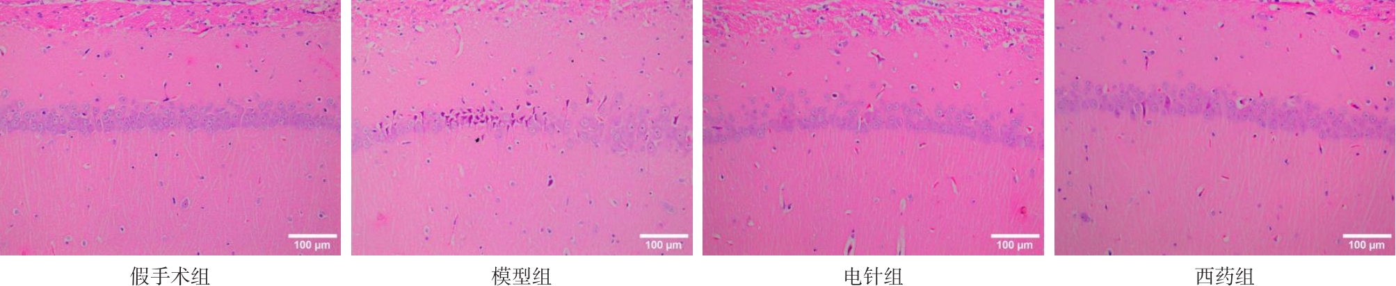
Fig.1
Morphological changes in the hippocampus of rats in each group"
Tab.3
Comparison of serum TNF - α and IL-1 β levels among different groups of rats"
| 组别 | TNF-α浓度 | IL-1β浓度 |
|---|---|---|
| 假手术组 | 38.71 ± 4.18 | 62.14 ± 7.93 |
| 模型组 | 165.64 ± 28.33▲ | 238.49 ± 35.60▲ |
| 电针组 | 125.52 ± 11.96? | 170.37 ± 20.63? |
| 西药组 | 93.88 ± 11.84??# | 142.19 ± 27.79?? |
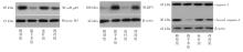
Fig.2
Comparison of NF?κB p65, NLRP3, and Caspase?1 protein expression in hippocampal tissues of rats in different groups"

Tab.4
Comparison of NF-κB p65, NLRP3, and Caspase-1 protein expression in hippocampal tissues of rats in different groups"
| 组别 | NF-κB p65 Protein level | NLRP3 Protein level | Caspase1Protein level |
|---|---|---|---|
| 假手术组 | 1.05 ± 0.06 | 1.07 ± 0.13 | 1.09 ± 0.05 |
| 模型组 | 5.16 ± 1.08▲ | 5.55 ± 0.79▲ | 7.42 ± 1.20▲ |
| 电针组 | 3.15 ± 0.01? | 3.83 ± 0.25? | 5.02 ± 0.59? |
| 西药组 | 1.96 ± 0.34?# | 2.65 ± 0.38?# | 2.03 ± 0.71?## |
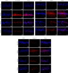
Fig.3
Comparison of positive expression of NF-κB p65, NLRP3, and Caspase-1 in the hippocampus of rats in each group"

Tab.5
Fluorescence expression of NF-κB p65, NLRP3, and Caspase-1 in hippocampal tissue of rats in each group"
| 组别 | NF-κB p65荧光强度 | NLRP3 荧光强度 | Caspase-1 荧光强度 |
|---|---|---|---|
| 假手术组 | 91 816.00 ± 13 678.32 | 91 092.67 ± 9 570.28 | 91 060.00 ± 18 668.42 |
| 模型组 | 1 545 102.56 ± 177 919.61▲ | 1 448 218.00 ± 280 592.01▲ | 1 677 210.78 ± 269 243.73▲ |
| 电针组 | 1 250 015.22 ± 189 131.27? | 1 050 356.00 ± 286 255.19 | 1 085 159.89 ± 133 167.58?? |
| 西药组 | 829 716.11 ± 100 050.21??# | 757 422.44 ± 156 653.38?? | 846 462.00 ± 92 857.19?? |
| 1 |
PASSERI E, ELKHOURY K, MORSINK M, et al. Alzheimer's Disease: Treatment Strategies and Their Limitations[J]. Int J Mol Sci, 2022,23(22):13954. doi:10.3390/ijms232213954
doi: 10.3390/ijms232213954 |
| 2 |
KLOSKE C M, DUGAN A J, WEEKMAN E M, et al. Inflammatory Pathways Are Impaired in Alzheimer Disease and Differentially Associated With Apolipoprotein E Status[J]. J Neuropathol Exp Neurol, 2021,80(10):922-932. doi:10.1093/jnen/nlab085
doi: 10.1093/jnen/nlab085 |
| 3 |
ROY E R, WANG B. WAN Y W,et al. Type I interferon response drives neuroinflammation and synapse loss in Alzheimer disease[J]. J Clin Invest, 2020,130(4):1912-1930. doi:10.1172/jci133737
doi: 10.1172/jci133737 |
| 4 |
PENNISI M, CRUPI R, DI PAOLA R, et al. Inflammasomes, hormesis, and antioxidants in neuroinflammation: Role of NRLP3 in Alzheimer disease[J]. J Neurosci Res, 2017,95(7):1360-1372. doi:10.1002/jnr.23986
doi: 10.1002/jnr.23986 |
| 5 | 潘芳芳,唐银杉,熊冰,等. 基于NF-κB/NLRP3/Caspase-1通路探究头穴透刺对阿尔茨海默病大鼠的干预效果[J]. 中华中医药学刊,2024,42(2):34-37+272. |
| 6 |
KONG Y, JIANG B, LUO X. Gut microbiota influences Alzheimer's disease pathogenesis by regulating acetate in Drosophila model[J]. Future Microbiol, 2018,13:1117-1128. doi:10.2217/fmb-2018-0185
doi: 10.2217/fmb-2018-0185 |
| 7 | 程美佳,袁常斌,鞠业涛,等. 涤痰汤通过调节IKB/NF-κB通路和细胞凋亡抑制阿尔茨海默病小鼠Tau蛋白过度磷酸化[J].时珍国医国药,2024,35(2):270-274. |
| 8 | 陈炜,李兴峰,陈业文,等. 温脾通络开窍方通过影响NF-κB通路改善阿尔兹海默症大鼠的认知功能研究[J]. 时珍国医国药,2021,32(10):2330-2333. |
| 9 | HE C, HUANG Z S, YU C C, et al. Preventive electroacupuncture ameliorates D-galactose-induced Alzheimer's disease-like inflammation and memory deficits, probably via modulating the microbiota-gut-brain axis[J]. Iran J Basic Med Sci, 2021,24(3):341-348. |
| 10 | 李真, 李梦醒, 覃云鹏, 等. 电针通过调控小胶质细胞改善阿尔茨海默病大鼠认知功能障碍的机制研究[J]. 针刺研究, 2023,48(11):1069-1078. |
| 11 |
陈虹茹,何川,黄重生,等. 电针联合重复经颅磁刺激对D-半乳糖诱导的阿尔茨海默病样模型大鼠学习记忆能力及神经炎症的影响 [J]. 实用医学杂志, 2021, 37(12): 1534-1538. doi:10.3969/j.issn.1006
doi: 10.3969/j.issn.1006 |
| 12 | 包新民,舒斯云. 大鼠脑立体定位图谱[M]. 北京:人民卫生出版社, 1991. |
| 13 | 李忠仁. 实验针灸学[M]. 北京:中国中医药出版社, 2007. |
| 14 | 姚春鹏. 黄帝内经[M]. 北京:中华书局, 2010. |
| 15 | 张静远, 赵娟, 虞鹤鸣, 等. 阿尔茨海默病脾虚痰饮病机探析[J]. 南京中医药大学学报, 2023,39(12):1174-1178. |
| 16 | 王杰,李凡,冯丽娜,等. 阿尔茨海默病的中医研究进展[J]. 中国中医基础医学杂志,2024,30(10):1790-1794. |
| 17 | 何川,黄重生,陈虹茹,等. 预针刺对AD样大鼠学习记忆能力及TLR4/NF-κB信号通路的影响[J]. 实用医学杂志,2020,36(18):2510-2514. |
| 18 |
YANG S, WANG G, MA Z F, et al. DietaryAdvancedGlycationEnd Products-InducedCognitive Impairment in Aged ICR Mice: Protective Role of Quercetin[J]. Mol Nutr Food Res, 2020,64(3):e1901019. doi:10.1002/mnfr.201901019
doi: 10.1002/mnfr.201901019 |
| 19 |
YANG Y, WANG L, ZHANG C, et al. Ginsenoside Rg1 improves Alzheimer's disease by regulating oxidative stress, apoptosis, and neuroinflammation through Wnt/GSK-3β/β-catenin signaling pathway[J]. Chem Biol Drug Des,2022,99(6):884-896. doi:10.1111/cbdd.14041
doi: 10.1111/cbdd.14041 |
| 20 |
EBERT S E, JENSEN P, OZENNE B, et al. Molecular imaging of neuroinflammation in patients after mild traumatic brain injury: A longitudinal (123) I-CLINDE single photon emission computed tomography study[J]. Eur J Neurol, 2019,26(12):1426-1432. doi:10.1111/ene.13971
doi: 10.1111/ene.13971 |
| 21 | 胡梦妮,张小蕾,荣臻,等. 电针对MPTP诱导帕金森病小鼠FoXO1/NLRP3通路介导神经炎症的影响[J]. 实用医学杂志,2024,40(11):1494-1499. |
| 22 |
XU L, SUN Y, LI M, et al. Dyrk2 mediated the release of proinflammatory cytokines in LPS-induced BV2 cells[J]. Int J Biol Macromol, 2018,109:1115-1124. doi:10.1016/j.ijbiomac.2017.11.095
doi: 10.1016/j.ijbiomac.2017.11.095 |
| 23 |
PAUDEL Y N, ANGELOPOULOU E, PIPERI C, et al. Impact of HMGB1, RAGE, and TLR4 in Alzheimer's Disease (AD): From Risk Factors to Therapeutic Targeting[J]. Cells, 2020,9(2):383. doi:10.3390/cells9020383
doi: 10.3390/cells9020383 |
| 24 |
SHAMIM D, LASKOWSKI M. Inhibition of inflammation mediated through the tumor necrosis factor α biochemical pathway can lead to favorable outcomes in Alzheimer disease[J]. J Cent Nerv Syst Dis, 2017,9:1-10. doi:10.1177/1179573517722512
doi: 10.1177/1179573517722512 |
| 25 |
HENEKA M T, KUMMER M P, STUTZ A, et al. NLRP3 is activated in Alzheimer's disease and contributes to pathology in APP/PS1 mice[J]. Nature, 2013,493(7434):674-678. doi:10.1038/nature11729
doi: 10.1038/nature11729 |
| 26 | HUANG L, GONG L, HUO X, et al. N-acetyldopamine dimer inhibits neuroinflammation through the TLR4/NF-kappaB and NLRP3/Caspase-1 pathways[J]. Acta Biochim Biophys Sin (Shanghai), 2022,55(1):23-33. |
| 27 |
KONG Y, JIANG B, LUO X. Gut microbiota influences Alzheimer's disease pathogenesis by regulating acetate in Drosophila model[J]. Future Microbiol, 2018,13:1117-1128. doi:10.2217/fmb-2018-0185
doi: 10.2217/fmb-2018-0185 |
| 28 | 宗堪堪,崔春爱. 小胶质细胞免疫特性的研究进展[J]. 实用医学杂志,2018,34(21):3638-3640. |
| [1] | Xingwei WU,Jianying WANG,Chengxiao GUO,Ziyi LIU,Chao SUN,Fei. YU. The effect of remimazolam on modulating the ROS/RAGE/NF-κB signaling pathway in LPS-induced microglial inflammation [J]. The Journal of Practical Medicine, 2025, 41(2): 153-161. |
| [2] | Bao WANG,Shulin MA,Xinhua YAO,Fan YANG,Kai WEN,Sijing LUO,Ying GAN,Yi. LU. Effect of intraoperative electroacupuncture analgesia on stress response to tracheal intubation in patients undergoing thyroid surgery under general anesthesia [J]. The Journal of Practical Medicine, 2024, 40(8): 1132-1136. |
| [3] | Zhe SHI,Xialin ZUO,Linhui PENG,Zhiwei LU,Kongping. LI. Effect of M1 microglial polarization on secondary damage in the thalamus after cerebral cortical infarction [J]. The Journal of Practical Medicine, 2024, 40(22): 3138-3145. |
| [4] | Mengni HU,Xiaolei ZHANG,Zhen RONG,Yao WANG,Ya′nan LI,Jun. MA. Effects of electroacupuncture on MPTP⁃induced FoXO1/NLRP3 pathway mediated neuroinflammation in mice with Parkinson′s disease [J]. The Journal of Practical Medicine, 2024, 40(11): 1494-1499. |
| [5] | Yuanyuan MAO,Jingjing. YUAN. Research advances of long noncoding RNA H19 in central nervous system diseases [J]. The Journal of Practical Medicine, 2023, 39(23): 3021-3026. |
| [6] | Ying GAN,Bao WANG,Jiayin YAO,Xinhua YAO,Dong LIU,Rong HUANG,Yi. LU. Analgesic effect of perioperative electroacupuncture stimulation on laparoscopic surgery [J]. The Journal of Practical Medicine, 2023, 39(20): 2612-2617. |
| [7] |
ZHONG Peirui, HE Xiaoyan, LIAO Ying, SUN Guanghua, LIU Jing, ZHOU Jun, LI Shuzhi, LIU Yuan, QU Mengjian..
Study on the mechanism of P53/P21 pathway in electroacupuncture inhibiting osteoblast aging in osteopo⁃ rotic rats [J]. The Journal of Practical Medicine, 2023, 39(2): 192-197. |
| [8] | DENG Xiren , ZENG Daojun, ZHANG Guanpeng, DUAN Xiaoxia. . The effect of baicalin on cognitive function of cerebral ischemia-reperfusion injury in mice through PGE2 [J]. The Journal of Practical Medicine, 2023, 39(15): 1881-1887. |
| [9] |
HUANG Xiarong, ZHOU Jun, SUN Guanghua, PENG Xinke, LIAO Yuan, LIU Jing, LUO Fu, ZHONG Peirui, PENG Ting, HU Lizhi..
Effect of electroacupuncture on articular cartilage and subchondral polarization⁃related protein expression in aged rats [J]. The Journal of Practical Medicine, 2023, 39(12): 1473-1479. |
| [10] |
LI Hongy⁃ing , SHEN Yuan, WU Qiaofeng, XIE Lushuang, YU Shuguang..
Role of microglia polarization signaling pathways in neuroinflammation:A literature review
[J]. The Journal of Practical Medicine, 2022, 38(14): 1838-1846.
|
| [11] |
LI Hanzhang, QI Ling, ZHANG Xiaolei, CHEN Xianglin, GUO Lei, GUO Shuqin, MA Jun..
Effect of electroacupuncture on GLP⁃1R⁃mediated PI3K/AKT/GSK⁃3β pathway in mice with Parkinson′s disease [J]. The Journal of Practical Medicine, 2021, 37(21): 2712-2716. |
| [12] |
GUO Zhuang, ZHOU Lijun..
Dual role of astrocyte⁃microglia crosstalk in neuroinflammation [J]. The Journal of Practical Medicine, 2021, 37(18): 2432-2436. |
| Viewed | ||||||
|
Full text |
|
|||||
|
Abstract |
|
|||||