

The Journal of Practical Medicine ›› 2024, Vol. 40 ›› Issue (15): 2148-2153.doi: 10.3969/j.issn.1006-5725.2024.15.018
• Medical Examination and Clinical Diagnosis • Previous Articles Next Articles
Yue LÜ,Hujing LU,Juanjuan. ZHANG
Received:2024-02-01
Online:2024-08-10
Published:2024-07-30
CLC Number:
Yue LÜ,Hujing LU,Juanjuan. ZHANG. Value of MRI-DWI combined with attenuation imaging in diagnosis of focal nodular lesions < 2 cm in fatty liver[J]. The Journal of Practical Medicine, 2024, 40(15): 2148-2153.
Tab.1
Comparison of clinical data of patients with different focal nodular lesions"
| 组别 | 例数 | TG(mmol/L) | 年龄(岁) | HDL-C(mmol/L) | 性别[例(%)] | TC(mmol/L) | LDL-C(mmol/L) | |
|---|---|---|---|---|---|---|---|---|
| 男 | 女 | |||||||
| 良性结节组 | 41 | 2.16 ± 0.33 | 49.87 ± 9.36 | 1.25 ± 0.18 | 24(58.54) | 17(41.46) | 5.43 ± 1.52 | 2.97 ± 0.38 |
| 恶性结节组 | 27 | 2.22 ± 0.35 | 48.14 ± 9.73 | 1.29 ± 0.17 | 18(66.67) | 9(33.33) | 5.20 ± 1.57 | 2.81 ± 0.42 |
| t/χ2值 | 0.716 | 0.734 | 0.916 | 0.456 | 0.603 | 1.629 | ||
| P值 | 0.476 | 0.465 | 0.363 | 0.500 | 0.549 | 0.108 | ||
Tab.2
Comparison of clinical data of patients with different degrees of fatty liver disease"
| 组别 | 例数 | TG(mmol/L) | 年龄(岁) | HDL-C(mmol/L) | 性别[例(%)] | TC(mmol/L) | LDL-C(mmol/L) | |
|---|---|---|---|---|---|---|---|---|
| 男 | 女 | |||||||
| 轻度组 | 17 | 2.17 ± 0.32 | 49.72 ± 10.15 | 1.27 ± 0.14 | 10(58.82) | 7(41.18) | 5.41 ± 1.39 | 2.79 ± 0.38 |
| 中度组 | 27 | 2.23 ± 0.34 | 48.20 ± 9.42 | 1.30 ± 0.16 | 16(59.26) | 11(40.74) | 5.10 ± 1.46 | 2.62 ± 0.34 |
| 重度组 | 24 | 2.20 ± 0.35 | 49.35 ± 9.98 | 1.29 ± 0.15 | 16(66.67) | 8(33.33) | 5.28 ± 1.32 | 2.68 ± 0.37 |
| F/χ2值 | 0.167 | 0.151 | 0.205 | 0.378 | 0.272 | 1.159 | ||
| P值 | 0.847 | 0.860 | 0.815 | 0.828 | 0.763 | 0.320 | ||
Tab.3
Comparison of ADC and AC values in patients with different focal nodular lesions"
| 组别 | 例数 | ADC值(×10-3mm2/s) | AC值(dB·cm-1·MHz-1) | ||
|---|---|---|---|---|---|
| b = 100 s/mm2 | b = 500 s/mm2 | b = 1 000 s/mm2 | |||
| 良性结节组 | 41 | 2.32 ± 0.36 | 2.18 ± 0.27 | 2.11 ± 0.24 | 0.52 ± 0.13 |
| 恶性结节组 | 27 | 1.78 ± 0.19 | 1.68 ± 0.16 | 1.60 ± 0.15 | 0.97 ± 0.18 |
| t值 | 7.153 | 8.660 | 9.835 | 11.970 | |
| P值 | < 0.001 | < 0.001 | < 0.001 | < 0.001 | |
Tab.4
Comparison of ADC and AC values in patients with different degrees of fatty liver disease"
| 组别 | 例数 | ADC值(×10-3mm2/s) | AC值(dB·cm-1·MHz-1) | ||
|---|---|---|---|---|---|
| b = 100 s/mm2 | b = 500 s/mm2 | b = 1 000 s/mm2 | |||
| 轻度组 | 17 | 2.06 ± 0.29 | 1.96 ± 0.27 | 1.89 ± 0.25 | 0.59 ± 0.17 |
| 中度组 | 27 | 1.83 ± 0.23? | 1.78 ± 0.21? | 1.72 ± 0.19? | 0.91 ± 0.24? |
| 重度组 | 24 | 1.41 ± 0.18?# | 1.35 ± 0.16?# | 1.28 ± 0.12?# | 1.25 ± 0.33?# |
| F值 | 42.878 | 47.187 | 51.177 | 32.076 | |
| P值 | < 0.001 | < 0.001 | < 0.001 | < 0.001 | |
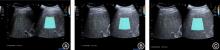
Fig.1
Comparison of ADC and AC values in patients with different degrees of fatty liver disease"

Tab.5
Analysis of the correlation between ADC and AC values under different b values and the degree of fatty liver disease, TG, HDL-C, TC and LDL-C"
| 指标 | 脂肪肝病变程度 | TG | HDL-C | TC | LDL-C | |||||
|---|---|---|---|---|---|---|---|---|---|---|
| r值 | P值 | r值 | P值 | r值 | P值 | r值 | P值 | r值 | P值 | |
| ADC值(b = 100 s/mm2) | -0.528 | < 0.001 | -0.082 | 0.543 | 0.128 | 0.361 | 0.320 | 0.414 | 0.074 | 0.453 |
| ADC值(b = 500 s/mm2) | -0.571 | < 0.001 | -0.145 | 0.267 | 0.194 | 0.265 | 0.189 | 0.265 | 0.098 | 0.324 |
| ADC值(b = 1 000 s/mm2) | -0.494 | < 0.001 | -0.268 | 0.321 | 0.172 | 0.297 | 0.274 | 0.306 | 0.128 | 0.139 |
| AC值 | 0.465 | < 0.001 | 0.129 | 0.207 | -0.274 | 0.012 | 0.074 | 0.572 | -0.041 | 0.742 |
Tab.6
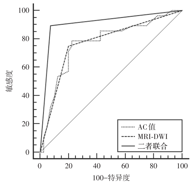
Analysis of the diagnostic value of MMRI -DWI combined with AC value in the malignant transformation of fatty liver with focal nodular disease < 2 cm"
| 指标 | 最佳截断点 | 灵敏度(%) | 特异度(%) | P值 | AUC值 | 95%CI |
|---|---|---|---|---|---|---|
| MRI-DWI | - | 75.00 | 80.00 | < 0.001 | 0.775 | 0.658 ~ 0.867 |
| AC值 | 0.99 dB·cm-1·MHz-1 | 78.57 | 77.50 | < 0.001 | 0.773 | 0.655 ~ 0.866 |
| 二者联合 | - | 89.29 | 92.50 | < 0.001 | 0.909 | 0.814 ~ 0.965 |
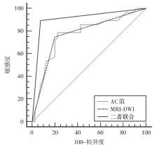
Fig.2
ROC curve of MMRI DWI combined with AC value in the diagnosis of fatty liver disease with focal nodular disease < 2 cm into malignancy"
| 1 |
王蕾,贵瑶瑶,寇丽. 艾塞那肽联合多烯磷脂酰胆碱对酒精性脂肪肝患者血清HMGB1、APN水平的影响[J]. 实用医学杂志,2023,39(9):1164-1168. doi:10.3969/j.issn.1006-5725.2023.09.018
doi: 10.3969/j.issn.1006-5725.2023.09.018 |
| 2 |
LEGOUT J D, BOLAN C W, BOWMAN A W, et al. Focal nodular hyperplasia and focal nodular hyperplasia-like lesions[J]. Radiographics, 2022,42(4):1043-1061. doi:10.1148/rg.210156
doi: 10.1148/rg.210156 |
| 3 |
BHANDARI A, KOPPEN J, WASTNEY T, et al. A systematic review and meta-analysis of spectral CT to differentiate focal liver lesions[J]. Clin Radiol, 2023,78(6):430-436. doi:10.1016/j.crad.2023.02.017
doi: 10.1016/j.crad.2023.02.017 |
| 4 |
VERNUCCIO F, CANNELLA R, BARTOLOTTA T V, et al. Advances in liver US, CT, and MRI: moving toward the future[J]. Eur Radiol Exp, 2021,5(1):52. doi:10.1186/s41747-021-00250-0
doi: 10.1186/s41747-021-00250-0 |
| 5 |
ALWALID O, WANG Y, FAN W, et al. Value of gadoxetic acid-enhanced MR imaging and DWI in classification, characterization and confidence in diagnosis of solid focal liver lesions[J]. Scand J Gastroenterol, 2021,56(1):72-80. doi:10.1080/00365521.2020.1847314
doi: 10.1080/00365521.2020.1847314 |
| 6 |
LEE D H. Quantitative assessment of fatty liver using ultrasound attenuation imaging[J]. J Med Ultrason (2001), 2021,48(4):465-470. doi:10.1007/s10396-021-01132-z
doi: 10.1007/s10396-021-01132-z |
| 7 |
中国研究型医院学会肝病专业委员会,中国医师协会脂肪性肝病专家委员会,中华医学会肝病学分会脂肪肝与酒精性肝病学组,等. 中国脂肪性肝病诊疗规范化的专家建议(2019年修订版)[J]. 中华肝脏病杂志,2019,27(10):748-753. doi:10.3760/cma.j.issn.1007-3418.2019.10.003
doi: 10.3760/cma.j.issn.1007-3418.2019.10.003 |
| 8 |
中华医学会肝病学分会脂肪肝和酒精性肝病学组,中国医师协会脂肪性肝病专家委员会. 非酒精性脂肪性肝病防治指南(2018年更新版)[J]. 临床肝胆病杂志,2018,34(5):947-957. doi:10.3969/j.issn.1001-5256.2018.05.007
doi: 10.3969/j.issn.1001-5256.2018.05.007 |
| 9 |
SEMALTI K, KILAMBI R, PAL S, et al. Benign hepatic nodules in patients with primary extrahepatic portal vein obstruction: clinical and magnetic resonance imaging features[J]. J Clin Exp Hepatol, 2022,12(5):1301-1309. doi:10.1016/j.jceh.2022.04.015
doi: 10.1016/j.jceh.2022.04.015 |
| 10 |
SAAKE M, SEUSS H, RIEXINGER A, et al. Image quality and detection of small focal liver lesions in diffusion-weighted imaging: comparison of navigator tracking and free-breathing acquisition[J]. Invest Radiol, 2021,56(9):579-590. doi:10.1097/rli.0000000000000776
doi: 10.1097/rli.0000000000000776 |
| 11 |
SON J S, PARK H S, PARK S, et al. Motion-corrected versus conventional diffusion-weighted magnetic resonance imaging of the liver using non-rigid registration[J]. Diagnostics (Basel), 2023,13(6)1008. doi:10.3390/diagnostics13061008
doi: 10.3390/diagnostics13061008 |
| 12 |
KIM D H, KIM B, LEE H S, et al. Deep learning-accelerated liver diffusion-weighted imaging: intraindividual comparison and additional phantom study of free-breathing and respiratory-triggering acquisitions[J]. Invest Radiol, 2023,58(11):782-790. doi:10.1097/rli.0000000000000988
doi: 10.1097/rli.0000000000000988 |
| 13 |
MURTZ P, MESROPYAN N, SPRINKART A M, et al. Simplified intravoxel incoherent motion diffusion-weighted MRI of liver lesions: feasibility of combined two-colour index maps[J]. Eur Radiol Exp, 2021,5(1):33. doi:10.1186/s41747-021-00233-1
doi: 10.1186/s41747-021-00233-1 |
| 14 | 王晓丽,刘晓莹,谢靖红. 多b值DWI序列ADC值联合肿瘤标志物检测对肝脏良恶性肿瘤诊断中的临床价值[J]. 中国CT和MRI杂志,2020,18(1):101-104. |
| 15 |
BAO J, LV Y, WANG K, et al. A comparative study of ultrasound attenuation imaging, controlled attenuation parameters, and magnetic resonance spectroscopy for the detection of hepatic steatosis[J]. J Ultrasound Med, 2023,42(7):1481-1489. doi:10.1002/jum.16158
doi: 10.1002/jum.16158 |
| 16 |
POUWELS S, SAKRAN N, GRAHAM Y, et al. Non-alcoholic fatty liver disease (NAFLD): a review of pathophysiology, clinical management and effects of weight loss[J]. BMC Endocr Disord, 2022,22(1):63. doi:10.1186/s12902-022-00980-1
doi: 10.1186/s12902-022-00980-1 |
| 17 |
TOKUSHIGE K, IKEJIMA K, ONO M, et al. Evidence-based clinical practice guidelines for nonalcoholic fatty liver disease/nonalcoholic steatohepatitis 2020[J]. J Gastroenterol, 2021,56(11):951-963. doi:10.1007/s00535-021-01796-x
doi: 10.1007/s00535-021-01796-x |
| 18 |
THIAGARAJAN P, BAWDEN S J, AITHAL G P. Metabolic imaging in non-alcoholic fatty liver disease: applications of magnetic resonance spectroscopy[J]. J Clin Med, 2021,10(4):632. doi:10.3390/jcm10040632
doi: 10.3390/jcm10040632 |
| 19 |
IDILMAN I S, YIDIZ A E, KARAOSMANOGLU A D, et al. Proton density fat fraction: magnetic resonance imaging applications beyond the liver[J]. Diagn Interv Radiol, 2022,28(1):83-91. doi:10.5152/dir.2021.21845
doi: 10.5152/dir.2021.21845 |
| 20 |
LEE S J, KIM Y R, LEEY H, et al. US attenuation imaging for the evaluation and diagnosis of fatty liver disease[J]. J Korean Soc Radiol, 2023,84(3):666-675. doi:10.3348/jksr.2022.0053
doi: 10.3348/jksr.2022.0053 |
| 21 |
MAKHIJA N, VIKRAM N K, SRIVASTAVA D N, et al. Role of diffusion-weighted magnetic resonance imaging in the diagnosis and grading of hepatic steatosis in patients with non-alcoholic fatty liver disease: comparison with ultrasonography and magnetic resonance spectroscopy[J]. J Clin Exp Hepatol, 2021,11(6):654-660. doi:10.1016/j.jceh.2021.02.008
doi: 10.1016/j.jceh.2021.02.008 |
| 22 |
杜丹,余卫中,甘洪颖. 磁共振弥散加权成像表观弥散系数联合肝脾体积比值对非酒精性脂肪肝合并肝纤维化的诊断价值研究[J]. 中国医学装备, 2021,18(2):32-36. doi:10.3969/J.ISSN.1672-8270.2021.02.009
doi: 10.3969/J.ISSN.1672-8270.2021.02.009 |
| 23 |
张记,闫艳,李金燕. 超声声衰减成像对不同程度非酒精性脂肪肝的诊断价值[J]. 临床超声医学杂志,2023,25(9):718-722. doi:10.3969/j.issn.1008-6978.2023.09.010
doi: 10.3969/j.issn.1008-6978.2023.09.010 |
| 24 |
包静文,朱宇莉,徐庆玥,等. 超声声衰减成像评估代谢相关脂肪性肝病肝脂肪变程度的应用价值[J]. 中华超声影像学杂志,2021,30(10):868-873. doi:10.3760/cma.j.cn131148-20210421-00280
doi: 10.3760/cma.j.cn131148-20210421-00280 |
| Viewed | ||||||
|
Full text |
|
|||||
|
Abstract |
|
|||||