| [1] |
Wei HE,Liping LIU,Jingwei ZHUO,Xiaodong ZHANG,Tong YANG,Jubin. FENG.
CCR5 blockade reduces tumor growth by inducing apoptosis and impairing immunosuppression of tumor microenvironment
[J]. The Journal of Practical Medicine, 2024, 40(9): 1204-1210.
|
| [2] |
Wenxin LI,Minjun LU,Li LIN,Yueqin LIU,Xiaolan. ZHU.
circRAF1 regulates the proliferation and apoptosis of human ovarian granulosa cells
[J]. The Journal of Practical Medicine, 2024, 40(7): 910-917.
|
| [3] |
Zhen YANG,Shaoru JIANG,Xiaoyan CHEN,Xiaolin CHEN,Weimin DENG,Xinyu. GUO.
Effect of Jinghou Zengzhi Granules on ovarian GDF9 secretion and granulosa cells apoptosis in controlled ovarian hyperstimulation rats
[J]. The Journal of Practical Medicine, 2024, 40(7): 918-923.
|
| [4] |
Runwei MA,Chunjie MU,Wenting GUI,Yao DENG,Minzhang ZHAO,Min LIU,Yi SONG.
LncRNA SENCR targeted miR⁃206 regulates proliferation and apoptosis of human vascular smooth muscle cells of aortic dissection tissues
[J]. The Journal of Practical Medicine, 2024, 40(3): 302-308.
|
| [5] |
Lihong DING,Shijia GENG,Yujie WANG.
Effects of wedelobata on apoptosis and secretion of inflammatory cytokines in the alveolar epithelium infected by Streptococcus pneumonia
[J]. The Journal of Practical Medicine, 2024, 40(3): 316-320.
|
| [6] |
Bing′er WU,Qing LI,Kerong YANG,Jian ZHANG,Yi YU,Lei LEI,Bo. HU.
Impact of spermidine on proliferation and apoptosis in diffuse large B⁃cell lymphoma cell lines
[J]. The Journal of Practical Medicine, 2024, 40(22): 3130-3137.
|
| [7] |
Chunmei ZHANG,Xinhui HUANG,Jinqiu HU,Xiaoyan BI,Fuli. YA.
Sulforaphane protects human platelets from high glucose⁃induced cellular apoptosis through down-regulating PI3K/Akt signaling pathway
[J]. The Journal of Practical Medicine, 2024, 40(18): 2530-2536.
|
| [8] |
Rao LÜ,Jiadi YU,Liuzhen LI,Chulan ZHAN,Liyue ZHAO,Yueliang LI,Jun DONG,Jiao. LI.
Molecular mechanism of young Sca⁃1 bone marrow stem cell on old cardiac fibroblast cell apoptosis in aging mice
[J]. The Journal of Practical Medicine, 2024, 40(17): 2369-2374.
|
| [9] |
LI Xiaoping, ZHOU Hongjian, GAO Fangfang, LI Wei..
Tspan1 antagonizes oxaliplatin⁃induced apoptosis in colorectal cancer cells by inducing cellular autophagy
[J]. The Journal of Practical Medicine, 2023, 39(9): 1072-1078.
|
| [10] |
ZHANG Zhuoer, GAO Xiaoya, YU Juan, WANG Yuli..
Experimental study of lncRNA KCNQ1OT1 targeting miR⁃129⁃5p to regulate inflammation and apoptosis in a Parkinson′s disease cell model
[J]. The Journal of Practical Medicine, 2023, 39(6): 672-678.
|
| [11] |
LI Mingjuan, LI Xuexin, LIU Li..
Effect of short⁃duration mechanical ventilation on diaphragm function in septic rats
[J]. The Journal of Practical Medicine, 2023, 39(5): 579-590.
|
| [12] |
LIU Xueqing, GONG Xiuqun, WANG Xia, WANG Qiwei, LU Jun..
Therapeutic mechanism of edaravone dexboeol on vascular dementia
[J]. The Journal of Practical Medicine, 2023, 39(4): 505-509.
|
| [13] |
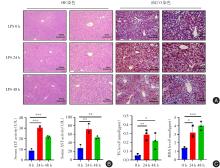
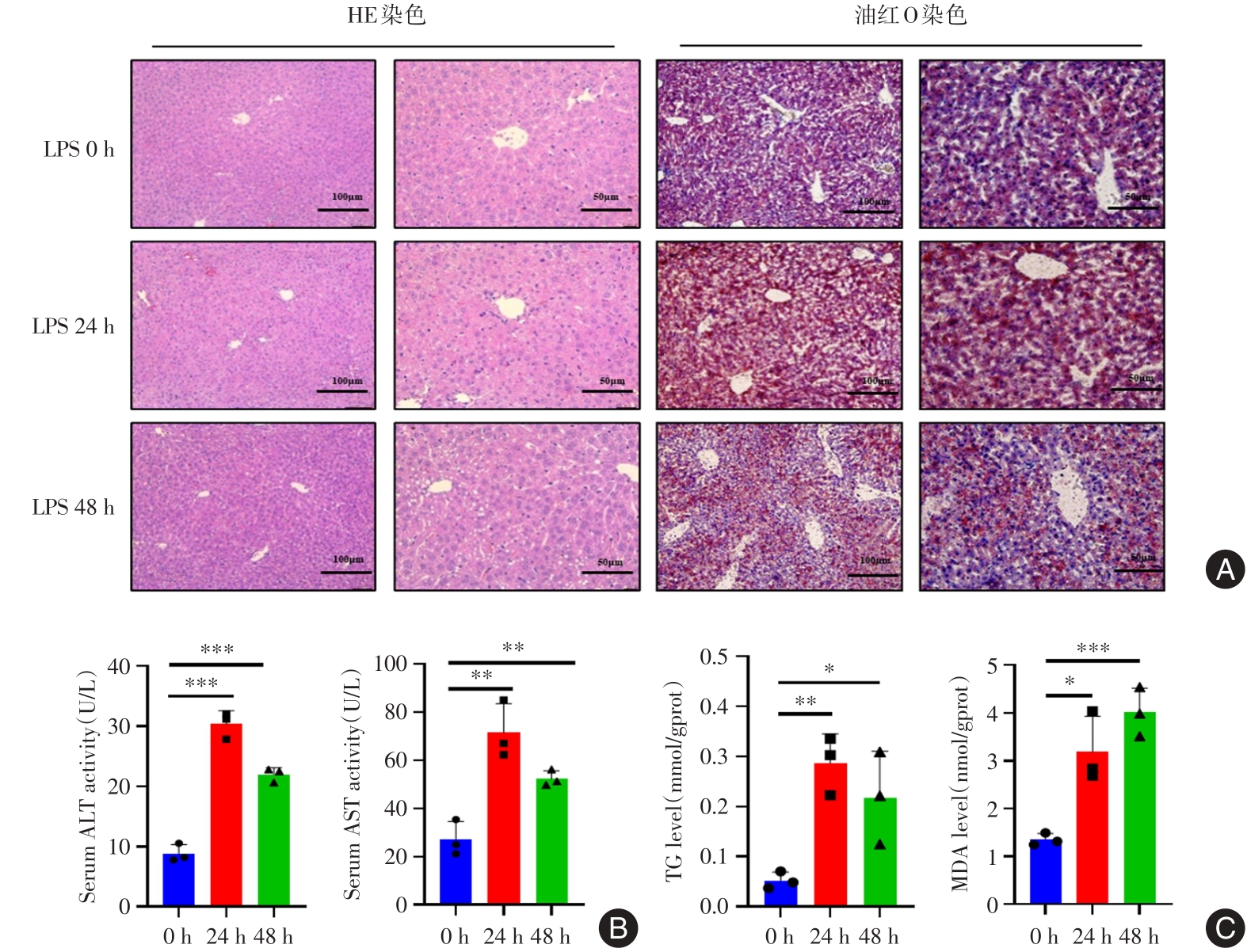
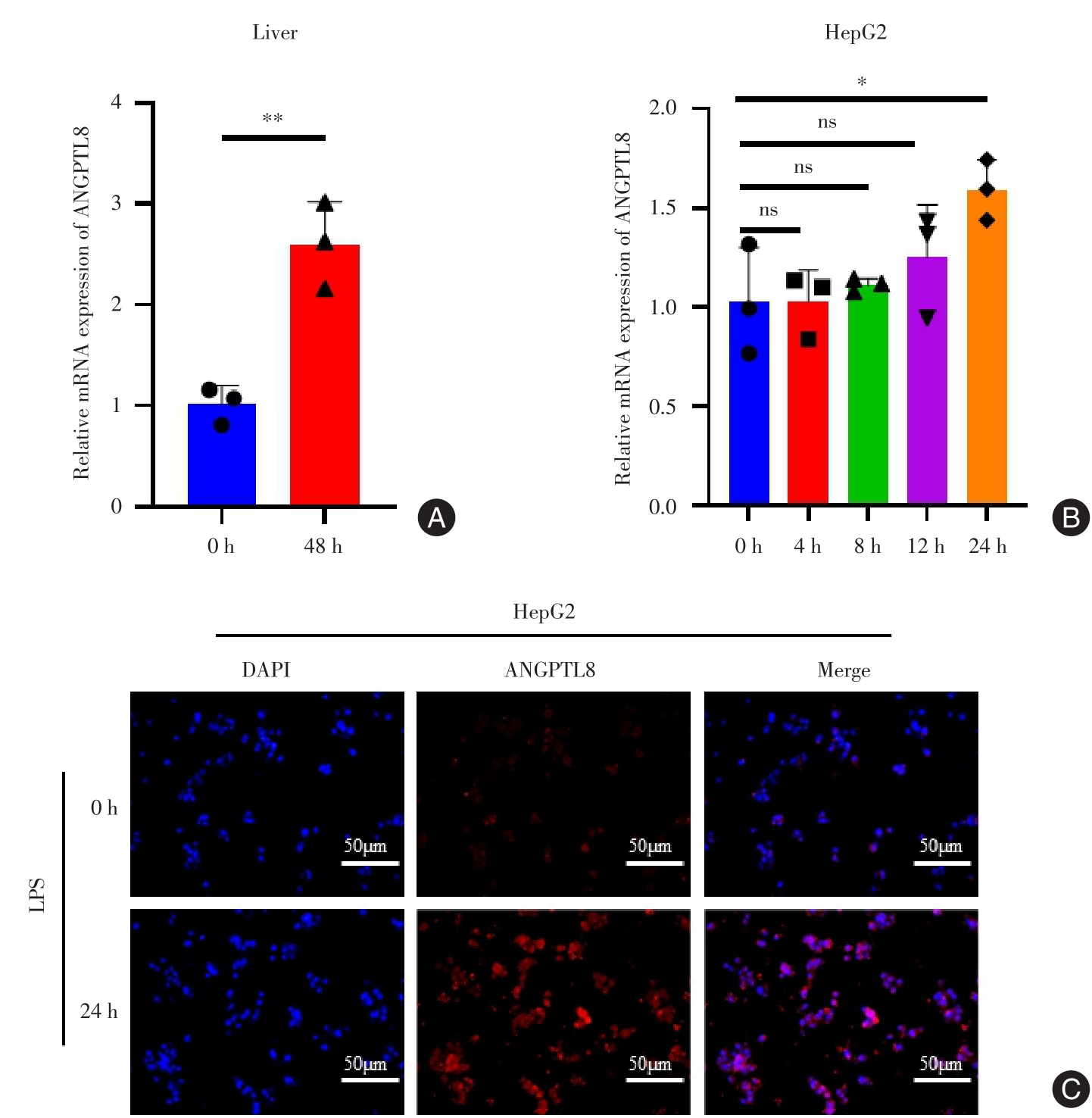
GAO Yujiu, HU Rong, FANG Chen, LI Panpan, MENG Xiang, GUO Xingrong, FENG Ying..
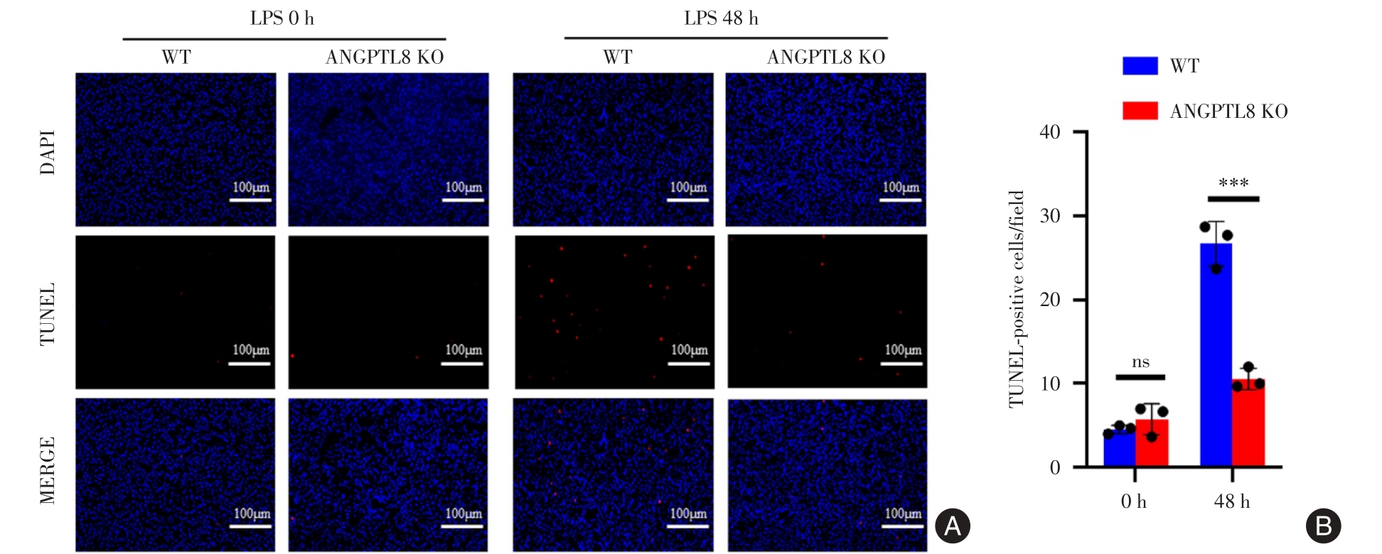
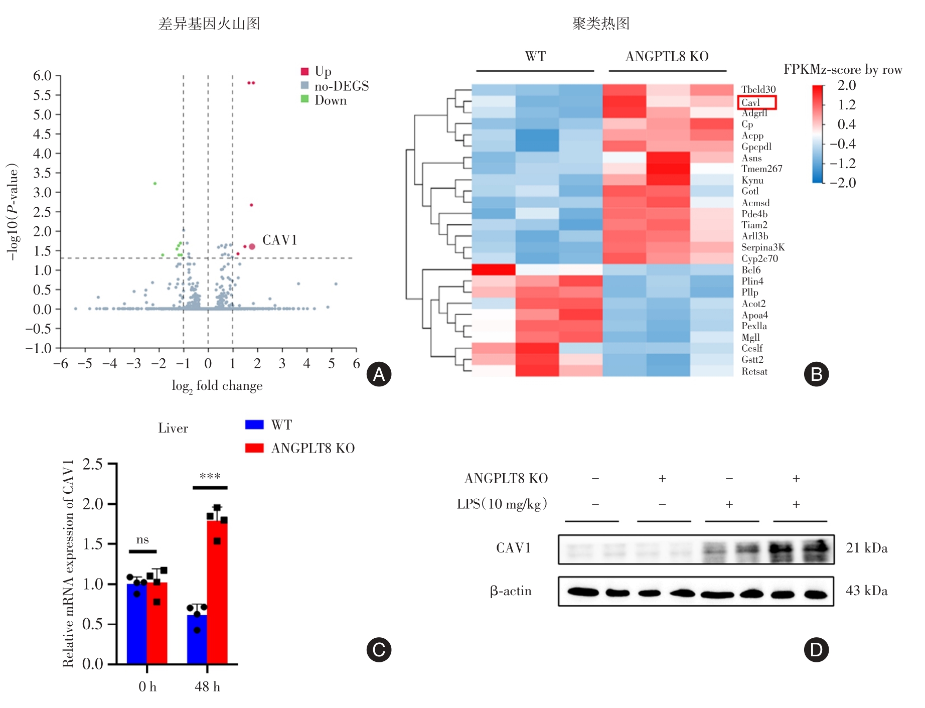
ANGPTL8 knockout alleviates DEN⁃induced acute liver injury
[J]. The Journal of Practical Medicine, 2023, 39(3): 278-284.
|
| [14] |
Yanchao LI,Qiaoli. REN.
IL⁃17A secreted by Th17 cells recruited by CXCL16 against B⁃ALL cell apoptosis
[J]. The Journal of Practical Medicine, 2023, 39(24): 3163-3168.
|
| [15] |
Wanchao WU,Yuhuan HAN,Lijie. LI.
Impacts of tanshinone IIA on lipopolysaccharide induced proliferation and apoptosis of dental pulp stem cells by regulating the Fas/FasL signaling pathway
[J]. The Journal of Practical Medicine, 2023, 39(24): 3182-3187.
|


 )
)