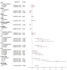
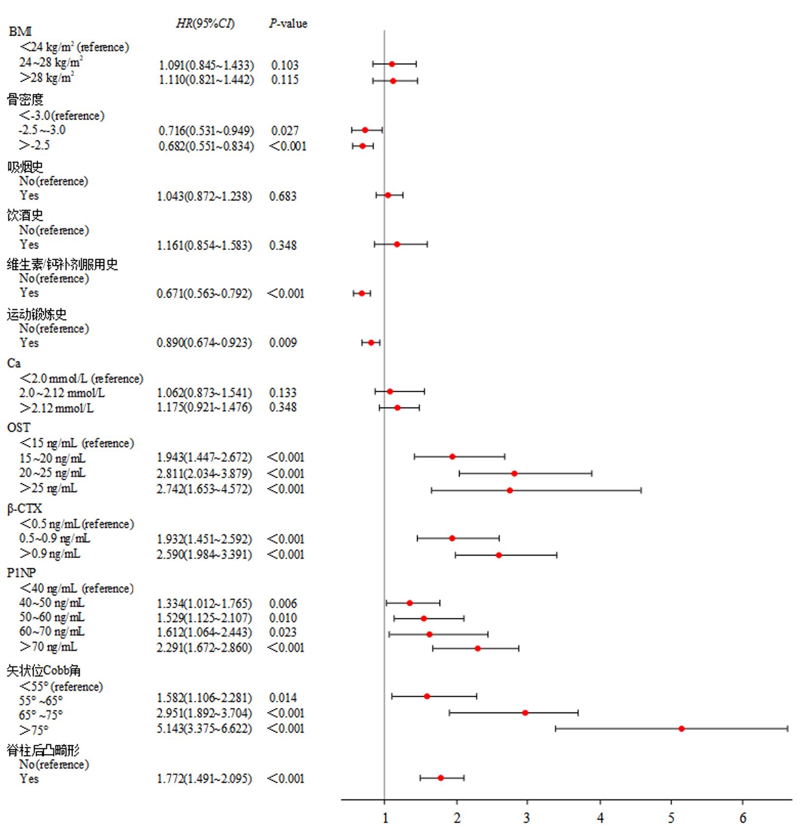
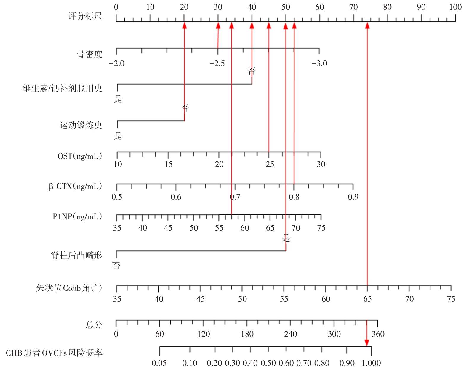
| 1 |
KAHRAMAN R, ŞAHIN A, ÖZTÜRK O, et al. Effects of Long-Term Tenofovir and Entecavir Treatment on Bone Mineral Density in Patients with Chronic Hepatitis B[J]. Turk J Gastroenterol, 2022, 33(1): 35-43.
|
| 2 |
PENG Y, XI S, HUANG R. Association between hepatitis B virus infection and risk of osteoporosis: A systematic review and meta-analysis[J]. Asian J Surg, 2023, 46(10):4598-4600.
|
| 3 |
TAO J, YAN Z, HUANG W, et al. Seropositive for hepatitis B and C viruses is associated with the risk of decreased bone mineral density in adults: An analysis of studies from the NHANES database[J]. Front Med (Lausanne), 2023, 10:1120083.
|
| 4 |
YANG Z, XING D, DONG Z, et al. Epidemiological Investigation of 387 Individuals Over 65 Years Old With Osteoporotic Fractures[J]. Altern Ther Health Med, 2023, 29(3): 207-211.
|
| 5 |
LI C, LAI X M, LIU N, et al. Correlation analysis of the vertebral compression degree and CT HU value in elderly patients with osteoporotic thoracolumbar fractures[J]. J Orthop Surg Res, 2023, 18(1):457.
|
| 6 |
BADR S, DAPVRIL H, LOMBARDO D, et al. Myosteatosis and bone marrow adiposity are not associated among postmenopausal women with fragility fractures[J]. Front Endocrinol (Lausanne), 2023, 14:1178464.
|
| 7 |
LIU Z, ZHANG Y, HUANG D, et al. Quantitative Study of Vertebral Body and Paravertebral Muscle Degeneration Based on Dual-Energy Computed Tomography: Correlation With Bone Mineral Density[J]. J Comput Assist Tomogr, 2023, 47(1): 86-92.
|
| 8 |
CAPRARIU R, OPREA M, POPA I, et al. Cohort study on the relationship between morphologic parameters of paravertebral muscles, BMI and lumbar lordosis on the severity of lumbar stenosis[J]. Eur J Orthop Surg Traumatol, 2023, 33(6): 2435-2443.
|
| 9 |
陈胜,毛渊青. MRI在鉴别骨质疏松性椎体骨折及转移性肿瘤所致椎骨骨折中的应用价值分析[J]. 中国CT和MRI杂志,2021,19(12):160-162.
|
| 10 |
HUANG W, NAGAO M, YONEMOTO N, et al. Evaluation of the efficacy and safety of romosozumab (evenity) for the treatment of osteoporotic vertebral compression fracture in postmenopausal women: A systematic review and meta-analysis of randomized controlled trials (CDM-J)[J]. Pharmacoepidemiol Drug Saf, 2023, 32(6): 671-684.
|
| 11 |
贺宝荣,郝定均. 急性症状性骨质疏松性胸腰椎压缩骨折椎体强化术临床指南[J]. 中华创伤杂志,2022,38(5):481-489.
|
| 12 |
CHENG T, LI G, MAO X, et al. Impact of Hepatitis B virus infection on postoperative complications and length of stay in elderly patients with hip fracture: A retrospective cohort study[J]. Injury, 2023, 54(7):110812.
|
| 13 |
ZHANG Y, GAO X, LIU T, et al. Association between osteoporosis and hepatitis B cirrhosis: a case-control study[J]. Afr Health Sci, 2020, 20(4): 1610-1616.
|
| 14 |
BANERJEE A, ATHALYE S, KHARGEKAR N, et al. Chronic Hepatitis B and Related Liver Diseases Are Associated with Reduced 25-Hydroxy-Vitamin D Levels: A Systematic Review and Meta-Analysis[J]. Biomedicines, 2023, 11(1):135.
|
| 15 |
VIETH R. Vitamin D supplementation: cholecalciferol, calcifediol, and calcitriol[J]. Eur J Clin Nutr, 2020, 74(11): 1493-1497.
|
| 16 |
BIVER E, HERROU J, LARID G, et al. Dietary recommendations in the prevention and treatment of osteoporosis[J]. Joint Bone Spine, 2023, 90(3):105521.
|
| 17 |
GRILI P P D F, VIDIGAL C V, CRUZ G F D, et al. Nutrient Patterns and Risk of Osteopenia in Postmenopausal Women[J]. Nutrients, 2023, 15(7):1670.
|
| 18 |
COUCE M L, SAENZ D E PIPAON M. Bone Mineralization and Calcium Phosphorus Metabolism[J]. Nutrients, 2021, 13(11):3692.
|
| 19 |
SUN L, WANG Z, ZHENG M, et al. Mineral and bone disorder after kidney transplantation: a single-center cohort study[J]. Ren Fail, 2023, 45(1):2210231.
|
| 20 |
LI F, YE X, YANG G, et al. Relationships between blood bone metabolic biomarkers and anemia in patients with chronic kidney disease[J]. Ren Fail, 2023, 45(1):2210227.
|
| 21 |
BEKÇIBAŞı M, DEVECI Ö, OĞUZ A, et al. Serum TNF-α, IL-1β, and IL-6 levels in chronic HBV-infected patients[J]. Int J Clin Pract, 2021, 75(8):e14292.
|
| 22 |
TAKEUCHI T, YOSHIDA H, TANAKA S. Role of interleukin-6 in bone destruction and bone repair in rheumatoid arthritis[J]. Autoimmun Rev, 2021, 20(9):102884.
|
| 23 |
李小海,谢光友,梁力嵩,等. 贵州中老年人腰椎QCT骨密度及脊柱脆性骨折的骨密度分析[J]. 实用医学杂志,2023,39(1):60-65.
|
| 24 |
赵锦胜,曹汉岐,杨寒石. 经伤椎与跨伤椎椎弓根螺钉内固定对胸腰椎爆裂性骨折患者的疗效及生物力学特征[J]. 实用医学杂志,2022,38(12):1465-1469.
|
| 25 |
LEBOFF M S, GREENSPAN S L, INSOGNA K L, et al. The clinician's guide to prevention and treatment of osteoporosis[J]. Osteoporos Int, 2022, 33(10): 2049-2102.
|
| 26 |
AZRIL, HUANG K Y, HOBLEY J, et al. Correlation of the degenerative stage of a disc with magnetic resonance imaging, chemical content, and biomechanical properties of the nucleus pulposus[J]. J Biomed Mater Res A, 2023, 111(7): 1054-1066.
|
| 27 |
JEON I, KIM S W, YU D. Paraspinal muscle fatty degeneration as a predictor of progressive vertebral collapse in osteoporotic vertebral compression fractures[J]. Spine J, 2022, 22(2): 313-320.
|
| 28 |
INCESOY M A, SELUK S, TURK O I, et al. Is Lenke type V adolescent idiopathic scoliosis associated with different muscular morphometry?[J]. J Pediatr Orthop B, 2023, 32(4): 363-368.
|
| 29 |
CHEN J, HUANG Y, ZENG X, et al. Functional magnetic resonance imaging reveals dysfunction of the paraspinal muscles in patients with chronic low back pain: a cross-sectional study[J]. Quant Imaging Med Surg, 2023, 13(6): 3416-3427.
|
| 30 |
OKUWAKI S, FUNAYAMA T, IKUMI A, et al. Risk factors affecting vertebral collapse and kyphotic progression in postmenopausal osteoporotic vertebral fractures[J]. J Bone Miner Metab, 2022, 40(2): 301-307.
|
| 31 |
AIVAZOGLOU L U, GUIMARÃES J B, COSTA M A F, et al. Whole-Body MRI in Limb Girdle Muscular Dystrophy Type R1/2A: Correlation With Clinical Scores[J]. Muscle Nerve, 2022, 66(4): 471-478.
|
| 32 |
XU Z, YOU W, CHEN W, et al. Single-cell RNA sequencing and lipidomics reveal cell and lipid dynamics of fat infiltration in skeletal muscle[J]. J Cachexia Sarcopenia Muscle, 2021, 12(1): 109-129.
|
| 33 |
MIURA K, KADONE H, ASADA T, et al. The fatty degeneration of the lumbar erector spinae muscles affects dynamic spinal compensation ability during gait in adult spinal deformity[J]. Sci Rep, 2021, 11(1):18088.
|