实用医学杂志 ›› 2024, Vol. 40 ›› Issue (11): 1526-1530.doi: 10.3969/j.issn.1006-5725.2024.11.010

• 基础研究 • 上一篇    下一篇

达格列净抑制血管紧张素Ⅱ诱导的心肌细胞肥大和凋亡

邵双印1,蔡欣桐2,肖莉丽2,高路2()   

  1. 1.河南省胸科医院心外科 (郑州 450008 )
    2.郑州大学第一附属医院心血管内科 (郑州 450052 )
  • 收稿日期:2023-08-02 出版日期:2024-06-10 发布日期:2024-06-13
  • 通讯作者: 高路 E-mail:gaomei1215@163.com
  • 基金资助:
    国家自然科学基金项目(81970201)

Dapagliflozin inhibits angiotensin Ⅱ⁃induced cardiomyocyte hypertrophy and apoptosis

Shuangyin SHAO1,Xintong CAI2,Lili XIAO2,Lu. GAO2()   

  1. *.Department of Cardiac Surgery,He′nan Thoracic Hospital,Zhengzhou 450008,China
  • Received:2023-08-02 Online:2024-06-10 Published:2024-06-13
  • Contact: Lu. GAO E-mail:gaomei1215@163.com

摘要:

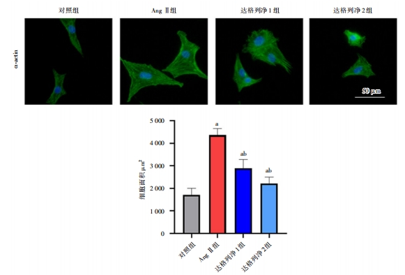
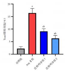
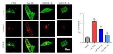
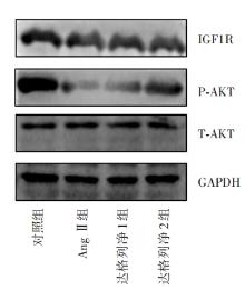
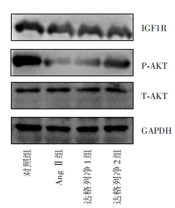
目的 探讨达格列净对血管紧张素Ⅱ(Ang Ⅱ)诱导的心肌细胞肥大反应和凋亡的影响。 方法 分离培养原代大鼠乳鼠心肌细胞,将其随机分为4组:对照组,Ang Ⅱ组、达格列净1组(0.5 μmol/L),达格列净2组(2 μmol/L)。采用α-actin染色检测细胞面积,采用qPCR检测胚胎基因的转录,采用Tunel染色检测细胞凋亡水平,采用caspase3试剂盒检测caspase3活性,采用免疫印迹检测经典信号分子。 结果 Ang Ⅱ组细胞面积明显大于对照组(P < 0.05);达格列净1组、达格列净2组细胞面积低于Ang Ⅱ组(P < 0.05)。qPCR结果显示Ang Ⅱ组胚胎基因转录明显高于对照组(P<0.05);达格列净1组,达格列净2组胚胎基因转录低于Ang Ⅱ组(P < 0.05)。tunel染色结果显示:Ang Ⅱ组细胞凋亡数量高于对照组(P<0.05);达格列净1组,达格列净2组细胞凋亡数量低于Ang Ⅱ组(P < 0.05)。Ang Ⅱ组细胞caspase3活性高于对照组(P < 0.05);达格列净1组,达格列净2组细胞caspase3活性低于Ang Ⅱ组(P < 0.05)。免疫印迹检测结果显示Ang Ⅱ组细胞胰岛素样生长因子1受体(IGF1R)和Akt激活程度低于对照组(P < 0.05);达格列净1组,达格列净2组细胞IGF1R和Akt激活高于Ang Ⅱ组(P < 0.05)。 结论 达格列净可直接作用于心肌细胞,保护其免受Ang Ⅱ诱导的损伤。

关键词: 达格列净, SGLT2抑制剂, 血管紧张素Ⅱ, 心肌细胞, 胰岛素样生长因子1受体

Abstract:

Objective This study aimed to investigate the effect of Dapagliflozin on the hypertrophy and apoptosis of cardiomyocytes induced by angiotensinⅡ(Ang Ⅱ). Methods Primary rat neonatal cardiomyocytes were isolated, cultured and randomly divided into 4 groups: control group, Ang Ⅱ group, dapagliflozin group 1 (0.5 μmol/L), and dapagliflozin group 2 (2 μmol/L). α-actin staining was used to detect cell area. qPCR was applied to detect embryonic gene transcription. Tunel staining was adopted to detect cell apoptosis level. The caspase3 kit was used to detect caspase3 activityand western blotting was used to detect classical signal molecules. Results The cell surface area of the Ang Ⅱ group was significantly larger than that of the control group (P < 0.05). The cell surface area of the dapagliflozin group 1 and the dapagliflozin group 2 was significantly lower than that of the Ang Ⅱ group (P < 0.05). The results of qPCR showed that the fetal gene transcription of Ang Ⅱ group was significantly higher than that of the control group (P < 0.05); the fetal gene transcription of dapagliflozin group 1 and dapagliflozin group 2 was lower than that of Ang Ⅱ group (P < 0.05). Tunel staining showed that the number of apoptosis in the Ang Ⅱ group was higher than that in the control group (P < 0.05); the number of apoptosis in the dapagliflozin group 1 and the Dapagliflozin group 2 was lower than that in the Ang Ⅱ group (P < 0.05). The caspase3 activity of the cells in the Ang Ⅱ group was higher than that of the control group (P < 0.05)but lower in dapagliflozin group 1 and the dapagliflozin group 2 (P < 0.05). The results of Western blotting showed that the activation of insulin-like growth factor 1 receptor (IGF1R) and Akt in the Ang Ⅱ group was lower than that in the control group (P < 0.05)but increased in the dapagliflozin group 1 and 2 cells (P < 0.05). Conclusion Dapagliflozin could directly act on cardiomyocytes to protect them from Ang Ⅱ-induced damage.

Key words: dapagliflozin, SGLT2 inhibitor, angiotensin Ⅱ, cardiomyocytes, insulin-like growth factor 1 receptor

中图分类号: