

The Journal of Practical Medicine ›› 2026, Vol. 42 ›› Issue (14): 2488-2504.doi: 10.3969/j.issn.1006-5725.2026.14.002
• Cardiovascular and Cerebrovascular Diseases Column • Previous Articles
Xiaolu JIANG1,Hongfu WEN1,Yuan SHEN1,Jun ZHANG1,Lini LI1,Renlu XU1,Wen ZHANG1,Xiaoming WANG2,Yifei JI3,Weidong LI4( )
)
Received:2026-04-22
Online:2026-07-25
Published:2026-08-05
Contact:
Weidong LI
E-mail:lwd10000@126.com
CLC Number:
Xiaolu JIANG,Hongfu WEN,Yuan SHEN,Jun ZHANG,Lini LI,Renlu XU,Wen ZHANG,Xiaoming WANG,Yifei JI,Weidong LI. Mechanism of FBXL4 inhibiting oxidative stress and apoptosis, and preventing brain injury after cardiopulmonary resuscitation by regulating Drp1-mediated mitochondrial dynamics[J]. The Journal of Practical Medicine, 2026, 42(14): 2488-2504.
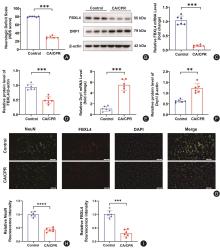
Fig.1
Changes in nerve function deficit induced by CA/CPR and expressions of FBXL4 and Drp1 in brain tissues"
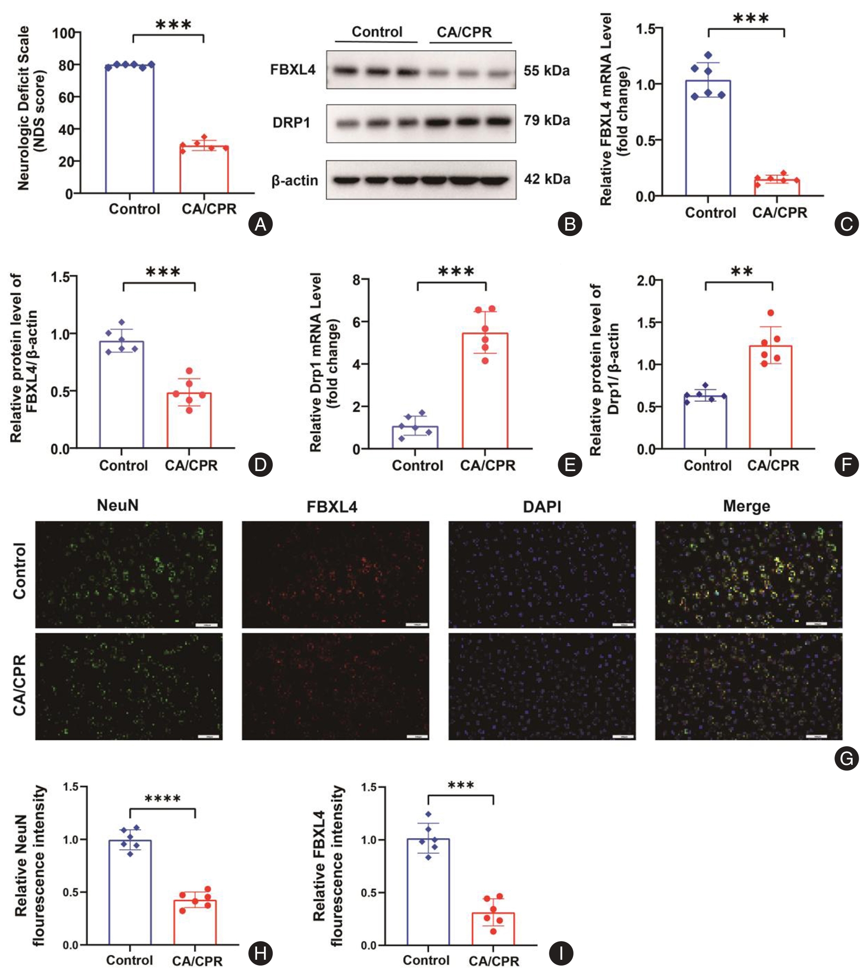
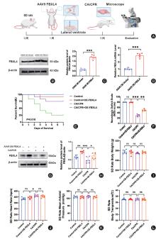
Fig.2
Effects of FBXL4 overexpression in brain tissues on survival rate and nerve function in CA/CPR rats"
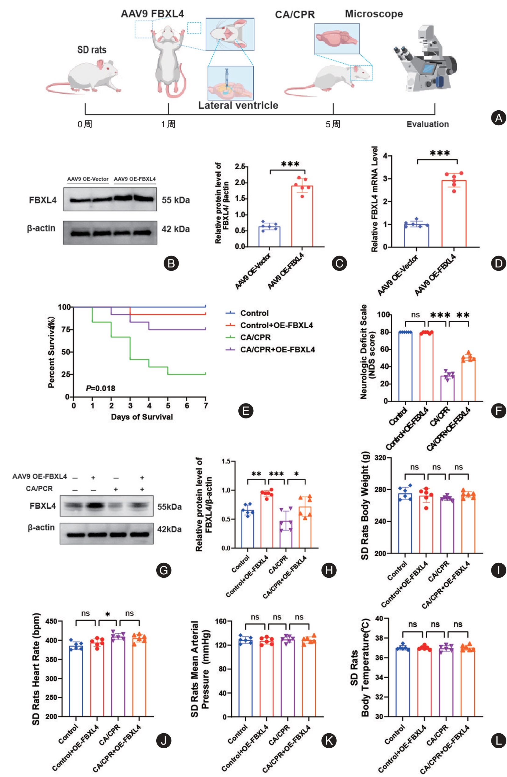
Fig.3
Overexpression of FBXL4 alleviating pathological injury of neurons in brain tissues of CA/CPR rats (HE staining)"
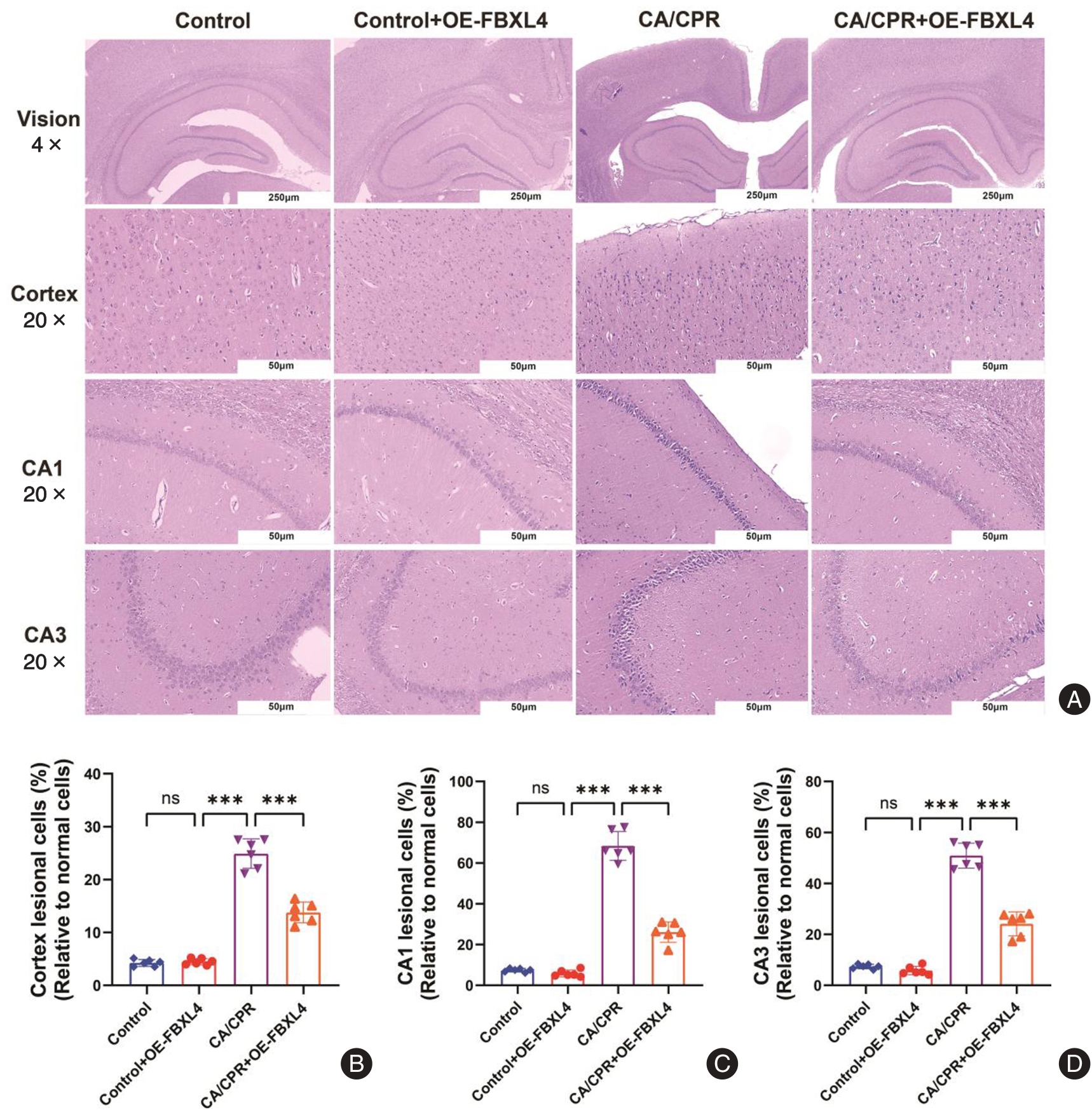
Fig.4
Overexpression of FBXL4 alleviating pathological injury of neurons in brain tissues of CA/CPR rats (Nissl staining)"
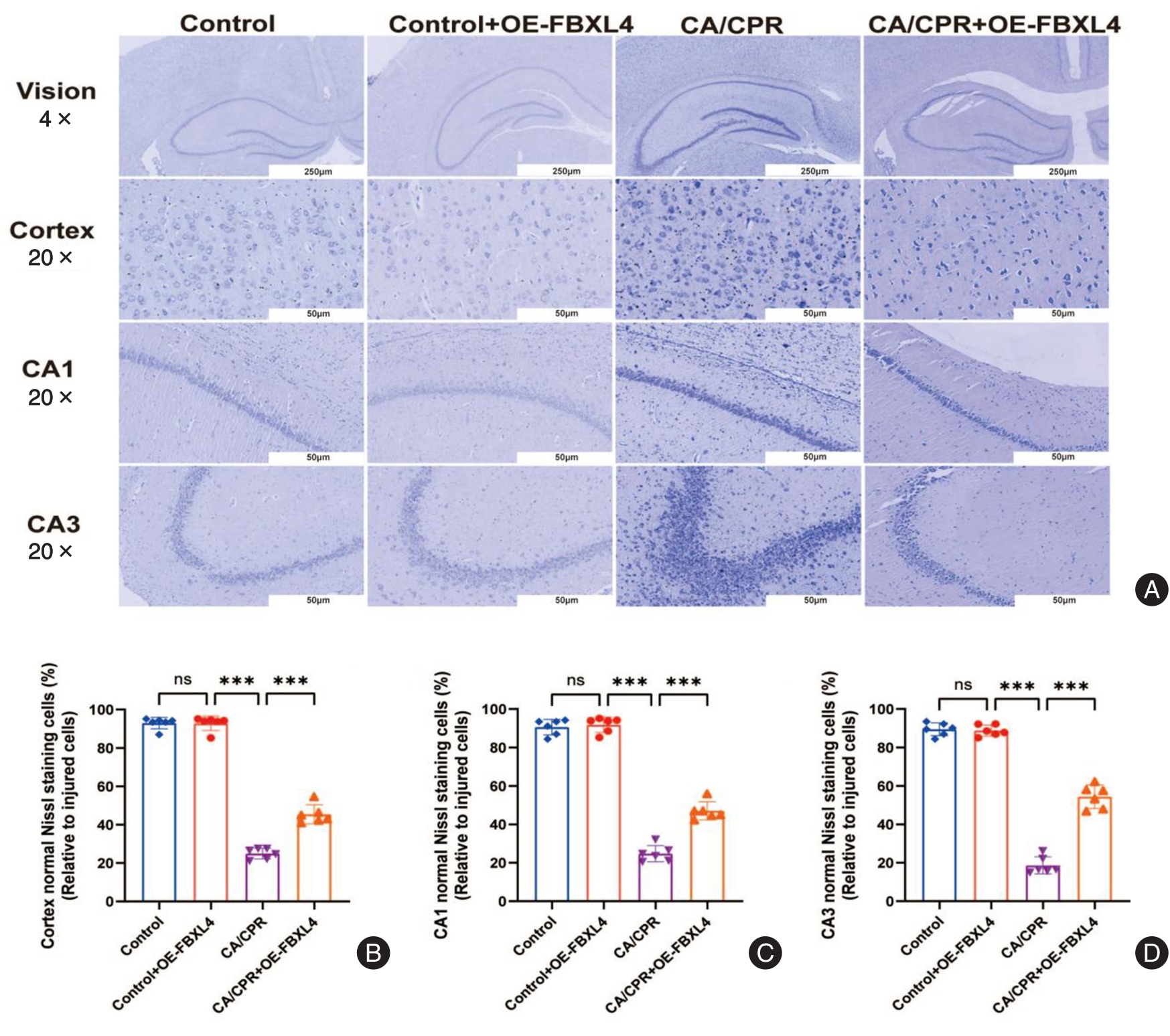
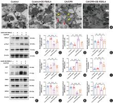
Fig.5
Overexpression of FBXL4 remodeling mitochondrial dynamics balance by regulating Drp1/Fis1/MFF axis"
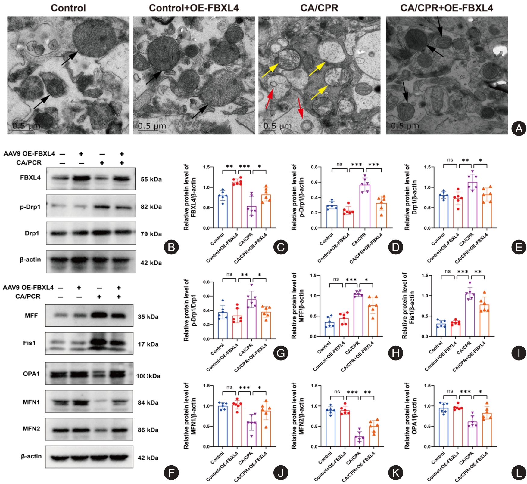
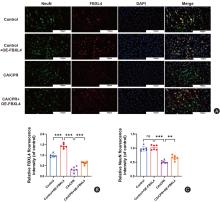
Fig.6
Localization expression of FBXL4 in cortical neurons and neuroprotective effect after overexpression"
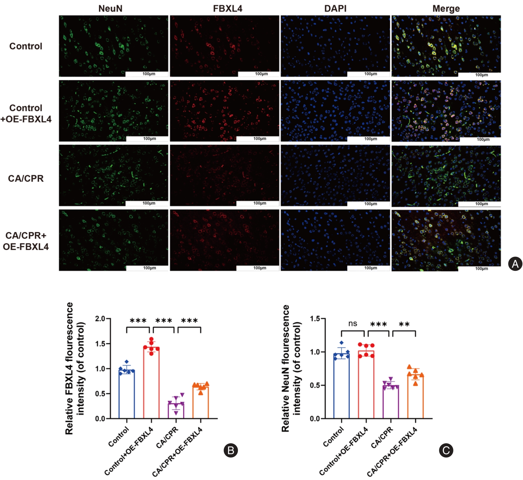
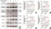
Fig.7
Effects of FBXL4 overexpression on expressions of apoptosis-related proteins in brain tissues of CA/CPR rats"
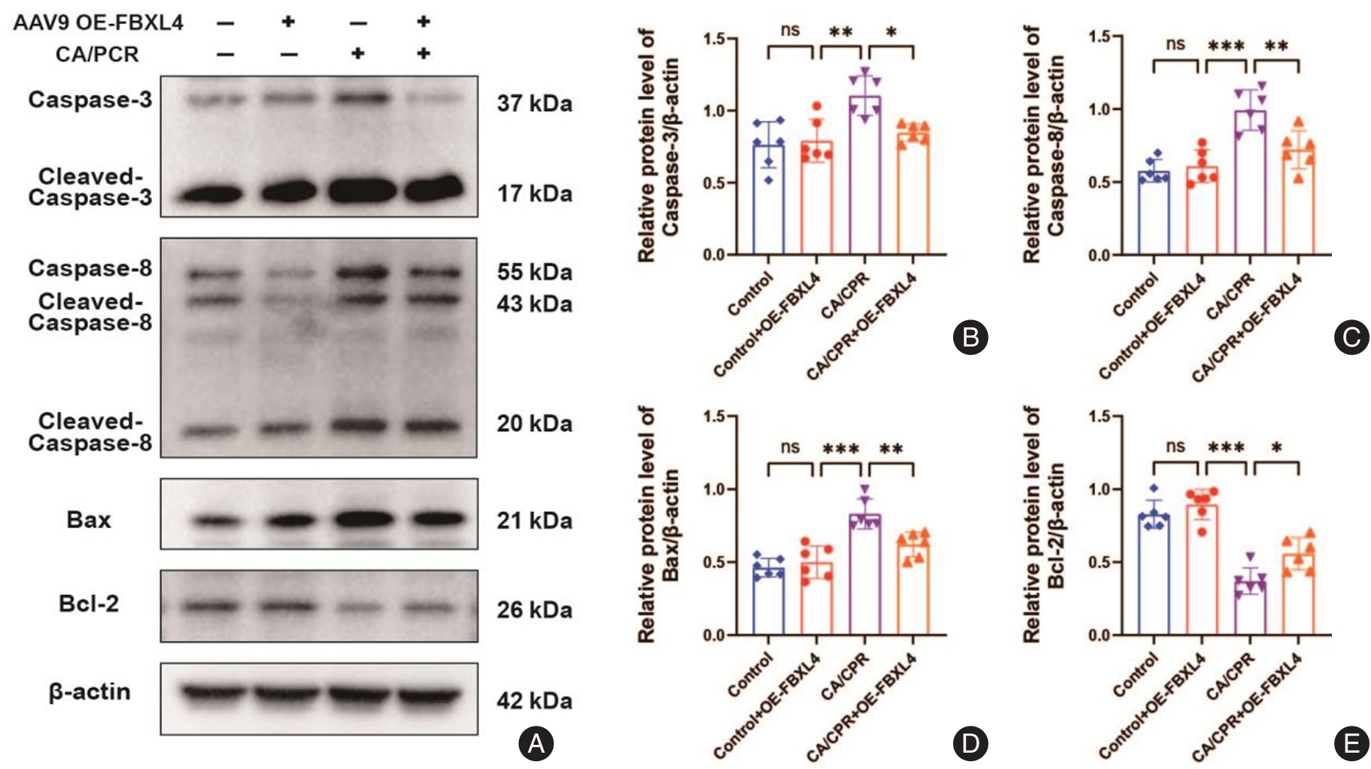
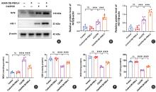
Fig.8
Effects of FBXL4 on oxidative stress indexes in brain tissues and Nrf2/HO-1 pathways in CA/CPR rats"
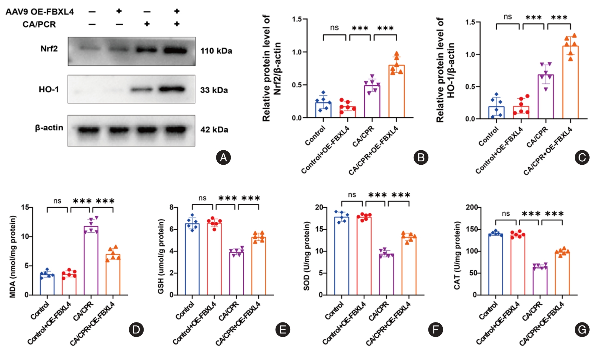
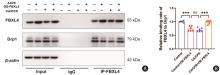
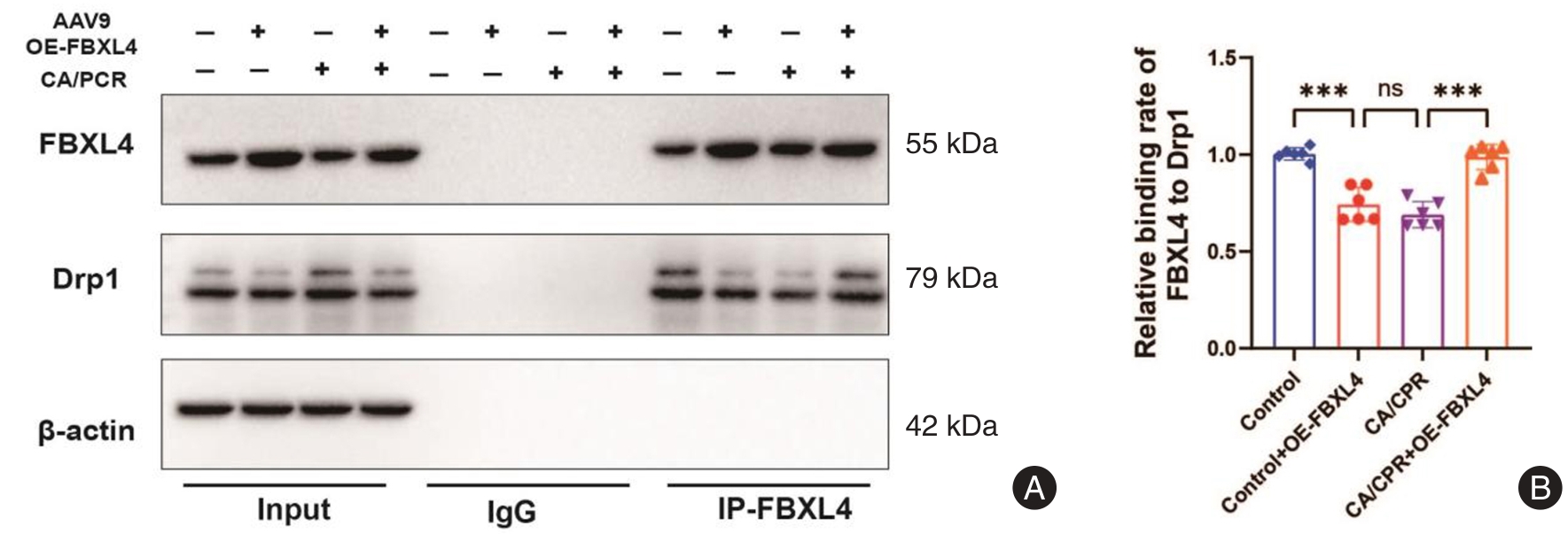
Fig.9
The confirmation of interaction between FBXL4 and Drp1 in brain tissues by Co-IP experiment"
| [1] |
ANDERSEN L W, HOLMBERG M J, BERG K M,et al. In-Hospital Cardiac Arrest: A Review[J]. JAMA,2019,321(12):1200-1210. doi:10.1001 /jama.2019.1696 .
doi: 10.1001 /jama.2019.1696 |
| [2] |
SANDRONI C, D'ARRIGO S, NOLAN J P. Prognostication after cardiac arrest[J]. Crit Care,2018,22(1):150. doi:10.1186/s13054-018-2060-7 .
doi: 10.1186/s13054-018-2060-7 |
| [3] |
MEHAFFEY J H, MONEY D, CHARLES E J,et al. Adenosine 2A Receptor Activation Attenuates Ischemia Reperfusion Injury During Extracorporeal Cardiopulmonary Resuscitation[J].Ann Surg,2019,269(6):1176-1183. doi:10.1097/sla. 0000000000002685 .
doi: 10.1097/sla. 0000000000002685 |
| [4] |
TUO Q Z, ZHANG S T, LEI P. Mechanisms of neuronal cell death in ischemic stroke and their therapeutic implications[J].Med Res Rev,2022,42(1):259-305. doi:10.1002/med.21817 .
doi: 10.1002/med.21817 |
| [5] |
MAO R, ZONG N, HU Y,et al. Neuronal Death Mechanisms and Therapeutic Strategy in Ischemic Stroke[J].Neurosci Bull,2022,38(10):1229-1247. doi:10.1007/s12264-022-00859-0 .
doi: 10.1007/s12264-022-00859-0 |
| [6] |
GUAN R, ZOU W, DAI X,et al. Mitophagy, a potential therapeutic target for stroke[J]. J Biomed Sci,2018,25(1):87. doi:10.1186/s12929-018-0487-4 .
doi: 10.1186/s12929-018-0487-4 |
| [7] |
LIANG W, HUANG X, CHEN W. The Effects of Baicalin and Baicalein on Cerebral Ischemia: A Review[J]. Aging Dis,2017,8(6):850-867. doi:10.14336/ad.2017.0829 .
doi: 10.14336/ad.2017.0829 |
| [8] |
QIU Z, WEI Y, SONG Q,et al. The Role of Myocardial Mitochondrial Quality Control in Heart Failure[J].Front Pharmacol,2019,10(1):1404. doi:10.3389/ fphar.2019.01404 .
doi: 10.3389/ fphar.2019.01404 |
| [9] |
DUAN X, LIU R, LAN W,et al. The Essential Role of Mitochondrial Dynamics in Viral Infections[J]. Int J Mol Sci,2025,26(5):1955. doi:10.3390/ijms 26051955 .
doi: 10.3390/ijms 26051955 |
| [10] |
WOODS L C, BERBUSSE G W, NAYLOR K. Microtubules Are Essential for Mitochondrial Dynamics-Fission, Fusion, and Motility-in Dictyostelium discoideum[J].Front Cell Dev Biol,2016,4(1):19. doi:10.3389/fcell. 2016.00019 .
doi: 10.3389/fcell. 2016.00019 |
| [11] |
SUÁREZ-RIVERO J M, VILLANUEVA-PAZ M, DE LA CRUZ-OJEDA P,et al. Mitochondrial Dynamics in Mitochondrial Diseases[J].Dig Dis,2016,5(1):1. doi:10.3390/diseases5010001 .
doi: 10.3390/diseases5010001 |
| [12] |
WANG N, WANG X, LAN B,et al. DRP1, fission and apoptosis[J].Cell Death Discov,2025,11(1):150. doi:10.1038/s41420-025-02458-0 .
doi: 10.1038/s41420-025-02458-0 |
| [13] |
ISHIHARA N, NOMURA M, JOFUKU A,et al. Mitochondrial fission factor Drp1 is essential for embryonic development and synapse formation in mice[J]. Nat Cell Biol,2009,11(8):958-966. doi:10.1038/ncb1907 .
doi: 10.1038/ncb1907 |
| [14] |
YU R, JIN S B, ANKARCRONA M,et al. The Molecular Assembly State of Drp1 Controls its Association With the Mitochondrial Recruitment Receptors Mff and MIEF1/2[J]. Front Cell Dev Biol,2021,9(1):706687. doi:10.3389/fcell. 2021.706687 .
doi: 10.3389/fcell. 2021.706687 |
| [15] |
SATO H, HOSHI M, IKEDA F,et al. Downregulation of mitochondrial biogenesis by virus infection triggers antiviral responses by cyclic GMP-AMP synthase[J].PLoS Pathog,2021,17(10):e1009841.doi:10.1371/journal.ppat. 1009841 .
doi: 10.1371/journal.ppat. 1009841 |
| [16] |
MURATA D, YAMADA T, TOKUYAMA T,et al. Mitochondrial Safeguard: A stress response that offsets extreme fusion and protects respiratory function via flickering-induced Oma1 activation[J]. EMBO J,2020,39(24):e105074. doi:10.15252/embj. 2020105074 .
doi: 10.15252/embj. 2020105074 |
| [17] |
CATANESE L, TARSIA J, FISHER M. Acute Ischemic Stroke Therapy Overview[J]. Circ Res,2017,120(3):541-558. doi:10.1161/circresaha. 116.309278 .
doi: 10.1161/circresaha. 116.309278 |
| [18] |
ZHAO Y X, CUI M, CHEN S F,et al. Amelioration of ischemic mitochondrial injury and Bax-dependent outer membrane permeabilization by Mdivi-1[J]. CNS Neurosci Ther,2014,20(6):528-538. doi:10.1111/cns.12266 .
doi: 10.1111/cns.12266 |
| [19] |
FLIPPO K H, LIN Z, DICKEY A S,et al. Deletion of a Neuronal Drp1 Activator Protects against Cerebral Ischemia[J]. J Neurosci,2020,40(15):3119-3129. doi:10.1523/jneurosci.1926-19.2020 .
doi: 10.1523/jneurosci.1926-19.2020 |
| [20] |
SHIELDS L Y, KIM H, ZHU L,et al. Dynamin-related protein 1 is required for normal mitochondrial bioenergetic and synaptic function in CA1 hippocampal neurons[J]. Cell Death Dis,2015,6(4):e1725. doi:10.1038/cddis.2015.94 .
doi: 10.1038/cddis.2015.94 |
| [21] |
PU Z X, WANG J L, LI Y Y,et al. A Bacterial Platform for Studying Ubiquitination Cascades Anchored by SCF-Type E3 Ubiquitin Ligases[J]. Biomolecules,2024,14(10):1209. doi:10.3390/biom14101209 .
doi: 10.3390/biom14101209 |
| [22] |
HWANG J, LAUINGER L, KAISER P. Distinct Stress Regulators in the CRL Family: Emerging Roles of F-Box Proteins: Cullin-RING Ligases and Stress-Sensing[J]. BioEssays,2025,47(5):e202400249. doi:10.1002/bies. 202400249 .
doi: 10.1002/bies. 202400249 |
| [23] |
CHEN Y, JIAO D, LIU Y,et al. FBXL4 mutations cause excessive mitophagy via BNIP3/BNIP3L accumulation leading to mitochondrial DNA depletion syndrome[J]. Cell Death Differ,2023,30(10):2351-2363. doi:10.1038/s41418- 023-01205-1 .
doi: 10.1038/s41418- 023-01205-1 |
| [24] |
PRASUN P. FBXL4-Related Mitochondrial Depletion Syndrome Underscores the role of Mitophagy in Stem Cell Differentiation during Embryogenesis[J]. Stem Cell Rep,2025,21(3):897-899. doi:10.1007/s12015 -025-10854-3 .
doi: 10.1007/s12015 -025-10854-3 |
| [25] |
BONNEN P E, YARHAM J W, BESSE A,et al. Mutations in FBXL4 cause mitochondrial encephalopathy and a disorder of mitochondrial DNA maintenance[J]. Am J Hum Genet,2013,93(3):471-481. doi:10.1016/j.ajhg. 2013.07.017 .
doi: 10.1016/j.ajhg. 2013.07.017 |
| [26] |
GAI X, GHEZZI D, JOHNSON M A,et al. Mutations in FBXL4, encoding a mitochondrial protein, cause early-onset mitochondrial encephalomyopathy[J]. Am J Hum Genet,2013,93(3):482-495. doi:10.1016/j.ajhg.2013.07.016 .
doi: 10.1016/j.ajhg.2013.07.016 |
| [27] |
ABUDUREYIMU M, LUO X, JIANG L,et al. FBXL4 protects against HFpEF through Drp1-Mediated regulation of mitochondrial dynamics and the downstream SERCA2a[J]. Redox Biol,2024,70(1):103081. doi:10.1016/j.redox. 2024.103081 .
doi: 10.1016/j.redox. 2024.103081 |
| [28] |
YU S, WU C, ZHU Y,et al. Rat model of asphyxia-induced cardiac arrest and resuscitation[J]. Front Neurosci,2022,16(1):1087725. doi:10.3389/fnins. 2022.1087725 .
doi: 10.3389/fnins. 2022.1087725 |
| [29] |
KATZ L, EBMEYER U, SAFAR P,et al. Outcome model of asphyxial cardiac arrest in rats[J]. J Cereb Blood Flow Metab,1995,15(6):1032-1039. doi:10.1038/jcbfm.1995.129 .
doi: 10.1038/jcbfm.1995.129 |
| [30] |
PERKINS G D, JACOBS I G, NADKARNI V M,et al. Cardiac arrest and cardiopulmonary resuscitation outcome reports: update of the Utstein Resuscitation Registry Templates for Out-of-Hospital Cardiac Arrest: a statement for healthcare professionals from a task force of the International Liaison Committee on Resuscitation (American Heart Association, European Resuscitation Council, Australian and New Zealand Council on Resuscitation, Heart and Stroke Foundation of Canada, InterAmerican Heart Foundation, Resuscitation Council of Southern Africa, Resuscitation Council of Asia); and the American Heart Association Emergency Cardiovascular Care Committee and the Council on Cardiopulmonary, Critical Care, Perioperative and Resuscitation[J]. Circulation,2015,132(13):1286-1300. doi:10.1161/cir. 0000000000000144 .
doi: 10.1161/cir. 0000000000000144 |
| [31] |
ADHIKARI A, LERNER T N, FINKELSTEIN J,et al. Basomedial amygdala mediates top-down control of anxiety and fear[J]. Nature,2015,527(7577): 179-185. doi:10.1038/nature15698 .
doi: 10.1038/nature15698 |
| [32] |
WANG P, YAO L, ZHOU L L,et al. Carbon Monoxide Improves Neurologic Outcomes by Mitochondrial Biogenesis after Global Cerebral Ischemia Induced by Cardiac Arrest in Rats[J]. Int J Biol Sci,2016,12(8):1000-1009. doi:10.7150/ijbs.13222 .
doi: 10.7150/ijbs.13222 |
| [33] |
CHEN M, LIU J, WU W,et al. SIRT1 restores mitochondrial structure and function in rats by activating SIRT3 after cerebral ischemia/reperfusion injury[J]. Cell Biol Toxicol,2024,40(1):31. doi:10.1007/s10565-024-09869-2 .
doi: 10.1007/s10565-024-09869-2 |
| [34] |
VONGSFAK J, PRATCHAYASAKUL W, APAIJAI N,et al. The Alterations in Mitochondrial Dynamics Following Cerebral Ischemia/Reperfusion Injury[J]. Antioxidants (Basel),2021,10(9):1384. doi:10.3390/antiox10091384 .
doi: 10.3390/antiox10091384 |
| [35] |
FLIPPO K H, STRACK S. Mitochondrial dynamics in neuronal injury, development and plasticity[J]. J Cell Sci,2017,130(4):671-681. doi:10.1242 /jcs.171017 .
doi: 10.1242 /jcs.171017 |
| [36] |
CHEN Y, GUO S, TANG Y,et al. Mitochondrial Fusion and Fission in Neuronal Death Induced by Cerebral Ischemia-Reperfusion and Its Clinical Application: A Mini-Review[J]. Med Sci Monit,2020,26(1):e928651. doi:10.12659/msm. 928651 .
doi: 10.12659/msm. 928651 |
| [37] |
HUANG J, CHEN L, YAO Z M,et al. The role of mitochondrial dynamics in cerebral ischemia-reperfusion injury[J].Biomed Pharmacother,2023,162(1): 114671. doi:10.1016/j.biopha.2023. 114671 .
doi: 10.1016/j.biopha.2023. 114671 |
| [38] |
TAN Y, ZHANG J, GE Q,et al. Ketone Body Improves Neurological Outcomes after Cardiac Arrest by Inhibiting Mitochondrial Fission in Rats[J]. Oxid Med Cell Longev,2022,2022(1):7736416. doi:10.1155/2022/7736416 .
doi: 10.1155/2022/7736416 |
| [39] |
CAI P, LI W, XU Y,et al. Drp1 and neuroinflammation: Deciphering the interplay between mitochondrial dynamics imbalance and inflammation in neurodegenerative diseases[J]. Neurobiol Dis, 2024,198(1):106561. doi:10.1016/j.nbd.2024.106561 .
doi: 10.1016/j.nbd.2024.106561 |
| [40] |
ZHANG J, WANG S, ZHANG H,et al. Drp1 acetylation mediated by CDK5-AMPK-GCN5L1 axis promotes cerebral ischemic injury via facilitating mitochondrial fission[J]. Mol Med, 2024,30(1):173. doi:10.1186/ s10020-024-00948-y .
doi: 10.1186/ s10020-024-00948-y |
| [41] |
GIACOMELLO M, PYAKUREL A, GLYTSOU C,et al. The cell biology of mitochondrial membrane dynamics[J]. Nat Rev Mol Cell Biol,2020,21(4): 204-224. doi:10.1038/s41580-020-0210-7 .
doi: 10.1038/s41580-020-0210-7 |
| [42] |
TILOKANI L, NAGASHIMA S, PAUPE V,et al. Mitochondrial dynamics: overview of molecular mechanisms[J]. Essays in biochemistry,2018, 62(3):341-360. doi:10.1042/ebc20170104 .
doi: 10.1042/ebc20170104 |
| [43] |
HAO S, HUANG H, MA R Y,et al. Multifaceted functions of Drp1 in hypoxia/ischemia-induced mitochondrial quality imbalance: from regulatory mechanism to targeted therapeutic strategy[J]. Mil Med Res,2023,10(1):46. doi:10.1186/s40779-023-00482-8 .
doi: 10.1186/s40779-023-00482-8 |
| [44] |
LIU M, YIN Z, LI B,et al. Targeting Drp1 in Cerebral Ischemia-Reperfusion Injury: Mechanisms and Therapeutic Implications[J]. CNS Neurosci Ther,2025,31(8):e70590. doi:10.1111/cns.70590 .
doi: 10.1111/cns.70590 |
| [45] |
SANDERSON T H, REYNOLDS C A, KUMAR R,et al. Molecular mechanisms of ischemia-reperfusion injury in brain: pivotal role of the mitochondrial membrane potential in reactive oxygen species generation[J]. Cell Mol Neurobiol,2013,47(1):9-23. doi:10.1007/s12035- 012-8344-z .
doi: 10.1007/s12035- 012-8344-z |
| [46] |
WANG P, LI Y, YANG Z,et al. Inhibition of dynamin-related protein 1 has neuroprotective effect comparable with therapeutic hypothermia in a rat model of cardiac arrest[J]. Transl Res,2018,194(1):68-78. doi:10.1016/j.trsl. 2018.01.002 .
doi: 10.1016/j.trsl. 2018.01.002 |
| [47] |
CHO B, CHO H M, KIM H J,et al. CDK5-dependent inhibitory phosphorylation of Drp1 during neuronal maturation[J]. Exp Mol Med,2014, 46(7):e105. doi:10.1038/emm.2014.36 .
doi: 10.1038/emm.2014.36 |
| [48] |
FLIPPO K H, GNANASEKARAN A, PERKINS G A,et al. AKAP1 Protects from Cerebral Ischemic Stroke by Inhibiting Drp1-Dependent Mitochondrial Fission[J]. J Neurosci,2018,38(38):8233-8242.doi:10.1523/jneurosci.0649-18. 2018 .
doi: 10.1523/jneurosci.0649-18. 2018 |
| [49] |
NHU N T, LI Q, LIU Y,et al. Effects of Mdivi-1 on Neural Mitochondrial Dysfunction and Mitochondria-Mediated Apoptosis in Ischemia-Reperfusion Injury After Stroke: A Systematic Review of Preclinical Studies[J]. Front Mol Neurosci,2021,14(1):778569. doi:10.3389/fnmol.2021.778569 .
doi: 10.3389/fnmol.2021.778569 |
| [50] |
TACCONE F S, CRIPPA I A, DELL'ANNA A M,et al. Neuroprotective strategies and neuroprognostication after cardiac arrest[J].Best Pract Res Clin Anaesthesiol,2015,29(4):451-464. doi:10.1016/j.bpa.2015.08.005 .
doi: 10.1016/j.bpa.2015.08.005 |
| [51] |
NOLAN J P, SANDRONI C, BÖTTIGER B W,et al. European Resuscitation Council and European Society of Intensive Care Medicine guidelines 2021: post-resuscitation care[J]. Intensive Care Med,2021,47(4):369-421. doi:10.1007/s00134-021-06368-4 .
doi: 10.1007/s00134-021-06368-4 |
| Viewed | ||||||
|
Full text |
|
|||||
|
Abstract |
|
|||||
