

The Journal of Practical Medicine ›› 2026, Vol. 42 ›› Issue (1): 45-55.doi: 10.3969/j.issn.1006-5725.2026.01.006
• Oncology: Diagnosis, Treatment and Prevention • Previous Articles Next Articles
Sheng NONG1,Zhanxiong LI2,Qi ZHANG3,Zhendong LU4,Minping HONG5,Wubiao CHEN4,Zilin LIU4( )
)
Received:2025-09-10
Online:2026-01-10
Published:2026-01-14
Contact:
Zilin LIU
E-mail:2262830331@qq.com
CLC Number:
Sheng NONG,Zhanxiong LI,Qi ZHANG,Zhendong LU,Minping HONG,Wubiao CHEN,Zilin LIU. Predictive performance of an interpretable BPNN model for axillary lymph node burden in breast cancer patients with 1 ~ 2 sentinel lymph node positive[J]. The Journal of Practical Medicine, 2026, 42(1): 45-55.
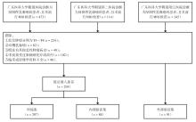
Fig.1
Flow diagram of patient enrollment and exclusion"
Tab.1
MRI scanning parameters at the three participating centers"
| 序列 | 参数项 | 广东医科大学附属医院(3.0T) | 广东医科大学附属阳江医院(3.0T) | 广东医科大学附属第二医院(1.5T) |
|---|---|---|---|---|
| T2WI-FS | 重复时间/ms | 5 139 | 3 500 | 517 |
| 回波时间 /ms | 85 | 55 | 98.4 | |
| FOV/mm2 | 320 × 320 | 360 × 360 | 300 × 300 | |
| 矩阵 | 320 × 320 | 400 × 400 | 352 × 128 | |
| 层厚/mm | 4.00 | 4.00 | 4.00 | |
| DWI | 重复时间/ms | 5 500 | 3 500 | 3 549.9 |
| 回波时间/ms | 60.6 | 55 | 55 | |
| FOV/mm2 | 320 × 320 | 204 × 340 | 320 × 320 | |
| 矩阵 | 320 × 320 | 113 × 189 | 320 × 320 | |
| 层厚/mm | 4.00 | 5.00 | 4.00 | |
| DCE-MRI | 重复时间/ms | 4.43 | 4.50 | 4.51 |
| 回报时间/ms | 1.5 | 1.61 | 1.61 | |
| FOV/mm2 | 340 × 340 | 360 × 360 | 370 × 370 | |
| 矩阵 | 448 × 336 | 400 × 400 | 256 × 160 | |
| 层厚/mm | 1.20 | 1.50 | 1.20 |
Tab.2
Patients′ baseline"
| 临床影像特征 | 水平 | 训练集(n = 207) | 内部验证集(n = 88) | 外部验证集(n = 91) | |||||||||
|---|---|---|---|---|---|---|---|---|---|---|---|---|---|
腋窝淋巴 结低负荷(n = 118) | 腋窝淋巴 结高负荷(n = 89) | χ2/t/ Z值 | P值 | 腋窝淋巴 结低负荷(n = 50) | 腋窝淋巴 结高负荷(n = 38) | χ2/t/ Z值 | P值 | 腋窝淋巴 结低负荷(n = 63) | 腋窝淋巴 结高负荷(n = 28) | χ2/t/ Z值 | P值 | ||
| 绝经状态 | 无 | 26 | 14 | 0.920 | 0.337 | 10 | 6 | 0.257 | 0.612 | 7 | 1 | 0.595 | 0.441 |
| 有 | 92 | 75 | 40 | 32 | 56 | 27 | |||||||
| HER-2 | 阴性 | 58 | 53 | 1.807 | 0.179 | 23 | 25 | 3.410 | 0.065 | 46 | 24 | 1.761 | 0.185 |
| 阳性 | 60 | 36 | 27 | 13 | 17 | 4 | |||||||
| ER | 阴性 | 25 | 20 | 0.003 | 0.959 | 9 | 14 | 3.971 | 0.046 | 19 | 9 | 0.036 | 0.85 |
| 阳性 | 93 | 69 | 41 | 24 | 44 | 19 | |||||||
| PR | 阴性 | 37 | 23 | 0.505 | 0.477 | 12 | 19 | 6.397 | 0.011 | 24 | 14 | 1.13 | 0.288 |
| 阳性 | 81 | 66 | 38 | 19 | 39 | 14 | |||||||
| Ki-67 | < 14% | 30 | 34 | 3.304 | 0.069 | 11 | 10 | 0.221 | 0.638 | 18 | 3 | 3.482 | 0.062 |
| ≥ 14% | 88 | 55 | 39 | 28 | 45 | 25 | |||||||
| 肿瘤数量 | 单发 | 98 | 60 | 6.026 | 0.014 | 42 | 28 | 1.412 | 0.235 | 48 | 24 | 1.064 | 0.302 |
| 多发 | 20 | 29 | 8 | 10 | 15 | 4 | |||||||
| 肿瘤形态 | 规则 | 54 | 30 | 2.578 | 0.108 | 20 | 17 | 0.199 | 0.656 | 14 | 4 | 0.769 | 0.380 |
| 不规则 | 64 | 59 | 30 | 21 | 49 | 24 | |||||||
| 强化模式 | 肿块型 | 92 | 60 | 2.379 | 0.123 | 43 | 30 | 0.759 | 0.383 | 51 | 22 | 0.069 | 0.792 |
| 非肿块型 | 26 | 29 | 7 | 8 | 12 | 6 | |||||||
| 强化特点 | 均匀 | 49 | 44 | 0.984 | 0.321 | 22 | 18 | 0.099 | 0.753 | 30 | 15 | 0.275 | 0.6 |
| 不均匀 | 69 | 45 | 28 | 20 | 33 | 13 | |||||||
| TIC曲线 | 流入/平台型 | 55 | 29 | 3.578 | 0.059 | 25 | 13 | 2.194 | 0.139 | 27 | 13 | 0.1 | 0.751 |
| 流出型 | 63 | 60 | 25 | 25 | 36 | 15 | |||||||
| 毛刺征 | 无 | 81 | 50 | 2.877 | 0.090 | 31 | 21 | 0.405 | 0.524 | 46 | 9 | 11.889 | <0.001 |
| 有 | 37 | 39 | 19 | 17 | 17 | 19 | |||||||
| BPE | 无或轻微强化 | 36 | 33 | 2.464 | 0.482 | 12 | 14 | 2.295 | 0.513 | 19 | 7 | 4.333 | 0.228 |
中度 强化 | 23 | 21 | 12 | 9 | 10 | 7 | |||||||
显著 强化 | 28 | 18 | 13 | 6 | 17 | 11 | |||||||
| 极度显著强化 | 31 | 17 | 13 | 9 | 17 | 3 | |||||||
| 乳腺腺体类型 | 脂肪型 | 11 | 4 | 2.598 | 0.458 | 4 | 5 | 2.793 | 0.425 | 6 | 1 | 3.032 | 0.388 |
| 少腺体型 | 36 | 34 | 21 | 10 | 23 | 12 | |||||||
多腺 体型 | 44 | 32 | 16 | 13 | 23 | 13 | |||||||
| 致密型 | 27 | 19 | 9 | 10 | 11 | 2 | |||||||
| 瘤周水肿评分 | BES评分1 ~ 2 | 90 | 35 | 27.425 | < 0.001 | 40 | 21 | 6.212 | 0.013 | 38 | 12 | 2.387 | 0.122 |
| BES评分3 ~ 4 | 28 | 54 | 10 | 17 | 25 | 16 | |||||||
| 肿瘤最大径 | < 2 cm | 41 | 13 | 9.653 | 0.002 | 18 | 9 | 1.540 | 0.215 | 23 | 3 | 6.319 | 0.012 |
| ≥ 2 cm | 77 | 76 | 32 | 29 | 40 | 25 | |||||||
| NLR | < 3 | 91 | 30 | 37.602 | < 0.001 | 34 | 5 | 24.140 | < 0.001 | 35 | 28 | 15.949 | < 0.001 |
| ≥ 3 | 27 | 59 | 16 | 33 | 28 | 0 | |||||||
腋窝淋巴结 皮质增厚 | 无 | 90 | 19 | 59.206 | < 0.001 | 39 | 9 | 23.547 | < 0.001 | 52 | 10 | 17.478 | < 0.001 |
| 有 | 28 | 70 | 11 | 29 | 11 | 18 | |||||||
腋窝淋巴结短径 [M(P25,P75)]/mm | 6.25 (4.43, 7.88) | 7.60 (5.40, 11.30) | -3.181 | 0.001 | 5.00(3.80, 7.47) | 11.30 (10.30, 12.80) | -4.44 | < 0.001 | 6.50 (6.00, 7.00) | 7.90 (6.77, 8.20) | -4.48 | < 0.001 | |
| 年龄( | 48.18 ± 10.92 | 50.15 ± 9.47 | -1.358 | 0.176 | 49.44 ± 12.35 | 48.42 ± 10.06 | 0.415 | 0.679 | 54.40 ± 9.81 | 53.04 ± 8.09 | 0.880 | 0.381 | |
ADC [M(P25,P75)]/ (×10-3mm2/s) | 829.00 (723.00, 906.00) | 812.00 (727.00, 912.00) | -0.497 | 0.620 | 828.00 (734.00, 960.00) | 798.00 (722.00, 920.00) | -0.518 | 0.604 | 832.00 (591.50, 1110.50) | 872.50 (752.25, 1098.50) | -1.803 | 0.279 | |
CA-153 [M(P25,P75)] | 54.55 (12.67, 114.42) | 18.80 (10.00, 85.70) | -2.064 | 0.039 | 45.80 (13.40, 114.30) | 33.45 (12.52, 118.05) | -0.514 | 0.607 | 42.00 (23.30, 59.15) | 72.90(50.55, 91.90) | -3.577 | < 0.001 | |
Tab.3
Univariate and multivariate logistic regression analyses"
| 因素 | 单因素 | 多因素 | 逐步回归OR | |||
|---|---|---|---|---|---|---|
| OR(95%CI) | P值 | OR(95%CI) | P值 | OR(95%CI) | P值 | |
| 肿瘤数量 | ||||||
| 单发 | ||||||
| 多发 | 2.99(1.47 ~ 6.09 ) | 0.002 | 2.29(0.94 ~ 5.56) | 0.068 | ||
| TIC曲线类型 | ||||||
| 流入型/平台型 | ||||||
| 流出型 | 1.99(1.07 ~ 3.71) | 0.030 | 1.68(0.75 ~ 3.77) | 0.209 | ||
| 瘤周水肿评分 | ||||||
| 1 ~ 2分 | ||||||
| 3 ~ 4分 | 4.95(2.55 ~ 9.63) | 0.001 | 5.57(2.43 ~ 12.73) | < 0.001 | 5.46(2.54 ~ 11.74) | < 0.001 |
| 肿瘤最大径 | ||||||
| < 2 cm | ||||||
| ≥ 2 cm | 3.52(1.57 ~ 7.87) | 0.002 | 2.05(0.75 ~ 5.62) | 0.164 | ||
| NLR | ||||||
| ≥ 3 | ||||||
| < 3 | 0.28(0.14 ~ 0.57) | < 0.001 | 0.25(0.10 ~ 0.60) | 0.002 | 0.26(0.11 ~ 0.60) | 0.002 |
| 腋窝淋巴结皮质增厚 | ||||||
| 有 | ||||||
| 无 | 0.17(0.08 ~ 0.32) | < 0.001 | 0.30(0.13 ~ 0.69) | 0.004 | 0.20(0.09 ~ 0.41) | < 0.001 |
| 强化特点 | ||||||
| 均匀 | ||||||
| 不均匀 | 0.50(0.27 ~ 0.94) | 0.030 | 0.59(0.27 ~ 1.28) | 0.180 | ||
| 腋窝淋巴结短径 | 1.09(1.01 ~ 1.18) | 0.030 | 1.08(0.98 ~ 1.18) | 0.128 | ||
Tab.4
Results of Kappa analysis for imaging features"
| MRI影像征象 | Kappa值 | P值 |
|---|---|---|
| 毛刺征 | 0.72 | < 0.001 |
| 强化模式 | 0.535 | < 0.001 |
| 瘤周水平评分 | 0.722 | < 0.001 |
| 背景实质强化 | 0.730 | < 0.001 |
| 腋窝淋巴结皮质增厚 | 0.714 | < 0.001 |
Tab.5
Performance of clinical imaging models"
| 队列 | 模型 | AUC(95%CI) | 准确度 | 敏感度 | 特异度 | 阳性预测值 | 阴性预测值 |
|---|---|---|---|---|---|---|---|
| 训练集 | LR | 0.776(0.706 ~ 0.835) | 0.739 | 0.742 | 0.737 | 0.680 | 0.791 |
| SVM | 0.817(0.750 ~ 0.876) | 0.826 | 0.753 | 0.881 | 0.827 | 0.825 | |
| RF | 0.851(0.791 ~ 0.903) | 0.812 | 0.753 | 0.856 | 0.798 | 0.821 | |
| BPNN | 0.874(0.819 ~ 0.918) | 0.807 | 0.831 | 0.788 | 0.747 | 0.861 | |
| 内部验证集 | LR | 0.748(0.639 ~ 0.848) | 0.705 | 0.605 | 0.780 | 0.676 | 0.722 |
| SVM | 0.735(0.622 ~ 0.836) | 0.716 | 0.579 | 0.820 | 0.710 | 0.719 | |
| RF | 0.803(0.697 ~ 0.894) | 0.761 | 0.632 | 0.860 | 0.774 | 0.754 | |
| BPNN | 0.823(0.728 ~ 0.906) | 0.795 | 0.816 | 0.780 | 0.738 | 0.848 | |
| 外部验证集 | LR | 0.660(0.530 ~ 0.780) | 0.681 | 0.533 | 0.754 | 0.516 | 0.767 |
| SVM | 0.703(0.583 ~ 0.811) | 0.692 | 0.467 | 0.803 | 0.538 | 0.754 | |
| RF | 0.663(0.525 ~ 0.781) | 0.703 | 0.500 | 0.803 | 0.556 | 0.766 | |
| BPNN | 0.793(0.690 ~ 0.880) | 0.769 | 0.700 | 0.803 | 0.636 | 0.845 |
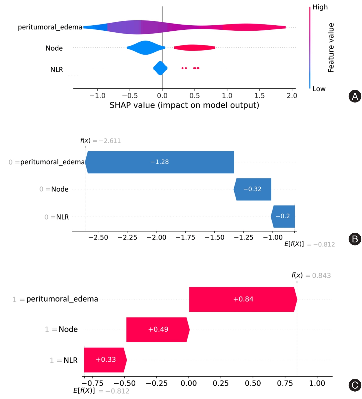
Fig.2
ROC curve of the clinical imaging model"
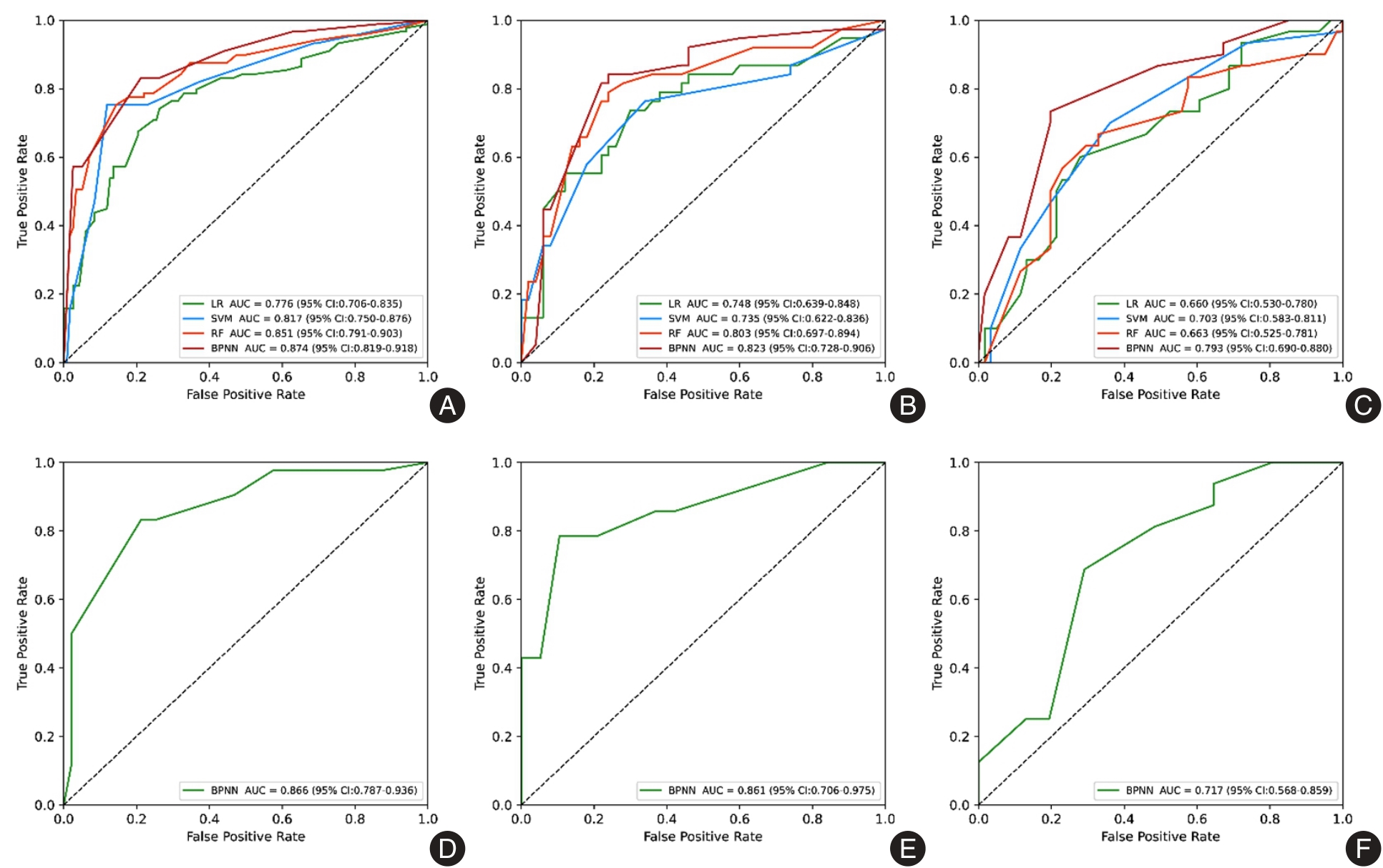

Fig.3
Interpretability analysis of the BPNN model"
Tab.6
Performance of BPNN across subgroups"
| 队列 | 模型 | AUC(95%CI) | 准确度 | 敏感度 | 特异度 | 阳性预测值 | 阴性预测值 |
|---|---|---|---|---|---|---|---|
| 训练集 | BPNN | 0.866(0.787 ~ 0.936) | 0.809 | 0.833 | 0.787 | 0.778 | 0.841 |
| 内部验证集 | BPNN | 0.861(0.706 ~ 0.975) | 0.848 | 0.786 | 0.895 | 0.846 | 0.850 |
| 外部验证集 | BPNN | 0.717(0.568 ~ 0.859) | 0.702 | 0.688 | 0.71 | 0.550 | 0.815 |
| [1] |
陈基明,朱浩雨,高静,等. 基于临床病理及常规和功能MRI影像组学模型预测乳腺癌腋窝淋巴结转移[J]. 中国医学影像技术, 2021, 37(6): 885-890.doi: 10.13929/j.issn.1003-3289. 2021.06.022 .
doi: 10.13929/j.issn.1003-3289. 2021.06.022 |
| [2] |
CHANG J M, LEUNG J W T, MOY L, et al. Axillary Nodal Evaluation in Breast Cancer: State of the Art[J]. Radiology, 2020, 295(3):500-515. doi: 10.1148/radiol.2020192534 .
doi: 10.1148/radiol.2020192534 |
| [3] |
WU T, LONG Q, ZENG L, et al. Axillary lymph node metastasis in breast cancer: From historical axillary surgery to updated advances in the preoperative diagnosis and axillary management[J]. BMC Surg, 2025, 25(1):81. doi: 10.1186/s12893-025-02802-2 .
doi: 10.1186/s12893-025-02802-2 |
| [4] |
丘海,归奕飞,刘媛. 术前腋窝超声正常的临床T1—2 N0乳腺癌患者发生前哨淋巴结转移的预测模型[J]. 实用医学杂志,2025,41(14):2143-2151. doi: 10.3969/j.issn.1006-5725. 2025. 14.004 .
doi: 10.3969/j.issn.1006-5725. 2025. 14.004 |
| [5] |
GIULIANO A E, BALLMAN K V, MCCALL L, et al. Effect of Axillary Dissection vs No Axillary Dissection on 10-Year Overall Survival Among Women With Invasive Breast Cancer and Sentinel Node Metastasis: The ACOSOG Z0011 (Alliance) Randomized Clinical Trial[J]. JAMA, 2017, 318(10):918-926. doi: 10.1001/jama.2017.11470 .
doi: 10.1001/jama.2017.11470 |
| [6] |
逯永晋,石志强,李彤,等. 乳腺癌前哨淋巴结阳性豁免腋窝清扫后区域淋巴结放疗的回顾性研究[J]. 中国癌症杂志, 2025, 35(2): 228-236. doi: 10.19401/j.cnki.1007-3639.2025.02.010 .
doi: 10.19401/j.cnki.1007-3639.2025.02.010 |
| [7] |
WEKKING D, PORCU M, DE SILVA P, et al. Breast MRI: Clinical Indications, Recommendations, and Future Applications in Breast Cancer Diagnosis[J]. Curr Oncol Rep, 2023, 25(4): 257-267. doi: 10.1007/s11912-023-01372-x .
doi: 10.1007/s11912-023-01372-x |
| [8] |
CASTELLOTE-HUGUET P, RUIZ-ESPANA S, GALAN-AUGE C, et al. Breast Cancer Diagnosis Using Texture and Shape Features in MRI[J]. Annu Int Conf IEEE Eng Med Biol Soc, 2023, 2023:1-4. doi: 10.1109/EMBC40787.2023.10340385 .
doi: 10.1109/EMBC40787.2023.10340385 |
| [9] |
赵杭菲, 姚劲草, 王立平, 等. 列线图预测T1-2期乳腺癌腋窝淋巴转移负荷的研究[J]. 中国超声医学杂志, 2023, 39(11): 1224-1226. doi: 10.3969/j.issn.1002-0101.2023.11.009 .
doi: 10.3969/j.issn.1002-0101.2023.11.009 |
| [10] |
吴怡雯, 周晓华, 陈菲, 等. 基于自动乳腺全容积扫查影像组学对预测乳腺癌腋窝淋巴结负荷的价值[J]. 中国超声医学杂志, 2023, 39(5):499-502. doi: 10.3969/j.issn.1002-0101. 2023.05.006 .
doi: 10.3969/j.issn.1002-0101. 2023.05.006 |
| [11] |
LIAO H, CHEN X, LU S, et al. MRI-Based Back Propagation Neural Network Model as a Powerful Tool for Predicting the Response to Induction Chemotherapy in Locoregionally Advanced Nasopharyngeal Carcinoma[J]. J Magn Reson Imaging, 2022, 56(2): 547-559. doi: 10.1002/jmri.28047 .
doi: 10.1002/jmri.28047 |
| [12] |
SATAKE H, ISHIGAKI S, ITO R, et al. Radiomics in breast MRI: current progress toward clinical application in the era of artificial intelligence[J]. Radiol Med, 2022, 127(1): 39-56. doi: 10.1002/jmri.28047 .
doi: 10.1002/jmri.28047 |
| [13] |
VAN DER VELDEN B H M, KUIJF H J, GILHUIJS K G A, et al. Explainable artificial intelligence (XAI) in deep learning-based medical image analysis[J]. Med Image Anal, 2022, 79:102470. doi: 10.1016/j.media.2022.102470 .
doi: 10.1016/j.media.2022.102470 |
| [14] |
MARTAINDALE S R. Breast MR Imaging: Atlas of Anatomy, Physiology, Pathophysiology, and Breast Imaging Reporting and Data Systems Lexicon[J]. Magn Reson Imaging Clin N Am, 2018, 26 (2):179-190. doi: 10.1016/j.mric.2017.12.001 .
doi: 10.1016/j.mric.2017.12.001 |
| [15] |
杨光飞,常瑞姣,武雅婷,等. 超声与MRI对乳腺BI-RADS 3~5类肿物的诊断分类一致性比较[J].中国医学影像学杂志, 2022, 30(10): 991-995. doi: 10.3969/j.issn.1005-5185. 2022. 10.004 .
doi: 10.3969/j.issn.1005-5185. 2022. 10.004 |
| [16] |
XU Z, DING Y, ZHAO K, et al. MRI characteristics of breast edema for assessing axillary lymph node burden in early-stage breast cancer: A retrospective bicentric study[J]. Eur Radiol, 2022, 32(12): 8213-8225. doi: 10.1007/s00330-022-08896-z .
doi: 10.1007/s00330-022-08896-z |
| [17] |
杨森,贾志莺,范清,等. 列线图可预测T1期乳腺癌前哨淋巴结转移:基于超声特征联合临床病理指标[J]. 分子影像学杂志, 2025, 48(7): 848-854. doi: 10.12122/j.issn.1674-4500. 2025. 07.09 .
doi: 10.12122/j.issn.1674-4500. 2025. 07.09 |
| [18] |
HARADA T L, UEMATSU T, NAKASHIMA K, et al. Evaluation of breast edema findings at T2-weighted breast MRI is useful for diagnosing occult inflammatory breast cancer and can predict prognosis after neoadjuvant chemotherapy[J]. Radiology, 2021, 299(1): 53-62. doi: 10.1148/radiol.2021202604 .
doi: 10.1148/radiol.2021202604 |
| [19] |
MALHAIRE C, SELHANE F, SAINT-MARTIN M J, et al. Exploring the added value of pretherapeutic MR descriptors in predicting breast cancer pathologic complete response to neoadjuvant chemotherapy[J]. Eur Radiol, 2023, 33(11): 8142-8154. doi: 10.1007/s00330-023-09797-5 .
doi: 10.1007/s00330-023-09797-5 |
| [20] |
KAISER C G, HEROLD M, KRAMMER J, et al. Prognostic value of “prepectoral edema” in MR-mammography[J]. Anticancer Res, 2017, 37(4): 1989-1995. doi: 10.21873/anticanres. 11542 .
doi: 10.21873/anticanres. 11542 |
| [21] |
CHEON H, KIM H J, KIM T H, et al. Invasive breast cancer: Prognostic value of peritumoral edema identified at preoperative MR imaging[J]. Radiology, 2018, 287(1): 68-75. doi: 10.1148/radiol.2017171157 .
doi: 10.1148/radiol.2017171157 |
| [22] |
PARK N J Y, JEONG J Y, PARK J Y, et al. Peritumoral edema in breast cancer at preoperative MRI: An interpretative study with histopathological review toward understanding tumor microenvironment[J]. Sci Rep, 2021, 11(1): 12992. doi: 10.1038/s41598-021-92283-z .
doi: 10.1038/s41598-021-92283-z |
| [23] |
YILMAZ R, AKPINAR Y, OZYAVUZ I, et al. Synchronous metastatic leiomyosarcoma and primer invasive ductal carcinoma tumors in the same breast: Mammography, ultrasonography, and magnetic resonance imaging findings[J]. Breast J, 2019, 25(2): 310-311. doi: 10.1111/tbj.13211 .
doi: 10.1111/tbj.13211 |
| [24] |
冯其柱,卢曼曼,孙杰,等. 新型全身性炎症指标对急性胰腺炎早期病情严重程度的预测价值[J]. 实用医学杂志, 2024, 40(14):1963-1968. doi: 10.3969/j.issn.1006-5725.2024.14.011 .
doi: 10.3969/j.issn.1006-5725.2024.14.011 |
| [25] |
KAWAGUCHI S, KINOWAKI K, TAMURA N, et al. High-accuracy prediction of axillary lymph node metastasis in invasive lobular carcinoma using focal cortical thickening on magnetic resonance imaging[J]. Breast Cancer, 2023, 30 (4): 637-646. doi: 10.1007/s12282-023-01457-2 .
doi: 10.1007/s12282-023-01457-2 |
| [26] |
史娜,王爽,马兆丽,等. 超声联合免疫炎症指标构建列线图预测早期浸润性导管癌淋巴结转移[J].中国超声医学杂志,2025,41(3):265-269. doi: 10.3969/j.issn.1002-0101. 2025. 03.006 .
doi: 10.3969/j.issn.1002-0101. 2025. 03.006 |
| [27] |
张伟娜,钟李长,师琳,等. 超声影像组学特征联合miRNA-34a表达水平对乳腺癌新辅助化疗患者病理完全缓解的预测能力[J]. 新医学,2025,56(7):645-653. doi: 10.12464/j.issn.0253-9802.2025-0023 .
doi: 10.12464/j.issn.0253-9802.2025-0023 |
| [28] |
BENEDETTO U, DIMAGLI A, SINHA S, et al. Machine learning improves mortality risk prediction after cardiac surgery: Systematic review and meta-analysis[J]. J Thorac Cardiovasc Surg, 2022, 163(6): 2075-2087. doi: 10.1016/j.jtcvs.2020.07.105 .
doi: 10.1016/j.jtcvs.2020.07.105 |
| [29] |
BELLE V, PAPANTONIS I. Principles and Practice of Explainable Machine Learning[J]. Front Big Data, 2021, 4:688969. doi: 10.3389/fdata.2021.688969 .
doi: 10.3389/fdata.2021.688969 |
| [1] | Yingchao WU,Liushan CHEN,Yuqi LIANG,Jieting CHEN,Junfeng HUANG,Qian ZUO,Qianjun CHEN. Development of an organoid⁃based pan⁃TKI precision screening platform to enhance therapeutic efficacy of ET+CDK4/6 inhibitors in HR+/HER2⁃low breast cancer [J]. The Journal of Practical Medicine, 2025, 41(18): 2786-2795. |
| [2] | Xinran ZHANG,Yan SHEN,Jiaojiao HU,Qingqing CHEN,Yangjie XIAO,Feng LU,Shasha YUAN,Xiaohong FU. Study on the applied value of combined clinical and ultrasound multiparameter constructed nomogram for predicting HER⁃2⁃positive breast cancer [J]. The Journal of Practical Medicine, 2025, 41(18): 2812-2819. |
| [3] | Jingshuo LI,Shoushi LIU,Hongwei. GUO. Advances in the mechanism and therapeutic potential of Erianin⁃induced apoptosis in breast cancer cells [J]. The Journal of Practical Medicine, 2025, 41(14): 2132-2137. |
| [4] | Hai QIU,Yifei GUI,Yuan. LIU. Predictors of sentinel lymph node metastasis in clinical T1⁃2 N0 breast cancer patients with preoperatively normal axillary ultrasound [J]. The Journal of Practical Medicine, 2025, 41(14): 2143-2151. |
| [5] | Yuling DUAN,Xuezhi ZHOU,Yongyi LI,Lixia MA,Desheng YANG,Jiao CHENG,Yan WU,Tao LIU,Guoyuan JIANG,Mei. WANG. Clinical value analysis of different MRI measurement methods in evaluating the efficacy of neoadjuvant therapy for breast cancer [J]. The Journal of Practical Medicine, 2025, 41(14): 2152-2159. |
| [6] | Lu JIANG,Weipeng LYU,Sijing CHEN,Yanhua FANG,Shanshan LIANG. Inhibitory effect of disitamab vedotin on breast cancer cells with different HER⁃2 expression levels in tumor organoid culture system [J]. The Journal of Practical Medicine, 2025, 41(12): 1808-1815. |
| [7] | Yaqian DENG,Wenxiao LI,Zelin XU,Jinmei MA,Tingting DU,Wen LIU,Jun LI. Predictive value of growth orientation quantification combined with S⁃Detect technique for axillary lymph node metastasis in breast cancer [J]. The Journal of Practical Medicine, 2025, 41(1): 100-107. |
| [8] | Zixu SONG,Guangzheng ZHU,Chenxu GUO,Jiaqi WU,Ligong ZHANG,Jun. QIAN. Expression of SLC35A2 and PFDN2 in breast cancer and its relationship with clinical observables and prognosis [J]. The Journal of Practical Medicine, 2024, 40(4): 496-502. |
| [9] | Fen LIU,Hui ZHAO,Limin. GUO. Relationship between multi⁃omics combined detection and clinicopathological characteristics, neoadjuvant chemotherapy effect of breast cancer [J]. The Journal of Practical Medicine, 2024, 40(24): 3539-3546. |
| [10] | Yinghua ZENG,Wenji LI,Li. ZHENG. The Impact of the timing of initial dressing change following PICC catheterization on postoperative breast cancer patients [J]. The Journal of Practical Medicine, 2024, 40(19): 2772-2777. |
| [11] | Che CHEN,Dehong LUO,Huangfei YU,Qin ZHANG,Xiaochi HU,Shenghua YU,Yajun. LI. Clinical Application of automatic delineation in whole breast radiotherapy with simultaneous integrated boost to the medial tumor beds [J]. The Journal of Practical Medicine, 2024, 40(17): 2406-2411. |
| [12] | Shaojin LI,Shipeng. ZHENG. Relevant preoperative imaging pathological features and tumor markers serve as predictive indicators for the risk of sentinel lymph node metastasis in breast cancer [J]. The Journal of Practical Medicine, 2024, 40(17): 2418-2424. |
| [13] | Lingyu FANG,Jinghua HU,Junfeng WEN,Shiqi HAN,Yali WANG,Lulan PU,Jingjia LI,Yi YANG,Shishan DENG,Lingmi HOU,Fangfang. ZHOU. Expression and significance of ubiquitin⁃specific proteases 20 and hypoxia inducible factor⁃1α in breast cancer [J]. The Journal of Practical Medicine, 2024, 40(16): 2270-2276. |
| [14] |
AI Yongbiao, HUANG Jun, YUAN Jie, LI Wenfang..
Androgen receptor expression in early triple negative breast cancer and its association with the clinicopath⁃ ological features and prognosis [J]. The Journal of Practical Medicine, 2023, 39(8): 975-979. |
| [15] | Xiran SHI,Heng WANG,Libing HE,Zhiqiang QIU,Hongjian LI,Xiaoxue. XU. Advance in predicting lymph node metastasis of breast cancer by multimodal MRI [J]. The Journal of Practical Medicine, 2023, 39(22): 2861-2865. |
| Viewed | ||||||
|
Full text |
|
|||||
|
Abstract |
|
|||||