

The Journal of Practical Medicine ›› 2025, Vol. 41 ›› Issue (16): 2447-2454.doi: 10.3969/j.issn.1006-5725.2025.16.002
• Feature Report: • Previous Articles
Chao WANG1,Qianqian XU1,Shujuan ZHANG1,Yanping ZHU2( )
)
Received:2025-04-28
Online:2025-08-25
Published:2025-08-28
Contact:
Yanping ZHU
E-mail:1119788145@qq.com
CLC Number:
Chao WANG,Qianqian XU,Shujuan ZHANG,Yanping ZHU. Effect of human umbilical cord mesenchymal stem cell-derived exosomes on microglial polarization in neonatal rats with white matter injury[J]. The Journal of Practical Medicine, 2025, 41(16): 2447-2454.
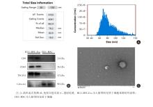
Fig.1
Characterization of HUC-MSC-Exo"

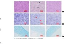
Fig.2
HUC-MSC-Exo improved pathological changes in neonatal rats with WMI"
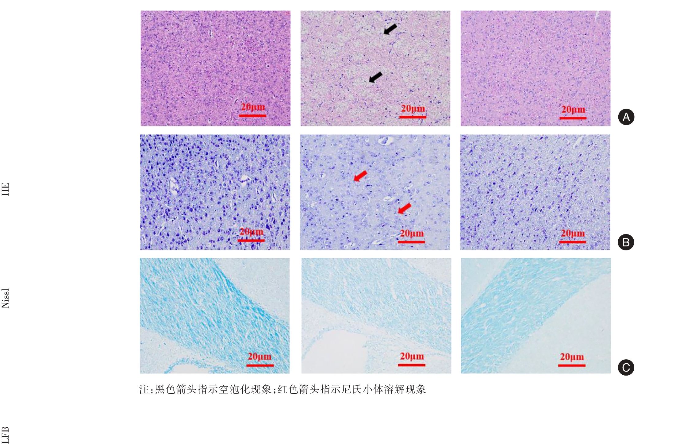
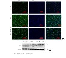
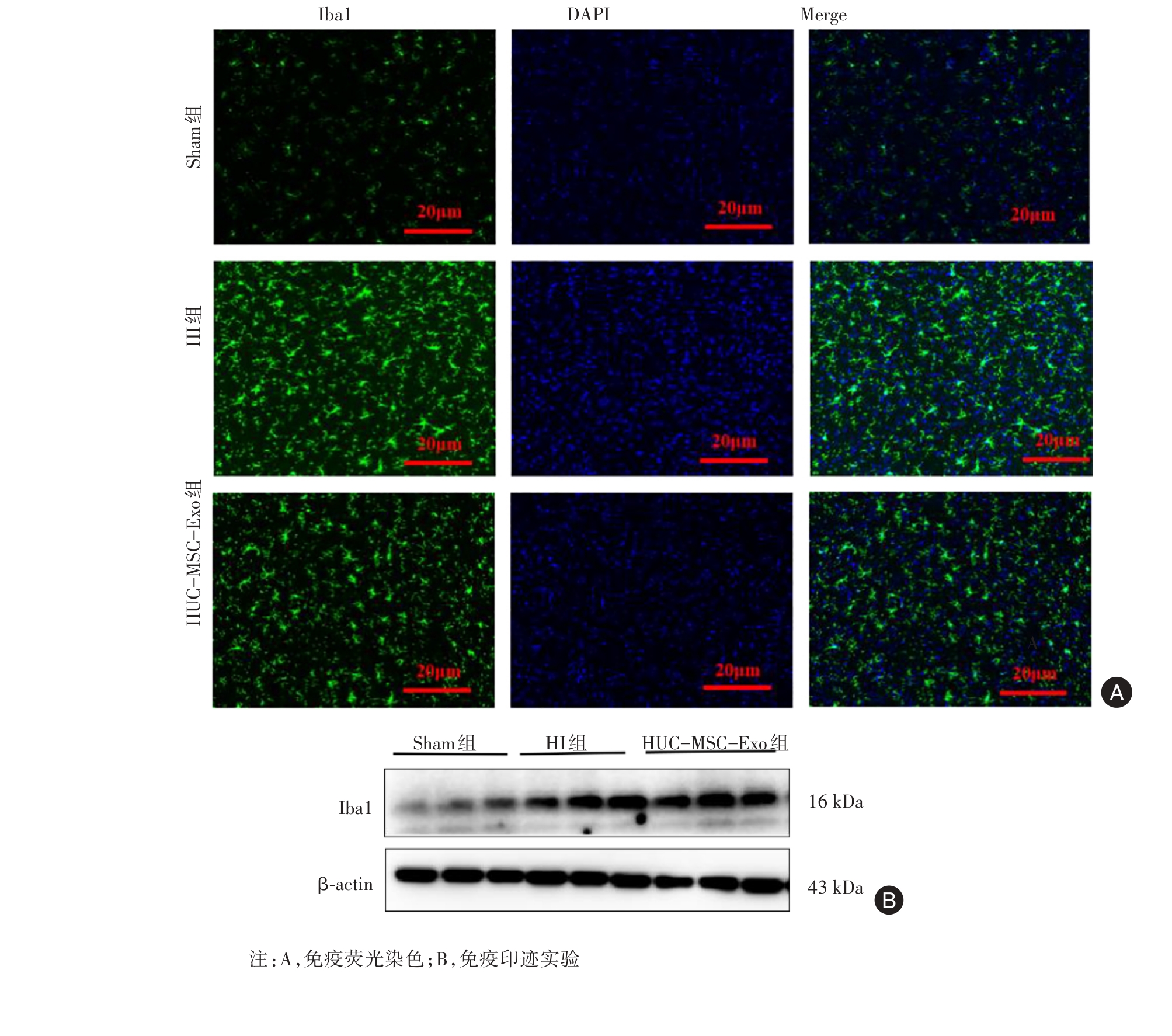
Fig.3
HUC-MSC-Exo suppressed microglial proliferation in neonatal rats with WMI"
Tab.1
Iba1 protein expression levels in the brain of rats in each group(n = 6) x ± s"
| 组别 | Iba1相对阳性区域/% | Iba1蛋白表达 |
|---|---|---|
| Sham组 | 4.03 ± 0.28 | 0.50 ± 0.03 |
| HI组 | 10.69 ± 0.37* | 1.65 ± 0.05* |
| HUC-MSC-Exo组 | 6.37 ± 0.22△ | 1.03 ± 0.04△ |
| F值 | 130.99 | 207.07 |
| P值 | < 0.001 | < 0.001 |
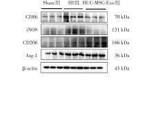
Fig.4
HUC-MSC-Exo suppressed the protein expression of CD86 and iNOS while promoting the protein expression of CD206 and Arg-1"
Tab.2
Comparison of CD86, iNOS, CD206 and Arg-1 protein expression levels in the brain of rats in each group(n = 6)"
| 组别 | CD86蛋白表达 | iNOS蛋白表达 | CD206蛋白表达 | Arg-1蛋白表达 |
|---|---|---|---|---|
| Sham组 | 0.54 ± 0.02 | 0.24 ± 0.03 | 0.31 ± 0.03 | 0.42 ± 0.02 |
| HI组 | 0.87 ± 0.05* | 0.58 ± 0.05* | 0.55 ± 0.01* | 0.74 ± 0.05* |
| HUC-MSC-Exo组 | 0.54 ± 0.06△ | 0.21 ± 0.03△ | 0.84 ± 0.04△ | 1.12 ± 0.08△ |
| F值 | 15.96 | 27.25 | 84.72 | 36.77 |
| P值 | < 0.001 | < 0.001 | < 0.001 | < 0.001 |
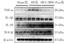
Fig.5
HUC-MSC-Exo suppressed the protein expression of TNF-α and IL-1β while promoting the protein expression of IL-10 and TGF-β"
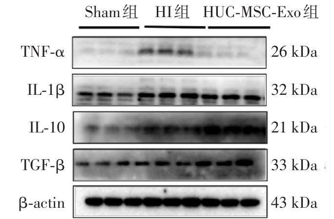
Tab.3
Comparison of TNF-α, IL-1β, IL-10 and TGF-β protein expression levels in the brain of rats in each group(n = 6)"
| 组别 | TNF-α蛋白表达 | IL-1β蛋白表达 | IL-10蛋白表达 | TGF-β蛋白表达 |
|---|---|---|---|---|
| Sham组 | 0.45 ± 0.03 | 0.49 ± 0.04 | 0.41 ± 0.04 | 0.40 ± 0.02 |
| HI组 | 0.89 ± 0.05* | 0.92 ± 0.03* | 0.75 ± 0.06* | 0.68 ± 0.03* |
| HUC-MSC-Exo组 | 0.66 ± 0.02△ | 0.46 ± 0.06△ | 1.17 ± 0.09△ | 1.03 ± 0.05△ |
| F值 | 34.43 | 29.96 | 31.84 | 66.83 |
| P值 | < 0.001 | < 0.001 | < 0.001 | < 0.001 |
| [1] |
XIAO J. Role of the Gut Microbiota-Brain Axis in Brain Damage in Preterm Infants [J]. ACS Pharmacol Transl Sci, 2024, 7(5):1197-1204. doi:10.1021/acsptsci.3c00369
doi: 10.1021/acsptsci.3c00369 |
| [2] |
JANA A, GARG S, GHOSH S, et al. Generation of Functional Neurons from Mesenchymal Stem Cells Using Neural Differentiator and Engineered Peptide Hydrogel: Potential Therapeutic Lead for Traumatic Brain Injury [J]. ACS Appl Mater Interfaces, 2024, 16(47):64476-64493. doi:10.1021/acsami.4c12554
doi: 10.1021/acsami.4c12554 |
| [3] |
ZHANG X, ZHOU Y, YE Y, et al. Human umbilical cord mesenchymal stem cell-derived exosomal microRNA-148a-3p inhibits neointimal hyperplasia by targeting Serpine1[J]. Arch Biochem Biophys, 2022, 719:109155. doi:10.1016/j.abb.2022.109155
doi: 10.1016/j.abb.2022.109155 |
| [4] |
ABBASZADEH H, GHORBANI F, DERAKHSHANI M, et al. Human umbilical cord mesenchymal stem cell-derived extracellular vesicles: A novel therapeutic paradigm [J]. J Cell Physiol, 2020, 235(2):706-717. doi:10.1002/jcp.29004
doi: 10.1002/jcp.29004 |
| [5] | 陈珊, 朱俊德, 赵雪, 等.神经干细胞来源外泌体对脑缺血再灌注损伤大鼠星形胶质细胞TGF⁃β1信号转导与炎症因子的影响[J].实用医学杂志,2023, 39(13):1600-1605. |
| [6] |
LEE J, HAMANAKA G, LO E H, et al. Heterogeneity of microglia and their differential roles in white matter pathology [J]. CNS Neurosci Ther, 2019, 25(12):1290-1298. doi:10.1111/cns.13266
doi: 10.1111/cns.13266 |
| [7] |
ZHAO Y, GAN Y, XU G, et al. MSCs-Derived Exosomes Attenuate Acute Brain Injury and Inhibit Microglial Inflammation by Reversing CysLT2R-ERK1/2 Mediated Microglia M1 Polarization [J]. Neurochem Res, 2020, 45(5):1180-1190. doi:10.1007/s11064-020-02998-0
doi: 10.1007/s11064-020-02998-0 |
| [8] |
DONG C, CHEN M, CAI B, et al. Mesenchymal Stem Cell-Derived Exosomes Improved Cerebral Infarction via Transferring miR-23a-3p to Activate Microglia [J]. Neuromolecular Med, 2022, 24(3):290-298. doi:10.1007/s12017-021-08686-8
doi: 10.1007/s12017-021-08686-8 |
| [9] |
ZHANG Y, XIE Y, HAO Z, et al. Umbilical Mesenchymal Stem Cell-Derived Exosome-Encapsulated Hydrogels Accelerate Bone Repair by Enhancing Angiogenesis [J]. ACS Appl Mater Interfaces, 2021, 13(16):18472-18487. doi:10.1021/acsami.0c22671
doi: 10.1021/acsami.0c22671 |
| [10] |
HUANG Y, MA J, FAN Y, et al. Mechanisms of human umbilical cord mesenchymal stem cells-derived exosomal lncRNA GAS5 in alleviating EMT of HPMCs via Wnt/β-catenin signaling pathway [J]. Aging (Albany NY), 2023, 15(10):4144-4158. doi:10.18632/aging.204719
doi: 10.18632/aging.204719 |
| [11] |
CARLONI S, CRINELLI R, PALMA L, et al. The Synthetic Cannabinoid URB447 Reduces Brain Injury and the Associated White Matter Demyelination after Hypoxia-Ischemia in Neonatal Rats [J]. ACS Chem Neurosci, 2020, 11(9):1291-1299. doi:10.1021/acschemneuro.0c00047
doi: 10.1021/acschemneuro.0c00047 |
| [12] |
VANNUCCI S J, BACK S A. The Vannucci Model of Hypoxic-Ischemic Injury in the Neonatal Rodent: 40 years Later [J]. Dev Neurosci, 2022, 44(4/5):186-193. doi:10.1159/000523990
doi: 10.1159/000523990 |
| [13] |
BOBIS-WOZOWICZ S, KMIOTEK K, KANIA K, et al. Diverse impact of xeno-free conditions on biological and regenerative properties of hUC-MSCs and their extracellular vesicles [J]. J Mol Med (Berl), 2017, 95(2):205-220. doi:10.1007/s00109-016-1471-7
doi: 10.1007/s00109-016-1471-7 |
| [14] |
GAO X, GAO L F, ZHANG Y N, et al. Huc-MSCs-derived exosomes attenuate neuropathic pain by inhibiting activation of the TLR2/MyD88/NF-κB signaling pathway in the spinal microglia by targeting Rsad2 [J]. Int Immunopharmacol, 2023, 114:109505. doi:10.1016/j.intimp.2022.109505
doi: 10.1016/j.intimp.2022.109505 |
| [15] |
ZHAI X, CHEN K, YANG H, et al. Extracellular vesicles derived from CD73 modified human umbilical cord mesenchymal stem cells ameliorate inflammation after spinal cord injury [J]. J Nanobiotechnology, 2021, 19(1):274. doi:10.1186/s12951-021-01022-z
doi: 10.1186/s12951-021-01022-z |
| [16] |
EBRAHIM N, SAIHATI H A AL, ALALI Z, et al. Exploring the molecular mechanisms of MSC-derived exosomes in Alzheimer's disease: Autophagy, insulin and the PI3K/Akt/mTOR signaling pathway [J]. Biomed Pharmacother, 2024, 176:116836. doi:10.1016/j.biopha.2025.118042
doi: 10.1016/j.biopha.2025.118042 |
| [17] |
ZHANG J, WANG C, YANG G, et al. Olfactory mucosal mesenchymal stem cell-derived exosome Lnc A2M-AS1 ameliorates oxidative stress by regulating TP53INP1-mediated mitochondrial autophagy through interacting with IGF2BP1 in Parkinson's diseases [J]. Cell Biol Toxicol, 2025, 41(1):60. doi:10.1007/s10565-025-10009-7
doi: 10.1007/s10565-025-10009-7 |
| [18] |
ZHANG L, BAI W, PENG Y, et al. Human umbilical cord mesenchymal stem cell-derived exosomes provide neuroprotection in traumatic brain injury through the lncRNA TUBB6/Nrf2 pathway [J]. Brain Res, 2024, 1824:148689. doi:10.1016/j.brainres.2023.148689
doi: 10.1016/j.brainres.2023.148689 |
| [19] |
ZHANG Z, ZOU X, ZHANG R, et al. Human umbilical cord mesenchymal stem cell-derived exosomal miR-146a-5p reduces microglial-mediated neuroinflammation via suppression of the IRAK1/TRAF6 signaling pathway after ischemic stroke [J]. Aging (Albany NY), 2021, 13(2):3060-3079. doi:10.18632/aging.202466
doi: 10.18632/aging.202466 |
| [20] |
LI P, KASLAN M, LEE S H, et al. Progress in Exosome Isolation Techniques [J]. Theranostics, 2017, 7(3):789-804. doi:10.7150/thno.18133
doi: 10.7150/thno.18133 |
| [21] |
CHEN X, SAI Y, CUI W, et al. Human umbilical cord mesenchymal stem cell-derived exosomes combined with mouse nerve growth factor can more effectively ameliorate the motor disorder and brain pathological injury in mice with cerebral palsy [J]. Adv Clin Exp Med, 2025. DOI: 10.17219/acem/192773 .
doi: 10.17219/acem/192773 |
| [22] |
MARSTERS C M, NESAN D, FAR R, et al. Embryonic microglia influence developing hypothalamic glial populations [J]. J Neuroinflammation, 2020,17(1):146. doi:10.1186/s12974-020-01811-7
doi: 10.1186/s12974-020-01811-7 |
| [23] |
CUI L, LUO W, JIANG W, et al. Human umbilical cord mesenchymal stem cell-derived exosomes promote neurological function recovery in rat after traumatic brain injury by inhibiting the activation of microglia and astrocyte [J]. Regen Ther, 2022, 21:282-287. doi:10.1016/j.reth.2022.07.005
doi: 10.1016/j.reth.2022.07.005 |
| [24] |
RONALDSON P T, DAVIS T P. Regulation of blood-brain barrier integrity by microglia in health and disease: A therapeutic opportunity [J]. J Cereb Blood Flow Metab, 2020, 40():S6-S24. doi:10.1177/0271678x20951995
doi: 10.1177/0271678x20951995 |
| [25] |
YANG L, YU X, ZHANG Y, et al. Caffeine treatment started before injury reduces hypoxic-ischemic white-matter damage in neonatal rats by regulating phenotypic microglia polarization [J]. Pediatr Res, 2022, 92(6):1543-1554. doi:10.1038/s41390-021-01924-6
doi: 10.1038/s41390-021-01924-6 |
| [26] | ZONG L, HUANG P, SONG Q, et al. Bone marrow mesenchymal stem cells-secreted exosomal H19 modulates lipopolysaccharides-stimulated microglial M1/M2 polarization and alleviates inflammation-mediated neurotoxicity [J]. Am J Transl Res, 2021, 13(3):935-951. |
| [27] |
LIU W, RONG Y, WANG J, et al. Exosome-shuttled miR-216a-5p from hypoxic preconditioned mesenchymal stem cells repair traumatic spinal cord injury by shifting microglial M1/M2 polarization [J]. J Neuroinflammation, 2020, 17(1):47. doi:10.1186/s12974-020-1726-7
doi: 10.1186/s12974-020-1726-7 |
| [28] |
WANG J, WEI Q, YANG Y, et al. Small extracellular vesicles derived from four dimensional-culture of mesenchymal stem cells induce alternatively activated macrophages by upregulating IGFBP2/EGFR to attenuate inflammation in the spinal cord injury of rats [J]. Front Bioeng Biotechnol, 2023, 11:1146981. doi:10.3389/fbioe.2023.1146981
doi: 10.3389/fbioe.2023.1146981 |
| [1] | Zhicong LIU,Daiyin LIU,Juntian LONG,Chixing CHENG,Jian HUANG. Exosomal lncRNA CIAT1 promotes collective invasion of bladder cancer [J]. The Journal of Practical Medicine, 2025, 41(9): 1299-1308. |
| [2] | Xingwei WU,Jianying WANG,Chengxiao GUO,Ziyi LIU,Chao SUN,Fei. YU. The effect of remimazolam on modulating the ROS/RAGE/NF-κB signaling pathway in LPS-induced microglial inflammation [J]. The Journal of Practical Medicine, 2025, 41(2): 153-161. |
| [3] | Huizhen LIU,Xiaonan LU,Ge LIU,Guanqing CAI,Pingan LI,Yingjian ZENG,Guangbin SHANG. The effects and mechanism of total flavonoids of Sarcandra glabra in modulating bone marrow mesenchymal stem cells and their exosomes to promote megakaryocyte differentiation [J]. The Journal of Practical Medicine, 2025, 41(11): 1618-1626. |
| [4] | Fangming WANG,Wenxuan SHANG,Jingwen ZHANG,Yingxiao JI,Litao. LI. Research advances on the regulation of microglia polarization by autophagy in ischemic stroke [J]. The Journal of Practical Medicine, 2024, 40(9): 1324-1330. |
| [5] | Fu CHEN,Bin LIU,Shuaijun HE,Yong ZHAO,Weizhou. WANG. The relationship between semen quality and trace element levels in seminal plasma and miR⁃184 levels in seminal vesicles of male infertility patients [J]. The Journal of Practical Medicine, 2024, 40(7): 930-935. |
| [6] | Li XIAO,Shumin LUO,Fang XU,Pengpeng LU,Enhong XING,Weihua. LI. Effect of culture time on immune⁃related membrane proteins of mouse dendritic cells and their exosomes [J]. The Journal of Practical Medicine, 2024, 40(7): 941-947. |
| [7] | Yuxin CHENG,Liang LIU,Shiyu DONG,Shengchao LI,Meng ZHANG. Research advances in exosomal proteins, mRNA and non⁃coding RNA regulation of Hepatocellular Carcinoma [J]. The Journal of Practical Medicine, 2024, 40(6): 748-755. |
| [8] | Shu CHEN,Jinglei ZHANG,Kang RONG,Nan ZHANG,Weiyi SUN. Research progress of exosomes in distant metastasis and drug resistance of gastric cancer [J]. The Journal of Practical Medicine, 2024, 40(6): 870-876. |
| [9] | Yan JIANG,Xiaoqin WANG,Hong MEI,Xinxin LIU,Zhenliang LIAO,Kun YU,Banghai FENG,Song QIN. Type Ⅱ alveolar epithelial cell⁃derived exosomal miR⁃21⁃5p targeting SKP2 alleviate bronchopulmonary dysplasia [J]. The Journal of Practical Medicine, 2024, 40(23): 3298-3305. |
| [10] | Zhe SHI,Xialin ZUO,Linhui PENG,Zhiwei LU,Kongping. LI. Effect of M1 microglial polarization on secondary damage in the thalamus after cerebral cortical infarction [J]. The Journal of Practical Medicine, 2024, 40(22): 3138-3145. |
| [11] | Qing LUO,Jinjin HUANG,Tingting REN,Ruihua ZHOU,Donghua XU,Zhenhua WANG,Guoying WANG. The effect of umbilical cord stem cell exosomes on the proliferation of dermal papilla cells [J]. The Journal of Practical Medicine, 2024, 40(20): 2828-2834. |
| [12] | Jiao XU,Min ZHANG,Guogang. XIE. Research progress of mast cell⁃derived exosomes in bronchial asthma [J]. The Journal of Practical Medicine, 2024, 40(15): 2194-2198. |
| [13] | Ruxue SUN,Mengli ZHU,Jingjing LIU,Fei. CHEN. The neuroprotective mechanism of RKIP signaling pathway expression on microglial polarization in cerebral hemorrhage rat [J]. The Journal of Practical Medicine, 2024, 40(14): 1935-1940. |
| [14] |
HOU Yongzhe, ZHANG Qin, ZHAO Xiaochen, HE Miao, YU Lingling, BAI Hai, WU Tao..
Research progress of extracellular vesicles derived from mesenchymal stem cells in treatment of acute lung injury [J]. The Journal of Practical Medicine, 2023, 39(3): 390-394. |
| [15] | Dan XIE,Shi. OUYANG. Effect of Yinchenhao Decoction combined with exosomes derived from umbilical cord mesenchymal stem cells on acute liver failure and hepatocyte pyroptosis [J]. The Journal of Practical Medicine, 2023, 39(23): 3034-3042. |
| Viewed | ||||||
|
Full text |
|
|||||
|
Abstract |
|
|||||