

The Journal of Practical Medicine ›› 2025, Vol. 41 ›› Issue (2): 264-270.doi: 10.3969/j.issn.1006-5725.2025.02.017
• Medical Examination and Clinical Diagnosis • Previous Articles
Wei LU1,Pan ZHANG1,Yushu. QIN2
Received:2024-07-25
Online:2025-01-25
Published:2025-01-26
CLC Number:
Wei LU,Pan ZHANG,Yushu. QIN. Clinical significance of CT perfusion imaging combined with artificial intelligence in evaluating reperfusion injury after cerebral infarction[J]. The Journal of Practical Medicine, 2025, 41(2): 264-270.
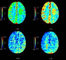
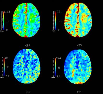
Fig.1
CT perfusion imaging"
Tab.1
Comparison of basic data between reperfusion injury group and non-perfusion injury group"
| 因素 | 再灌注损伤组(n = 31) | 无灌注损伤组(n = 75) | t/χ 2 值 | P值 |
|---|---|---|---|---|
| 性别/[例(%)] | 0.871 | 0.351 | ||
| 男 | 20(64.52) | 41(54.67) | ||
| 女 | 11(35.48) | 34(45.33) | ||
| 年龄≥ 60岁/[例(%)] | 19(61.29) | 36(48.00) | 1.552 | 0.213 |
| 基础疾病/[例(%)] | ||||
| 高血压 | 15(48.39) | 26(34.67) | 1.741 | 0.187 |
| 糖尿病 | 13(41.94) | 24(32.00) | 0.953 | 0.329 |
| 高脂血症 | 12(38.71) | 25(33.33) | 0.279 | 0.597 |
| 冠心病 | 4(12.90) | 7(9.33) | 0.301 | 0.584 |
| 吸烟史 | 12(38.71) | 25(33.33) | 0.279 | 0.597 |
| 饮酒史 | 6(19.35) | 12(16.00) | 0.175 | 0.676 |
| 房颤史 | 7(22.58) | 15(20.00) | 0.089 | 0.766 |
| 脑梗死部位/[例(%)] | 0.418 | 0.812 | ||
| 丘脑 | 7(22.58) | 13(17.33) | ||
| 脑叶 | 5(16.13) | 12(16.00) | ||
| 基底节 | 19(61.29) | 50(66.67) | ||
| TOAST分型/[例(%)] | 0.498 | 0.779 | ||
| 大动脉粥样硬型脑梗死 | 18(58.06) | 49(65.33) | ||
| 小血管闭塞型脑梗死 | 10(32.26) | 20(26.67) | ||
| 心源性脑梗死 | 3(9.68) | 6(8.00) | ||
| DNT/min | 134.36 ± 25.36 | 115.21 ± 20.04 | 4.131 | < 0.001 |
| 收缩压/mmHg | 148.95 ± 15.47 | 146.84 ± 13.96 | 0.686 | 0.494 |
| 舒张压/mmHg | 82.94 ± 10.26 | 83.29 ± 10.03 | 0.162 | 0.871 |
| TG/(mmoL/L) | 1.59 ± 0.26 | 1.52 ± 0.24 | 1.333 | 0.185 |
| TC/(mmoL/L) | 4.48 ± 0.62 | 4.31 ± 0.59 | 1.330 | 0.187 |
| 血肌酐/(μmoL /L) | 91.32 ± 10.51 | 89.27 ± 9.95 | 0.949 | 0.345 |
| 尿素氮/(mmol/L) | 4.52 ± 0.71 | 4.41 ± 0.68 | 0.748 | 0.456 |
| 纤维蛋白原/(g/L) | 3.15 ± 0.48 | 2.81 ± 0.42 | 3.634 | < 0.001 |
| WBC计数/(× 109/L) | 43.36 ± 8.15 | 42.94 ± 7.92 | 0.246 | 0.806 |
| PT/s | 15.01 ± 2.01 | 14.54 ± 1.95 | 1.119 | 0.266 |
| TT/s | 14.36 ± 2.15 | 13.99 ± 2.08 | 0.825 | 0.411 |
| NIHSS评分/分 | 9.01 ± 1.53 | 7.15 ± 1.29 | 6.388 | < 0.001 |
Tab.2
Comparison of CT perfusion imaging and AI parameters between the reperfusion injury group and the non-perfusion injury group"
| 组别 | 例数 | CBF/[mL/(100g·min)] | CBV/(mL/100 g) | MTT/s | TTP/s | 平均CT值 | 峰度 | 熵 |
|---|---|---|---|---|---|---|---|---|
| 再灌注损伤组 | 31 | 12.05 ± 1.76 | 1.23 ± 0.18 | 4.31 ± 0.65 | 26.28 ± 3.26 | 9.25 ± 1.49 | 5.23 ± 1.01 | 8.05 ± 1.23 |
| 无灌注损伤组 | 75 | 14.39 ± 1.92 | 1.01 ± 0.15 | 3.96 ± 0.61 | 24.51 ± 3.09 | 10.89 ± 1.58 | 4.06 ± 0.79 | 8.64 ± 1.36 |
| t值 | 5.844 | 6.471 | 2.636 | 2.640 | 4.941 | 6.377 | 2.087 | |
| P值 | < 0.001 | < 0.001 | 0.010 | 0.010 | < 0.001 | < 0.001 | 0.039 |
Tab.3
Analysis of factors affecting reperfusion injury after cerebral infarction"
| 因素 | β | SE | Wald χ2值 | P值 | OR | 95%CI |
|---|---|---|---|---|---|---|
| NIHSS评分 | 1.654 | 0.402 | 16.929 | < 0.001 | 5.228 | 2.151 ~ 12.705 |
| CBF | 1.329 | 0.317 | 17.576 | < 0.001 | 3.777 | 1.554 ~ 9.180 |
| CBV | 1.308 | 0.296 | 19.527 | < 0.001 | 3.699 | 1.522 ~ 9.989 |
| 平均CT值 | 1.417 | 0.245 | 33.451 | < 0.001 | 4.125 | 1.697 ~ 10.024 |
Tab.4
Value analysis of CBF, CBV and average CT values in predicting reperfusion injury in patients with cerebral infarction after thrombolytic therapy"
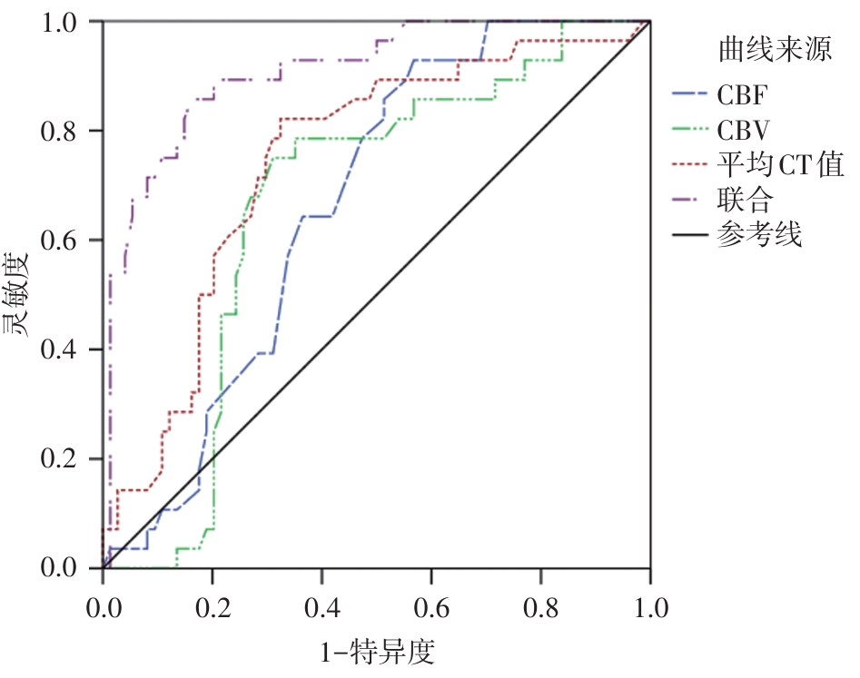
| 指标 | 最佳截断点 | 灵敏度/% | 特异度/% | AUC | 95%CI |
|---|---|---|---|---|---|
| CBF/[mL/(100g·min)] | 13.16 | 67.74 | 70.67 | 0.665 | 0.561 ~ 0.768 |
| CBV/(mL/100 g) | 1.14 | 70.97 | 74.67 | 0.667 | 0.555 ~ 0.778 |
| 平均CT值 | 9.95 | 77.42 | 77.33 | 0.744 | 0.639 ~ 0.849 |
| 联合 | - | 87.10 | 90.67 | 0.908 | 0.845 ~ 0.971 |
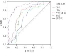
Fig.2
ROC curve of CBF, CBV and average CT values predicting reperfusion injury in cerebral infarction patients after thrombolytic therapy"
| 1 | 王立志,罗伟良,刘武,等. Percheron动脉闭塞致双侧丘脑梗死临床及影像学特征分析[J]. 实用医学杂志, 2020,36(4):553-556. |
| 2 |
SHANG W, ZHANG Y, XUE L, et al. Evaluation of collateral circulation and short-term prognosis of patients with acute cerebral infarction by perfusion-weighted MRI[J]. Ann Palliat Med, 2022, 11(4):1351-1359. doi:10.21037/apm-21-3589
doi: 10.21037/apm-21-3589 |
| 3 |
OCHIAI H, KANEMARU K, MATSUDA S, et al. A case of acute cerebral infarction with a favorable prognosis after rt-PA administration by a general physician with telestroke support[J]. J Rural Med, 2021, 16(2):119-122. doi:10.2185/jrm.2020-048
doi: 10.2185/jrm.2020-048 |
| 4 |
CHIHI M, DARKWAH OPPONG M, PIERSCIANEK D, et al. Analysis of brain natriuretic peptide levels after traumatic acute subdural hematoma and the risk of post-operative cerebral infarction[J]. J Neurotrauma, 2021, 38(22):3068-3076. doi:10.1089/neu.2021.0169
doi: 10.1089/neu.2021.0169 |
| 5 |
ANDE S R, GRYNSPAN J, AVIV R I, et al. Imaging for predicting hemorrhagic transformation of acute ischemic stroke-a narrative review[J]. Can Assoc Radiol J, 2022, 73(1):194-202. doi:10.1177/08465371211018369
doi: 10.1177/08465371211018369 |
| 6 | 秦霜,戴才文,王根强,等. CT灌注成像联合血清D-D、MMP-9对急性脑梗死患者溶栓后出血转化的预测价值研究[J]. 中国CT和MRI杂志,2023,21(6):24-27. |
| 7 |
WOUTERS A, ROBBEN D, CHRISTENSEN S, et al. Prediction of Stroke Infarct Growth Rates by Baseline Perfusion Imaging[J]. Stroke, 2022, 53(2):569-577. doi:10.1161/strokeaha.121.034444
doi: 10.1161/strokeaha.121.034444 |
| 8 |
RAMOS L A, VAN DER STEEN W E, SALES BARROS R, et al. Machine learning improves prediction of delayed cerebral ischemia in patients with subarachnoid hemorrhage[J]. J Neurointerv Surg, 2019, 11(5):497-502. doi:10.1136/neurintsurg-2018-014258
doi: 10.1136/neurintsurg-2018-014258 |
| 9 | 徐伟,李辉萍,贺国华,等. 利用人工智能系统预测脑梗死静脉溶栓后的出血转化[J]. 中国卫生统计,2021,38(2):250-253. |
| 10 |
MOROZOV A, TARATKIN M, BAZARKIN A, et al. A systematic review and meta-analysis of artificial intelligence diagnostic accuracy in prostate cancer histology identification and grading[J]. Prostate Cancer Prostatic Dis, 2023, 26(4):681-692. doi:10.1038/s41391-023-00673-3
doi: 10.1038/s41391-023-00673-3 |
| 11 |
MAKITIE A A, ALABI R O, NG S P, et al. Artificial Intelligence in Head and Neck Cancer: A Systematic Review of Systematic Reviews[J]. Adv Ther, 2023, 40(8):3360-3380. doi:10.1007/s12325-023-02527-9
doi: 10.1007/s12325-023-02527-9 |
| 12 |
中华医学会神经病学分会,中华医学会神经病学分会脑血管病学组. 中国急性缺血性脑卒中诊治指南2018[J]. 中华神经科杂志, 2018, 51(9):666-682. doi:10.3760/cma.j.issn.1006-7876.2018.09.004
doi: 10.3760/cma.j.issn.1006-7876.2018.09.004 |
| 13 |
KNIGHT-GREENFIELD A, NARIO J J Q, GUPTA A. Causes of acute stroke: A patterned approach[J]. Radiol Clin North Am, 2019, 57(6):1093-1108. doi:10.1016/j.rcl.2019.07.007
doi: 10.1016/j.rcl.2019.07.007 |
| 14 |
CHALOS V, VAN DER ENDE N A M, LINGSMA H F, et al. National institutes of health stroke scale: An alternative primary outcome measure for trials of acute treatment for ischemic stroke[J]. Stroke, 2020, 51(1):282-290. doi:10.1161/str.50.suppl_1.tmp6
doi: 10.1161/str.50.suppl_1.tmp6 |
| 15 | 李水仙,陈星宇,阳清伟,等. Y型双支架取栓治疗急性大脑中动脉M1段分叉部闭塞脑梗死7例[J]. 介入放射学杂志,2022,31(2):167-171. |
| 16 |
NG F C, CHURILOV L, YASSI N, et al. Prevalence and Significance of Impaired Microvascular Tissue Reperfusion Despite Macrovascular Angiographic Reperfusion (No-Reflow)[J]. Neurology, 2022, 98(8):790-801. doi:10.1212/wnl.0000000000013210
doi: 10.1212/wnl.0000000000013210 |
| 17 |
MOUGHAL S, TRIPPIER S, AL-MOUSA A, et al. Strokectomy for malignant middle cerebral artery infarction: Experience and meta-analysis of current evidence[J]. J Neurol, 2022, 269(1):149-158. doi:10.1007/s00415-020-10358-9
doi: 10.1007/s00415-020-10358-9 |
| 18 | 陈峰,王洁,甘解华,等. MR三维动脉自旋标记、双指数模型弥散加权成像评估急性前循环大血管闭塞患者再通后再灌注损伤风险[J]. 介入放射学杂志,2021,30(12):1210-1214. |
| 19 |
NOONE M L, MOIDEEN F, KRISHNA R B, et al. Mobile App Based Strategy Improves Door-to-Needle Time in the Treatment of Acute Ischemic Stroke[J]. J Stroke Cerebrovasc Dis, 2020,29(12):105319. doi:10.1016/j.jstrokecerebrovasdis.2020.105319
doi: 10.1016/j.jstrokecerebrovasdis.2020.105319 |
| 20 |
BAO L, ZHANG S, GONG X, et al. Trousseau Syndrome Related Cerebral Infarction: Clinical Manifestations, Laboratory Findings and Radiological Features[J]. J Stroke Cerebrovasc Dis, 2020,29(9):104891. doi:10.1016/j.jstrokecerebrovasdis.2020.104891
doi: 10.1016/j.jstrokecerebrovasdis.2020.104891 |
| 21 |
SHANG W, ZHANG Y, XUE L, et al. Evaluation of collateral circulation and short-term prognosis of patients with acute cerebral infarction by perfusion-weighted MRI[J]. Ann Palliat Med, 2022,11(4):1351-1359. doi:10.21037/apm-21-3589
doi: 10.21037/apm-21-3589 |
| 22 |
WU D, YIN L, ZHANG Y, et al. Evaluation of microcirculation in asymptomatic cerebral infarction with multi-parameter imaging of spectral CT[J]. Brain Res Bull, 2023,203(1):110775. doi:10.1016/j.brainresbull.2023.110775
doi: 10.1016/j.brainresbull.2023.110775 |
| 23 | 韩龙, 张海莲, 马琼. CT灌注成像联合miR-195在急性脑梗死诊断及预后分析中的应用[J]. 影像科学与光化学, 2022, 40(4):927-931. |
| 24 |
YUEN N, MLYNASH M, O'RIORDAN A, et al. Cerebral perfusion imaging predicts final infarct volume after basilar artery thrombectomy[J]. J Stroke Cerebrovasc Dis, 2023,32(1):106866. doi:10.1016/j.jstrokecerebrovasdis.2022.106866
doi: 10.1016/j.jstrokecerebrovasdis.2022.106866 |
| 25 |
ADAMOU A, BELTSIOS E T, BANIA A, al et, Papanagiotou P. Artificial intelligence-driven ASPECTS for the detection of early stroke changes in non-contrast CT: A systematic review and meta-analysis[J]. J Neurointerv Surg, 2023, 5(2):298-304. doi:10.1136/jnis-2022-019447
doi: 10.1136/jnis-2022-019447 |
| Viewed | ||||||
|
Full text |
|
|||||
|
Abstract |
|
|||||