

The Journal of Practical Medicine ›› 2025, Vol. 41 ›› Issue (2): 153-161.doi: 10.3969/j.issn.1006-5725.2025.02.001
• Basic Research •
Xingwei WU,Jianying WANG,Chengxiao GUO,Ziyi LIU,Chao SUN,Fei. YU( )
)
Received:2024-10-12
Online:2025-01-25
Published:2025-01-26
Contact:
Fei. YU
E-mail:yufei001@bzmc.edu.cn
CLC Number:
Xingwei WU,Jianying WANG,Chengxiao GUO,Ziyi LIU,Chao SUN,Fei. YU. The effect of remimazolam on modulating the ROS/RAGE/NF-κB signaling pathway in LPS-induced microglial inflammation[J]. The Journal of Practical Medicine, 2025, 41(2): 153-161.
Tab.1
Comparison of the effects of different concentrations of remimazolam on the cell viability of BV2 cells"
| 组别 | 细胞活性 |
|---|---|
| 对照组 | 100.00 ± 1.46 |
| Rema(10 μg/mL) | 100.56 ± 1.19 |
| Rema(20 μg/mL) | 99.03 ± 1.21 |
| Rema(50 μg/mL) | 98.59 ± 1.60 |
| Rema(100 μg/mL) | 99.27 ± 1.53 |
| Rema(200 μg/mL) | 98.70 ± 0.13 |
| Rema(500 μg/mL) | 87.45 ± 2.10# |
| F值 | 31.29 |
| P值 | < 0.001 |
Tab.2
Comparison of the effects of different treatments on the cell viability of BV2 cells"
| 组别 | 细胞活力 |
|---|---|
| 对照组 | 100 ± 7.71 |
| Rema组 | 98.97 ± 9.42 |
| 模型组 | 49.63 ± 10.01# |
| LPS + Rema(50 μg/mL)组 | 74.94 ± 5.04* |
| LPS + Rema(100 μg/mL)组 | 85.38 ± 3.79* |
| LPS + Rema(200 μg/mL)组 | 83.63 ± 9.76* |
| F值 | 20.95 |
| P值 | < 0.001 |
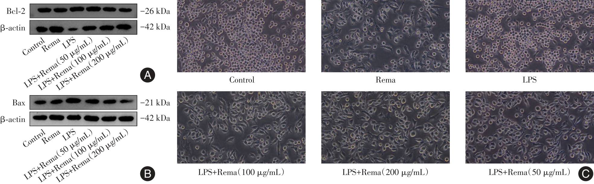
Fig.1
Effects of different concentrations of remimazolam and LPS on apoptotic protein expression and cell morphologyin BV2 cells"
Tab.3
Comparison of mRNA expression of inflammatory factors in BV2 cells μnder different treatment methods"
| 组别 | TNF-α | IL-6 | IL-1β |
|---|---|---|---|
| 对照组 | 1.01 ± 0.01 | 1.08 ± 0.09 | 1.01 ± 0.01 |
| Rema组 | 2.28 ± 0.35 | 1.21 ± 0.26 | 0.27 ± 0.06 |
| 模型组 | 50.77 ± 10.81# | 6 578.87 ± 808.00# | 866.69 ± 158.07# |
| LPS + Rema(50 μg/mL)组 | 33.99 ± 7.75* | 4 479.15 ± 920.42* | 601.24 ± 148.29* |
| LPS + Rema(100 μg/mL)组 | 31.30 ± 3.22* | 3 320.10 ± 693.78* | 479.18 ± 87.37* |
| LPS + Rema(200 μg/mL)组 | 26.49 ± 9.56* | 2 129.11 ± 410.63* | 299.96 ± 50.57* |
| F值 | 24.23 | 37.14 | 56.05 |
| P值 | < 0.001 | <0.001 | < 0.001 |
Tab.4
Comparison of TNF-α and IL-6 secretion and IL-1β protein expression in BV2 cells treated with different methods x ± s"
| 组别 | TNF-α | IL-6 | IL-1β |
|---|---|---|---|
| 对照组 | 31.33 ± 2.06 | 17.74 ± 4.84 | 0.22 ± 0.11 |
| Rema组 | 32.92 ± 1.72 | 8.06 ± 5.35 | 0.16 ± 0.04 |
| 模型组 | 2 335.67 ± 119.6# | 4 046.77 ± 142.68# | 1.25 ± 0.05# |
| LPS + Rema(50 μg/mL)组 | 1 572.65 ± 75.04* | 3 664.52 ± 133.06* | 0.96 ± 0.08* |
| LPS + Rema(100 μg/mL)组 | 1 458.66 ± 33.74* | 3 520.97 ± 127.87* | 0.87 ± 0.12* |
| LPS + Rema(200 μg/mL)组 | 1 113.46 ± 31.19* | 3 372.58 ± 64.86* | 0.73 ± 0.08* |
| F值 | 678.9 | 1 098 | 50.68 |
| P值 | < 0.001 | < 0.001 | < 0.001 |
Tab.5
Comparison of oxidative stress-related indicators in BV2 cells treated with different methods"
| 组别 | MDA含量 | SOD活性 | GSH活性 |
|---|---|---|---|
| Control组 | 1.54 ± 0.29 | 10.85 ± 0.65 | 70.67 ± 10.10 |
| Rema组 | 1.61 ± 0.28 | 10.46 ± 1.03 | 69.15 ± 5.57 |
| 模型组 | 5.39 ± 1.00# | 7.36 ± 0.72# | 41.19 ± 3.26# |
| LPS + Rema(50 μg/mL)组 | 2.32 ± 0.57* | 9.15 ± 1.01 | 59.69 ± 3.30* |
| LPS + Rema(100 μg/mL)组 | 1.66 ± 0.13* | 9.92 ± 1.60* | 70.02 ± 7.88* |
| LPS + Rema(200 μg/mL)组 | 1.51 ± 0.39* | 9.93 ± 0.89* | 81.72 ± 7.67* |
| F值 | 25.17 | 4.375 | 12.39 |
| P值 | < 0.001 | 0.008 8 | < 0.001 |
Fig.2
Detection of intracellular ROS levels in BV2 cells of different treatment groups using ROS fluorescent probes(100 ×)"
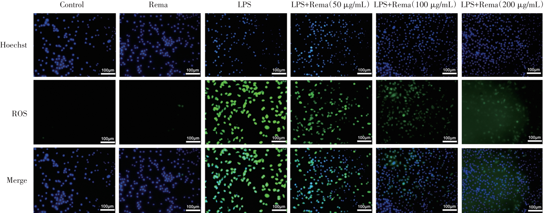
Fig.3
The effects of different treatment groups on the NF-κB pathway and nuclear translocation"
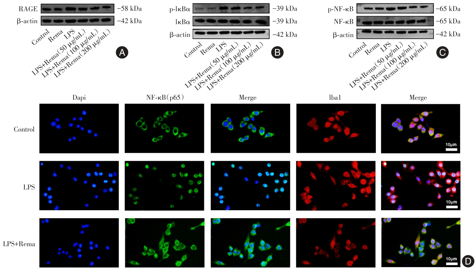
Tab.6
Effects of different treatment groups on the expression of Bax, Bcl-2 and NF-κB pathway-related proteins in BV2 cells x ± s"
| 组别 | Bax | Bcl-2 | RAGE | p-IκBα/IκBα | p-NF-κB/NF-κB |
|---|---|---|---|---|---|
| Control组 | 0.86 ± 0.02 | 0.96 ± 0.12 | 0.22 ± 0.11 | 0.68 ± 0.26 | 0.73 ± 0.21 |
| Rema组 | 1.03 ± 0.20 | 1.09 ± 0.04 | 0.16 ± 0.04 | 0.97 ± 0.49 | 0.94 ± 0.21 |
| 模型组 | 1.99 ± 0.38# | 0.45 ± 0.10# | 1.25 ± 0.05# | 2.36 ± 0.32# | 1.50 ± 0.04# |
| LPS + Rema(50 μg/mL)组 | 1.39 ± 0.15* | 0.84 ± 0.04* | 0.96 ± 0.08* | 1.37 ± 0.23* | 0.88 ± 0.25* |
| LPS + Rema(100 μg/mL)组 | 1.12 ± 0.25* | 1.07 ± 0.07* | 0.87 ± 0.12* | 1.18 ± 0.27* | 0.75 ± 0.23* |
| LPS + Rema(200 μg/mL)组 | 0.90 ± 0.36* | 1.21 ± 0.27* | 0.73 ± 0.08* | 0.59 ± 0.39* | 0.74 ± 0.17* |
| F值 | 5.355 | 8.212 | 7.353 | 7.224 | 4.462 |
| P值 | 0.008 1 | 0.001 4 | 0.002 3 | 0.002 5 | 0.015 8 |
Fig.4
Effects of different treatment groups on M1 and M2 polarization phenotypes of BV2 cells"
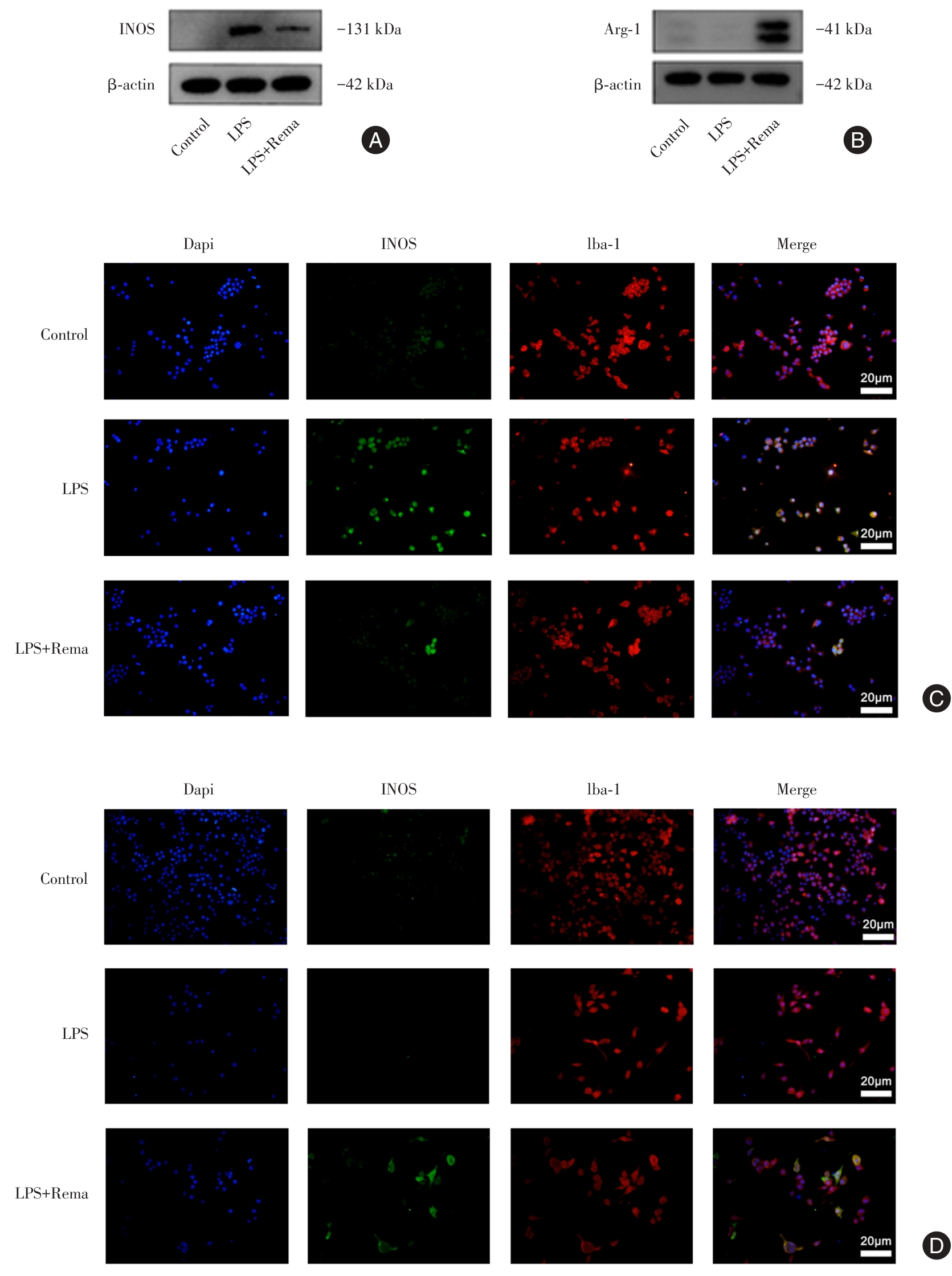
Tab.7
Effects of different treatment groups on the mRNA and protein expressions of M1/M2 polarization-related molecules"
| 组别 | INOS | Arg-1 | |||
|---|---|---|---|---|---|
| mRNA | 蛋白表达量 | mRNA | 蛋白表达量 | ||
| Control组 | 1.13 ± 0.38* | 0.33 ± 0.03* | 1.01 ± 0.10* | 0.26 ± 0.09 | |
| 模型组 | 2041.3 ± 356.19 | 1.33 ± 0.08 | 0.62 ± 0.07 | 0.21 ± 0.12 | |
| LPS + rema(200 μg/mL)组 | 849.01 ± 111.64* | 1.08 ± 0.09* | 1.67 ± 0.17* | 1.25 ± 0.28* | |
| F值 | 45.23 | 107.3 | 39.43 | 20.28 | |
| P值 | < 0.001 | < 0.001 | < 0.001 | 0.002 1 | |
| 1 |
KWON H S, KOH S H. Neuroinflammation in neurodegenerative disorders: The roles of microglia and astrocytes[J]. Transl Neurodegener, 2020, 9(1): 42. doi:10.1186/s40035-020-00221-2
doi: 10.1186/s40035-020-00221-2 |
| 2 |
HANISCH U K, KETTENMANN H. Microglia: Active sensor and versatile effector cells in the normal and pathologic brain[J]. Nat Neurosci, 2007, 10(11): 1387-1394. doi:10.1038/nn1997
doi: 10.1038/nn1997 |
| 3 |
ORIHUELA R, MCPHERSON C A, HARRY G J. Microglial M1/M2 polarization and metabolic states[J]. Br J Pharmacol, 2016, 173(4): 649-665. doi:10.1111/bph.13139
doi: 10.1111/bph.13139 |
| 4 |
WU Y G, SONG L J, YIN L J, et al. The effects and potential of microglial polarization and crosstalk with other cells of the central nervous system in the treatment of Alzheimer's disease[J]. Neural Regen Res, 2023, 18(5): 947-954. doi:10.4103/1673-5374.355747
doi: 10.4103/1673-5374.355747 |
| 5 |
XIAN X, CAI L L, LI Y, et al. Neuron secrete exosomes containing miR-9-5p to promote polarization of M1 microglia in depression[J]. J Nanobiotechnology, 2022, 20(1): 122. doi:10.1186/s12951-022-01332-w
doi: 10.1186/s12951-022-01332-w |
| 6 |
TAO W, HU Y, CHEN Z, et al. Magnolol attenuates depressive-like behaviors by polarizing microglia towards the M2 phenotype through the regulation of Nrf2/HO-1/NLRP3 signaling pathway[J]. Phytomedicine, 2021, 91: 153692. doi:10.1016/j.phymed.2021.153692
doi: 10.1016/j.phymed.2021.153692 |
| 7 |
HSU C H, PAN Y J, ZHENG Y T, et al. Ultrasound reduces inflammation by modulating M1/M2 polarization of microglia through STAT1/STAT6/PPARgamma signaling pathways[J]. CNS Neurosci Ther, 2023, 29(12): 4113-4123. doi:10.1111/cns.14333
doi: 10.1111/cns.14333 |
| 8 |
DING B, LIN C, LIU Q, et al. Tanshinone IIA attenuates neuroinflammation via inhibiting RAGE/NF-kappaB signaling pathway in vivo and in vitro[J]. J Neuroinflammation, 2020, 17(1): 302. doi:10.1186/s12974-020-01981-4
doi: 10.1186/s12974-020-01981-4 |
| 9 |
马小斐, 刘函晔, 王丹丹, 等. 紫云英苷通过NOX2/ROS/NF-κB信号通路抑制哮喘气道炎症 [J].中国药理学通报, 2021, 37(6): 797-802. doi:10.3969/j.issn.1001-1978.2021.06.011
doi: 10.3969/j.issn.1001-1978.2021.06.011 |
| 10 |
ZHOU Z, YANG Y, WEI Y, et al. Remimazolam Attenuates LPS-Derived Cognitive Dysfunction via Subdiaphragmatic Vagus Nerve Target alpha7nAChR-Mediated Nrf2/HO-1 Signal Pathway[J]. Neurochem Res, 2024, 49(5): 1306-1321. doi:10.1007/s11064-024-04115-x
doi: 10.1007/s11064-024-04115-x |
| 11 |
LIU X, LIN S, ZHONG Y, et al. Remimazolam Protects Against LPS-Induced Endotoxicity Improving Survival of Endotoxemia Mice[J]. Front Pharmacol, 2021, 12: 739603. doi:10.3389/fphar.2021.739603
doi: 10.3389/fphar.2021.739603 |
| 12 |
王静, 高煜茹, 蔡钱伟, 等. 瑞马唑仑通过调节肺泡巨噬细胞极化减轻脂多糖诱导的急性肺损伤[J]. 实用医学杂志, 2023, 39(9): 1092-1097. doi:10.3969/j.issn.1006-5725.2023.09.005
doi: 10.3969/j.issn.1006-5725.2023.09.005 |
| 13 |
GLASS C K, SAIJO K, WINNER B, et al. Mechanisms underlying inflammation in neurodegeneration[J]. Cell, 2010, 140(6): 918-934. doi:10.1016/j.cell.2010.02.016
doi: 10.1016/j.cell.2010.02.016 |
| 14 |
HE Y, LIU T, HE Q, et al. Microglia facilitate and stabilize the response to general anesthesia via modulating the neuronal network in a brain region-specific manner[J]. Elife, 2023, 12:RP92252. doi:10.7554/elife.92252.2.sa0
doi: 10.7554/elife.92252.2.sa0 |
| 15 |
ZHANG J, WANG X, ZHANG Q, et al. Application effects of remimazolam and propofol on elderly patients undergoing hip replacement[J]. BMC Anesthesiol, 2022, 22(1): 118. doi:10.1186/s12871-022-01641-5
doi: 10.1186/s12871-022-01641-5 |
| 16 | 段梅, 刘佳, 张静, 等. 瑞马唑仑对脑缺血再灌注损伤模型大鼠脑神经的保护作用及其机制 [J]. 精准医学杂志, 2023, 38(4): 345-349,355. |
| 17 | 蒋素芳, 万倩, 王雪吉, 等. 脂氧素A4对LPS诱导小胶质细胞活化的影响及SIRT1/NF-κB信号通路在其中的作用[J].中华麻醉学杂志, 2023, 43(10): 1177-1182. |
| 18 |
XIE H, LU F, LIU W, et al. Remimazolam alleviates neuropathic pain via regulating bradykinin receptor B1 and autophagy[J]. J Pharm Pharmacol, 2021, 73(12): 1643-1651. doi:10.1093/jpp/rgab080
doi: 10.1093/jpp/rgab080 |
| 19 |
ZHAO M, WANG Y, LI L, et al. Mitochondrial ROS promote mitochondrial dysfunction and inflammation in ischemic acute kidney injury by disrupting TFAM-mediated mtDNA maintenance[J]. Theranostics, 2021, 11(4): 1845-1863. doi:10.7150/thno.50905
doi: 10.7150/thno.50905 |
| 20 |
XU R, MA L, CHEN T, et al. Sophorolipid Suppresses LPS-Induced Inflammation in RAW264.7 Cells through the NF-κB Signaling Pathway[J]. Molecules, 2022, 27(15):5037. doi:10.3390/molecules27155037
doi: 10.3390/molecules27155037 |
| 21 |
COUGHLAN M T, COOPER M E, FORBES J M. Renal microvascular injury in diabetes: RAGE and redox signaling[J]. Antioxid Redox Signal, 2007, 9(3): 331-342. doi:10.1089/ars.2006.1469
doi: 10.1089/ars.2006.1469 |
| 22 |
WANG L, WU J, GUO X, et al. RAGE Plays a Role in LPS-Induced NF-kappaB Activation and Endothelial Hyperpermeability[J]. Sensors (Basel), 2017, 17(4):722. doi:10.3390/s17040722
doi: 10.3390/s17040722 |
| 23 |
GAOJIAN T, DINGFEI Q, LINWEI L, et al. Parthenolide promotes the repair of spinal cord injury by modulating M1/M2 polarization via the NF-kappaB and STAT 1/3 signaling pathway[J]. Cell Death Discov, 2020, 6(1): 97. doi:10.1038/s41420-020-00333-8
doi: 10.1038/s41420-020-00333-8 |
| 24 |
ZHOU Q, ZHANG Y, LU L, et al. Copper induces microglia-mediated neuroinflammation through ROS/NF-kappaB pathway and mitophagy disorder[J]. Food Chem Toxicol, 2022, 168: 113369. doi:10.1016/j.fct.2022.113369
doi: 10.1016/j.fct.2022.113369 |
| [1] | Lihong WU,Yan GUO,Jing CAO,Xiaoyan DU,Qingqing LIANG,Xiaocheng GAO,Yanru WANG,Yang DENG,Long GAO. Mechanism of neodymium oxide exposure causing brain tissue damage in mouse [J]. The Journal of Practical Medicine, 2025, 41(1): 30-34. |
| [2] | Junjie ZHANG,Xiaochun YANG,Zhuoyi LIU,Ruoqiu WU,Ping LI,Qulian GUO,E. WANG. A randomized controlled study of remimazolam tosilate under bispectral index and bypass EtCO2 monitoring in daytime hysteroscopic surgery [J]. The Journal of Practical Medicine, 2024, 40(8): 1047-1051. |
| [3] | Hongya CHEN,Huijun WANG,Yue WANG,Shaofei SU,Guyan. WANG. Remimazolam versus propofol on quality of recovery after general anesthesia in elders undergoing ocular fundus daytime surgery [J]. The Journal of Practical Medicine, 2024, 40(8): 1052-1057. |
| [4] | Lili TANG,Yue SUN,Xuesheng LIU,Yao. LU. The gender⁃specific effects on doses of remimazolam and quality of postoperative recovery in patients undergoing ambulatory arthroscopic surgery [J]. The Journal of Practical Medicine, 2024, 40(8): 1063-1068. |
| [5] | Ling XIAO,Chunlei GAO,Wei GUO,Ning WANG,Xuan ZHANG,Ming. LIU. Codonopsis polysaccharide protected LPS⁃induced acute lung injury by inhibiting MAPK/NF⁃κB signaling pathway in mice [J]. The Journal of Practical Medicine, 2024, 40(7): 948-954. |
| [6] | Kanglin CAI,Jinkai ZHANG,Liangdi RAN,Dajun HU,Zhitao FENG,Huilian. HUANG. Research progress on antidepressant pharmacological effects and mechanisms of Bupleuri Radix⁃Paeoniae Radix Alba herb⁃pair [J]. The Journal of Practical Medicine, 2024, 40(4): 447-452. |
| [7] | Chunhua HU,Guyan WANG,Huijun WANG,Chunhua XI,Congya ZHANG,Lili. WU. Effect of remimazolam combined with desflurane and flumazenil antagonism for anesthesia during ophthalmic day surgery [J]. The Journal of Practical Medicine, 2024, 40(4): 537-542. |
| [8] | Peng SUN,Zhaojin JIA,Xiuhua LI,Xiaowei CHEN,Runsheng WEI,Yantao JIN,Jiantao. JIN. The effects of different extracorporeal circulation temperature combined with dexmedetomidine on oxidative stress in patients undergoing cardiac surgery under cardiopulmonary bypass [J]. The Journal of Practical Medicine, 2024, 40(24): 3521-3526. |
| [9] | Zhe SHI,Xialin ZUO,Linhui PENG,Zhiwei LU,Kongping. LI. Effect of M1 microglial polarization on secondary damage in the thalamus after cerebral cortical infarction [J]. The Journal of Practical Medicine, 2024, 40(22): 3138-3145. |
| [10] | Tianyue YU,Qian GUO,Hao HU,Yujing SU,Jianhua CHEN. Advances in oxidative stress⁃related pathways with diagnostic and predictive value in schizophrenia [J]. The Journal of Practical Medicine, 2024, 40(20): 2935-2940. |
| [11] | Jieqiong LIU,Yali YAO,Qian SUI,Ke LI,Fang HUANG,Yongqing. CAO. Based on the novel anti-heart failure drug ARNI, the mechanism of prevention of cardiotoxicity caused by anthracycline antitumor drugs was discussed [J]. The Journal of Practical Medicine, 2024, 40(2): 188-194. |
| [12] | Ganggang LU,Shenglong LI,Yongqiang ZHAO,Yunpeng JIA,Yonglin LIANG,Yuanbo. ZHAO. Research progress on the correlation between oxidative stress and ferroptosis in diabetic impotence [J]. The Journal of Practical Medicine, 2024, 40(16): 2229-2235. |
| [13] | Yunlong SUN,Zhe MENG,Xijia WANG,Lu. GAO. Liraglutide ameliorates high glucose⁃induced endothelial cell injuryvia Nrf2 [J]. The Journal of Practical Medicine, 2024, 40(15): 2051-2055. |
| [14] | Jiaman LIN,Yongxin YE,Shanghang LI,Yunfei. CHAI. Effect of remifentanil fast⁃track anesthesia on enhancing postoperative recovery quality in patients undergoing cardiac valve surgery: a prospective randomized controlled trial [J]. The Journal of Practical Medicine, 2024, 40(14): 1988-1994. |
| [15] | Chun LONG,Hongying BI,Changzhen YANG,Jiakai WANG,Yan TANG,Xu. LIU. Emodin upregulates the Sirt2 to attenuate LPS-induced oxidative stress response in RAW264.7 cells [J]. The Journal of Practical Medicine, 2024, 40(13): 1785-1790. |
| Viewed | ||||||
|
Full text |
|
|||||
|
Abstract |
|
|||||