

The Journal of Practical Medicine ›› 2025, Vol. 41 ›› Issue (19): 3096-3105.doi: 10.3969/j.issn.1006-5725.2025.19.021
• Medical Examination and Clinical Diagnosis • Previous Articles
Xiaokun GAO1,2,Ziming XIE1,2,Guangyu TAO2,Yanbing SUN2,Hua REN2,Jiahui YU2,Lin ZHU2,Hong YU2,Qiming. NI2( )
)
Received:2025-07-16
Online:2025-10-10
Published:2025-10-10
Contact:
Qiming. NI
E-mail:13816011196@163.com
CLC Number:
Xiaokun GAO,Ziming XIE,Guangyu TAO,Yanbing SUN,Hua REN,Jiahui YU,Lin ZHU,Hong YU,Qiming. NI. The application value of multi⁃parameter quantitative analysis of spectral and perfusion CT in differentiating pathological types of lung cancer[J]. The Journal of Practical Medicine, 2025, 41(19): 3096-3105.

Fig.1
Inclusion and exclusion criteria for patients"
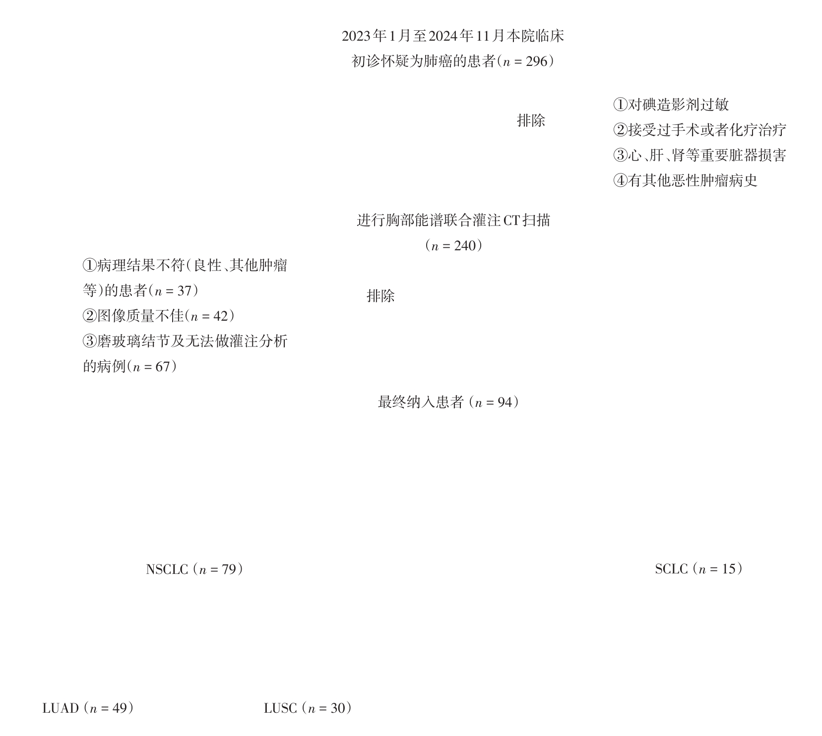
Fig.2
Spectral combined perfusion CT patient image"
Tab.1
Comparison of clinical data between LUSC group, LUAD group and SCLC group"
| 项目 | LUAD组 (n = 49) | LUSC组 (n = 30) | SCLC组 (n = 15) | P值 | |||
|---|---|---|---|---|---|---|---|
| 三组间P值 | LUAD组 vs. LUSC组 | LUAD组 vs. SCLC组 | LUSC组 vs. SCLC组 | ||||
| 性别 | < 0.001 | < 0.001 | 0.078 | 0.115 | |||
| 男 | 24(48.98) | 29(96.67) | 11(73.33) | ||||
| 女 | 25(51.02) | 1(3.33) | 4(26.67) | ||||
| 影像征象 | |||||||
| 分叶 | 47(95.92) | 25(83.33) | 15(100.00) | 0.059 | |||
| 毛刺 | 40(81.63) | 23(76.67) | 14(93.33) | 0.394 | |||
| 胸膜牵拉 | 38(77.55) | 8(26.67) | 6(40.00) | < 0.001 | < 0.001 | 0.011 | 0.399 |
| 肿瘤标志物 | |||||||
| 癌胚抗原(+) | 29(59.18) | 7(23.33) | 6(40.00) | 0.008 | 0.002 | 0.193 | 0.292 |
| 鳞癌特异性抗原(+) | 6(12.24) | 17(56.67) | 2(13.33) | < 0.001 | < 0.001 | 0.934 | 0.002 |
| 神经元特异性烯醇化酶(+) | 31(63.27) | 14(46.67) | 12(80.00) | 0.086 | |||
| 细胞角蛋白19片段(+) | 18(36.73) | 23(76.67) | 6(40.00) | 0.002 | 0.001 | 0.826 | 0.021 |
| 糖类抗原125(+) | 16(32.65) | 8(26.67) | 4(26.67) | 0.820 | |||
Tab.2
Comparison of quantitative spectral parameters between the LUSC group, the LUAD group and the SCLC group M[P25,P75]"
| 能谱参数 | LUAD组(n = 49) | LUSC组(n = 30) | SCLC组(n = 15) | P值 | |||
|---|---|---|---|---|---|---|---|
| 三组间P值 | LUAD组 vs. LUSC组 | LUAD组 vs. SCLC组 | LUSC组 vs. SCLC组 | ||||
| 动脉期 | |||||||
| K1 | 4.495(2.630,6.190) | 3.050(2.610,4.188) | 2.825(1.712,4.263) | 0.011 | 0.009 | 0.028 | 0.894 |
| K2 | 0.760(0.445,1.055) | 0.520(0.446,0.712) | 0.480(0.292,0.722) | 0.011 | 0.009 | 0.03 | 0.903 |
| K3 | 0.215(0.135,0.295) | 0.152(0.131,0.712) | 0.145(0.087,0.208) | 0.01 | 0.008 | 0.025 | 0.879 |
| K | 1.518(0.889,2.090) | 1.031(0.882,1.416) | 0.956(0.577,1.441) | 0.011 | 0.009 | 0.027 | 0.886 |
| ICL(x ± s) | 19.296 ± 7.474 | 13.962 ± 5.627 | 13.715 ± 7.516 | 0.017 | 0.015 | 0.032 | 0.83 |
| NIC(x ± s) | 0.153 ± 0.088 | 0.121 ± 0.078 | 0.110 ± 0.073 | 0.027 | 0.04 | 0.024 | 0.544 |
| WC/(mg/cm3) | 1 014(995,1 029) | 1 032(1 026,1 035) | 1 028(1 022,1 032) | < 0.001 | < 0.001 | 0.04 | 0.287 |
| Zeff(x ± s) | 8.772 ± 0.564 | 8.407 ± 0.342 | 8.409 ± 0.427 | 0.006 | 0.004 | 0.022 | 0.968 |
| 静脉期 | |||||||
| K1 | 4.485(3.390,5.565) | 3.370(2.337,3.904) | 3.160(2.288,4.537) | 0.002 | 0.001 | 0.032 | 0.702 |
| K2 | 0.755(0.580,0.950) | 0.577(0.401,0.664) | 0.540(0.390,0.765) | 0.003 | 0.001 | 0.034 | 0.686 |
| K3 | 0.205(0.165,0.260) | 0.168(0.120,0.189) | 0.155(0.120,0.217) | 0.004 | 0.002 | 0.051 | 0.644 |
| K | 1.516(1.147,1.906) | 1.140(0.789,1.320) | 1.068(0.773,1.534) | 0.002 | 0.001 | 0.031 | 0.701 |
| ICL(x ± s) | 19.703 ± 10.212 | 15.484 ± 12.546 | 15.183 ± 6.400 | 0.003 | 0.001 | 0.034 | 0.688 |
| NIC(x ± s) | 0.367 ± 0.124 | 0.291 ± 0.119 | 0.304 ± 0.130 | 0.001 | 0.001 | 0.025 | 0.674 |
| WC/(mg/cm3) | 1 019(1 002,1 026) | 1 030(1 023,1 037) | 1 030(1 023,1 032) | < 0.001 | < 0.001 | 0.002 | 0.703 |
| Zeff(x ± s) | 8.739 ± 0.401 | 8.428 ± 0.311 | 8.497 ± 0.342 | 0.001 | 0.001 | 0.025 | 0.674 |
Tab.3
Comparison of perfusion parameters between LUSC group, LUAD group and SCLC group"
| 灌注参数 | LUAD组 (n = 49) | LUSC组 (n = 30) | SCLC组 (n = 15) | P值 | |||
|---|---|---|---|---|---|---|---|
| 三组间P值 | LUAD组 vs. LUSC组 | LUAD组 vs. SCLC组 | LUSC组 vs. SCLC组 | ||||
| PI | 0.63 ± 3.31 | 0.16 ± 0.11 | 0.13 ± 0.09 | 0.498 | |||
| TTP/s | 24.34 ± 7.79 | 24.29 ± 7.67 | 25.2 ± 7.84 | 0.399 | |||
| BV/(mL/100 g) | 6.29 ± 5.31 | 7.43 ± 5.68 | 5.48 ± 4.38 | 0.177 | |||
| BF/[mL/(100 g·min)] | 120.48 ± 14 | 128.27 ± 13 | 95.64 ± 12 | 0.603 | |||
| PS/[mL/(100 g·min)] | 22.2 ± 4.93 | 19.1 ± 4.23 | 16.44 ± 3.6 | 0.014 | 0.001 | 0.032 | 0.164 |
| MTT/s | 6.86 ± 5.16 | 6.58 ± 3.82 | 7.40 ± 4.94 | 0.881 | |||
Tab.4
Comparison of ROC parameters between LUSC group and LUAD group"
| 参数类别 | AUC | 约登指数 | 敏感度/% | 特异度/% | ||||
|---|---|---|---|---|---|---|---|---|
| 训练组 | 测试组 | 训练组 | 测试组 | 训练组 | 测试组 | 训练组 | 测试组 | |
| 诊断模型1 | 0.81 | 0.77 | 0.55 | 0.72 | 100.0 | 100.0 | 54.8 | 72.4 |
| 诊断模型2 | 0.87 | 0.90 | 0.68 | 0.62 | 93.3 | 100.0 | 74.2 | 62.1 |
| 诊断模型3 | 0.88 | 0.88 | 0.72 | 0.68 | 100.0 | 83.3 | 72.4 | 85.0 |
| 诊断模型4 | 0.85 | 0.78 | 0.62 | 0.51 | 100.0 | 66.7 | 62.1 | 85.0 |
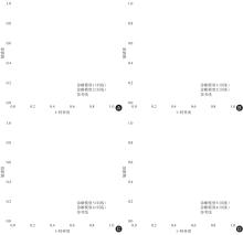
Fig.3
Model for differential diagnosis of lung adenocarcinoma or small cell lung cancer"
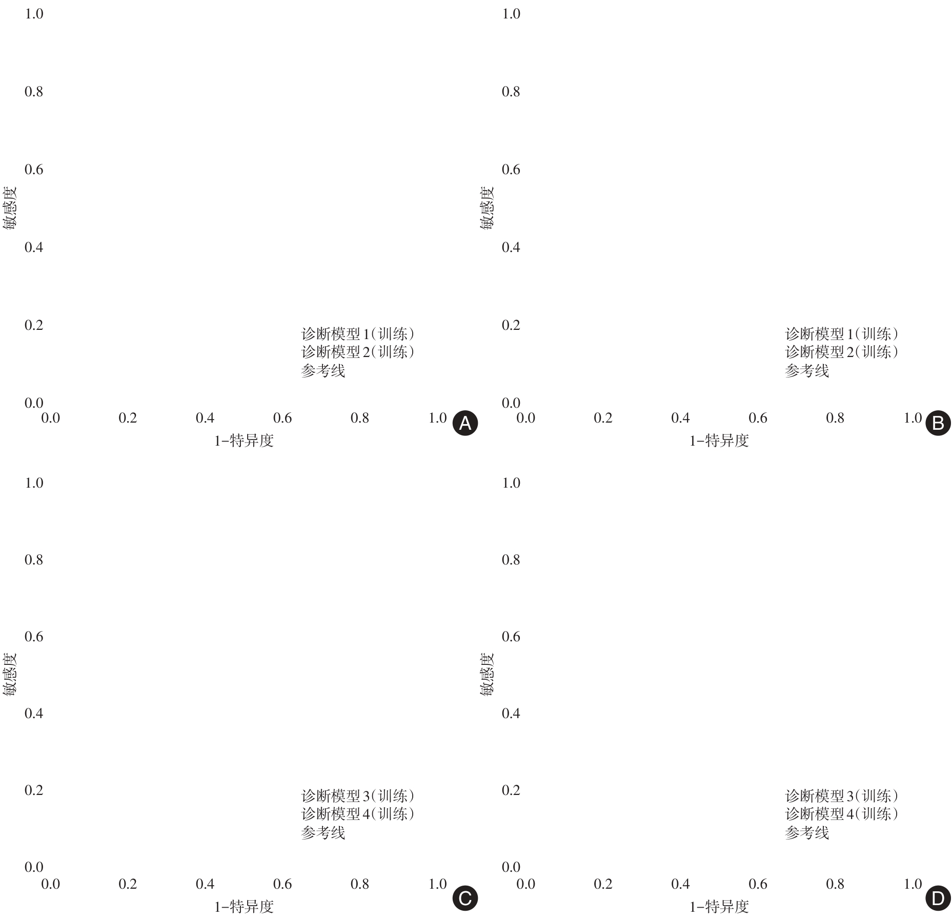
Fig.4
Calibration curve of diagnostic model"
| [1] |
SUNG H, FERLAY J, SIEGEL R L, et al. Global Cancer Statistics 2020: GLOBOCAN Estimates of Incidence and Mortality Worldwide for 36 Cancers in 185 Countries [J]. CA Cancer J Clin, 2021, 71(3): 209-249. doi:10.3322/caac.21660
doi: 10.3322/caac.21660 |
| [2] |
RUIZ-CORDERO R, DEVINE W P. Targeted Therapy and Checkpoint Immunotherapy in Lung Cancer [J]. Surgical Pathology Clinics, 2020, 13(1): 17-33. doi:10.1016/j.path.2019.11.002
doi: 10.1016/j.path.2019.11.002 |
| [3] | 熊丙万, 柯文飞, 江文洋. 分子靶向治疗在EGFR突变肺鳞癌中的研究进展 [J]. 中国肺癌杂志, 2024, (4): 283-290. |
| [4] |
MOLINA J R, YANG P, CASSIVI S D, et al. Non-small cell lung cancer: Epidemiology, risk factors, treatment, and survivorship [J]. Mayo Clin Proc, 2008, 83(5): 584-594. doi:10.1016/s0025-6196(11)60735-0
doi: 10.1016/s0025-6196(11)60735-0 |
| [5] |
KELLY K, CROWLEY J, BUNN P A, JR., et al. Randomized phase Ⅲ trial of paclitaxel plus carboplatin versus vinorelbine plus cisplatin in the treatment of patients with advanced non--small-cell lung cancer: A Southwest Oncology Group trial [J]. J Clin Oncol, 2001, 19(13): 3210-3218. doi:10.1200/jco.2001.19.13.3210
doi: 10.1200/jco.2001.19.13.3210 |
| [6] |
NGUYEN K T, SAKTHIVEL G, MILANO M T, et al. Oligoprogression in non-small cell lung cancer: A narrative review [J]. J Thorac Dis, 2022, 14(12): 4998-5011. doi:10.21037/jtd-22-536
doi: 10.21037/jtd-22-536 |
| [7] |
GONG L, XU L, YUAN Z, et al. Clinical outcome for small cell lung cancer patients with bone metastases at the time of diagnosis [J]. J Bone Oncol, 2019, 19: 100265. doi:10.1016/j.jbo.2019.100265
doi: 10.1016/j.jbo.2019.100265 |
| [8] |
DENG L, ZHANG G, LIN X, et al. Comparison of Spectral and Perfusion Computed Tomography Imaging in the Differential Diagnosis of Peripheral Lung Cancer and Focal Organizing Pneumonia [J]. Front Oncol, 2021, 11: 690254. doi:10.3389/fonc.2021.690254
doi: 10.3389/fonc.2021.690254 |
| [9] | 韩冬, 周洁丽, 于勇, 等. 基于能谱曲线斜率鉴别肾透明细胞癌WHO/ISUP简化分级 [J]. 临床放射学杂志, 2020, 39(6): 1133-1138. |
| [10] | 邓靓娜, 张国晋, 林晓强, 等. 能谱及灌注CT成像鉴别诊断周围型肺癌和局灶性机化性肺炎的对比研究 [J]. 中国医学影像学杂志, 2021, 29(12): 1206-1211. |
| [11] |
WANG Z, LI M, HUANG Y, et al. Clinical and radiological characteristics of central pulmonary adenocarcinoma: A comparison with central squamous cell carcinoma and small cell lung cancer and the impact on treatment response [J]. Onco Targets Ther, 2018, 11: 2509-2517. doi:10.2147/ott.s154385
doi: 10.2147/ott.s154385 |
| [12] |
KOENIGKAM SANTOS M, MULEY T, WARTH A, et al. Morphological computed tomography features of surgically resectable pulmonary squamous cell carcinomas: Impact on prognosis and comparison with adenocarcinomas [J]. Eur J Radiol, 2014, 83(7): 1275-1281. doi:10.1016/j.ejrad.2014.04.019
doi: 10.1016/j.ejrad.2014.04.019 |
| [13] |
JIA Y, XIAO X, SUN Q, et al. CT spectral parameters and serum tumour markers to differentiate histological types of cancer histology [J]. Clin Radiol, 2018, 73(12): 1033-1040. doi:10.1016/j.crad.2018.07.104
doi: 10.1016/j.crad.2018.07.104 |
| [14] |
XU X, SUI X, ZHONG W, et al. Clinical utility of quantitative dual-energy CT iodine maps and CT morphological features in distinguishing small-cell from non-small-cell lung cancer [J]. Clin Radiol, 2019, 74(4): 268-277. doi:10.1016/j.crad.2018.10.012
doi: 10.1016/j.crad.2018.10.012 |
| [15] |
LI J, XU Q, MAO C, et al. Correlation between Imaging Features and Pathological Stages of Primary Lung Tumors Based on Nanocontrast Agents [J]. Comput Math Methods Med, 2021, 2021: 2343299. doi:10.1155/2021/2343299
doi: 10.1155/2021/2343299 |
| [16] |
LIN L Y, ZHANG Y, SUO S T, et al. Correlation between dual-energy spectral CT imaging parameters and pathological grades of non-small cell lung cancer [J]. Clin Radiol, 2018, 73(4): 412.e1-e7. doi:10.1016/j.crad.2017.11.004
doi: 10.1016/j.crad.2017.11.004 |
| [17] |
WANG S, LIU G, FU Z, et al. Predicting Pathological Invasiveness of Lung Adenocarcinoma Manifesting as GGO-Predominant Nodules: A Combined Prediction Model Generated From DECT [J]. Acad Radiol, 2021, 28(4): 509-516. doi:10.1016/j.acra.2020.03.007
doi: 10.1016/j.acra.2020.03.007 |
| [18] |
LI Q, LI X, LI X Y, et al. Histological subtypes of solid-dominant invasive lung adenocarcinoma: Differentiation using dual-energy spectral CT [J]. Clin Radiol, 2021, 76(1): 77.e1-e7. doi:10.1016/j.crad.2020.08.034
doi: 10.1016/j.crad.2020.08.034 |
| [19] |
YUAN A, YU C J, KUO S H, et al. Vascular endothelial growth factor 189 mRNA isoform expression specifically correlates with tumor angiogenesis, patient survival, and postoperative relapse in non-small-cell lung cancer [J]. J Clin Oncol, 2001, 19(2): 432-441. doi:10.1200/jco.2001.19.2.432
doi: 10.1200/jco.2001.19.2.432 |
| [20] |
HONG S R, HUR J, MOON Y W, et al. Predictive factors for treatment response using dual-energy computed tomography in patients with advanced lung adenocarcinoma [J]. Eur J Radiol, 2018, 101: 118-123. doi:10.1016/j.ejrad.2018.02.019
doi: 10.1016/j.ejrad.2018.02.019 |
| [21] | 廖淑婷, 于向荣. 能谱CT和人工智能在甲状腺癌诊断中的应用 [J]. 实用医学杂志, 2022, (2): 129-133. |
| [22] |
梁百晖, 杨文, 刘岘, 等. 双层探测器光谱CT对直肠腺癌转移淋巴结的诊断价值 [J]. 实用医学杂志, 2023, 39(3): 374-380. doi:10.3969/j.issn.1006-5725.2023.03.020
doi: 10.3969/j.issn.1006-5725.2023.03.020 |
| [23] |
ZHONG L J, YU N, ZHOU X J, et al. Differentiating between pulmonary adenocarcinoma and squamous cell carcinoma by spectral CT volumetric quantitative analysis: A comparative study with conventional spectral analysis [J]. J Thorac Dis, 2023, 15(2): 679-689. doi:10.21037/jtd-23-115
doi: 10.21037/jtd-23-115 |
| [24] |
MU R, MENG Z, GUO Z, et al. Diagnostic value of dual-layer spectral detector CT in differentiating lung adenocarcinoma from squamous cell carcinoma [J]. Front Oncol, 2022, 12: 868216. doi:10.3389/fonc.2022.868216
doi: 10.3389/fonc.2022.868216 |
| [25] |
NASRULLAH N, SANG J, ALAM M S, et al. Automated Lung Nodule Detection and Classification Using Deep Learning Combined with Multiple Strategies [J]. Sensors (Basel), 2019, 19(17):3722. doi:10.3390/s19173722
doi: 10.3390/s19173722 |
| [26] |
CHU L L, KNEBEL R J, SHAY A D, et al. CT perfusion imaging of lung cancer: Benefit of motion correction for blood flow estimates [J]. Eur Radiol, 2018, 28(12): 5069-5075. doi:10.1007/s00330-018-5492-1
doi: 10.1007/s00330-018-5492-1 |
| [27] |
ZIELIŃSKI K W, KULIG A, ZIELIŃSKI J. Morphology of the microvascular bed in primary human carcinomas of lung. Part II. Morphometric investigations of microvascular bed of lung tumors [J]. Pathol Res Pract, 1984, 178(4): 369-377. doi:10.1016/s0344-0338(84)80029-1
doi: 10.1016/s0344-0338(84)80029-1 |
| [28] |
LUCCHI M, MUSSI A, FONTANINI G, et al. Small cell lung carcinoma (SCLC): The angiogenic phenomenon [J]. Eur J Cardiothorac Surg, 2002, 21(6): 1105-1110. doi:10.1016/s1010-7940(02)00112-4
doi: 10.1016/s1010-7940(02)00112-4 |
| [29] |
CHEN M L, WEI Y Y, LI X T, et al. Low-dose spectral CT perfusion imaging of lung cancer quantitative analysis in different pathological subtypes [J]. Transl Cancer Res, 2021, 10(6): 2841-2848. doi:10.21037/tcr-20-3479
doi: 10.21037/tcr-20-3479 |
| [30] | 胡蓉, 徐耀, 侯金鹏, 等. 能谱CT多参数定量分析对鉴别肺癌病理类型的价值 [J]. 实用放射学杂志, 2019, 35(3): 5. |
| Viewed | ||||||
|
Full text |
|
|||||
|
Abstract |
|
|||||