实用医学杂志 ›› 2025, Vol. 41 ›› Issue (9): 1327-1331.doi: 10.3969/j.issn.1006-5725.2025.09.008

• 基础研究 • 上一篇    

组织常驻记忆T细胞对咪喹莫特二次诱导银屑病样小鼠皮损的影响

周玉婵,郑荣昌,李华润,黄锦萍,秦思,李婷,卢镇宇,李思慧,李先文,李慕锦,温炬()   

  1. 广东省第二人民医院皮肤科 (广东 广州 510317 )
  • 收稿日期:2025-01-15 出版日期:2025-05-10 发布日期:2025-05-20
  • 通讯作者: 温炬 E-mail:wenju3139@163.com
  • 基金资助:
    广东省中医药局科研基金资助项目(20221014)

Expression of TRM cells in the lesions of imiquimod⁃induced models of psoriasis in mice

Yuchan ZHOU,Rongchang ZHENG,Huarun LI,Jinping HUANG,Si QIN,Ting LI,Zhenyu LU,Sihui LI,Xianwen LI,Mujin LI,Ju WEN()   

  1. Department of Dermatology and Venereology,Guangdong Second Provincial General Hospital,Guangzhou 510317,Guangdong,China
  • Received:2025-01-15 Online:2025-05-10 Published:2025-05-20
  • Contact: Ju WEN E-mail:wenju3139@163.com

摘要:

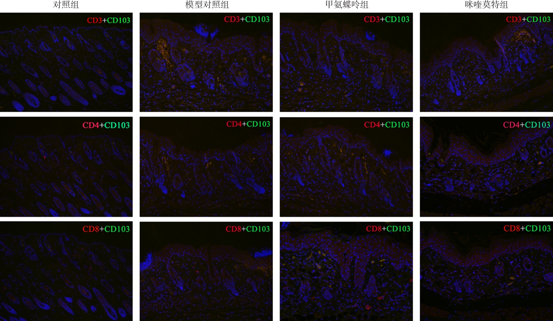
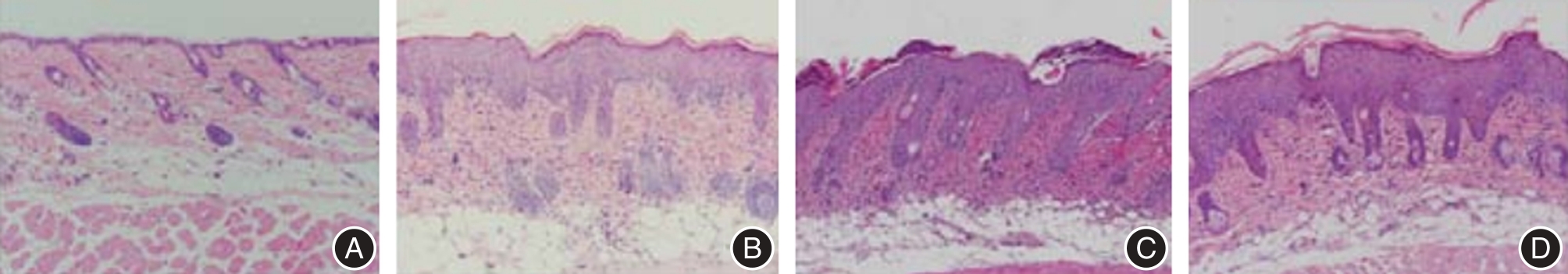
目的 探讨组织常驻记忆T细胞(TRM)对咪喹莫特二次诱导银屑病样小鼠皮损的影响。 方法 购置BALB/c雌性小鼠40只,随机取小鼠10只为空白对照组;剩余30只小鼠建立银屑病小鼠动物模型,建模成功后随机分为模型对照组、甲氨蝶呤组和咪喹莫特组各10只。空白对照组和模型对照组小鼠均匀涂抹凡士林干预,甲氨蝶呤组和咪喹莫特组涂抹5%咪喹莫特乳膏62.5 mg,甲氨蝶呤组按照 1 mg/kg剂量灌胃给药,各组灌胃体积为 10 mL/kg;模型对照组、空白对照组和咪喹莫特组灌胃等体积生理盐水。各组均完成6 d用药干预,比较各组银屑病面积和严重程度指数(PASI)、组织病理学结果、炎症因子及TRM细胞水平。 结果 (1)咪喹莫特组小鼠红斑、皮肤增厚及鳞屑评分[(2.54 ± 0.32)、(2.59 ± 0.25)、(2.52 ± 0.29)分]低于甲氨蝶呤组、模型对照组和对照组(P < 0.05);甲氨蝶呤组红斑、皮肤增厚及鳞屑评分低于模型对照组(P < 0.05);(2)HE染色结果表明,甲氨蝶呤组表皮逐渐变薄,角化不全细胞数量相对较少,且伴有毛囊增多;咪喹莫特组建模后细胞形态异常、白皮相对较厚;(3)咪喹莫特组TNF-α、IL-1β、IFN-γ和IL-23水平[(51.63 ± 4.39)、(35.53 ± 4.15)、(23.43 ± 3.41)、(15.24 ± 2.95)pg/mL]低于甲氨蝶呤组和模型对照组(P < 0.05);甲氨蝶呤组TNF-α、IL-1β、IFN-γ及IL-23低于模型对照组(P < 0.05);(4)咪喹莫特组CD8+ CD103+(15.39 ± 2.31)低于甲氨蝶呤组和模型对照组(P < 0.05);甲氨蝶呤组CD8+ CD103+水平低于模型对照组(P < 0.05)。 结论 咪喹莫特二次诱导银屑病样小鼠皮损更重、反应更快,且表皮增厚更多,CD8+ CD103+ TRM细胞及炎症因子可能参与银屑病的复发。

关键词: 组织常驻记忆T细胞, 咪喹莫特, 二次诱导, 银屑病, 病理学检查, 炎症因子

Abstract:

Objective To investigate the effect of tissue-resident memory T cells (TRM) on imiquimod-induced psoriatic-like skin lesions in mice, and to elucidate the underlying mechanisms of TRM involvement in this process. Methods Forty female BALB/c mice were procured and randomly allocated into four groups: ten in the blank control group, and thirty for the establishment of a psoriasis mouse model. Following successful modeling, the thirty mice were further randomized into three groups: the model control group, the methotrexate-treated group, and the imiquimod-treated group, with ten mice in each group. Mice in the blank control group and model control group were uniformly treated with Vaseline for intervention. The methotrexate group and the imiquimod group were treated with 62.5mg of 5% imiquimod cream. The methotrexate group was administered by gavage at a dose of 1 mg/kg, and the gavage volume of each group was 10 mL/kg.The model control group, blank group and imiquimod group were gavaged with the same volume of normal saline. Treatment was conducted over six consecutive days. Subsequently, comparisons were made across groups regarding the psoriasis area and severity index (PASI), histopathological findings, inflammatory cytokine levels, and TRM cell levels. Results (1) The imiquimod group exhibited significantly lower scores for erythema (2.54 ± 0.32), skin thickening (2.59 ± 0.25), and scaling (2.52 ± 0.29) compared to the methotrexate group, model control group, and blank control group (P < 0.05). Additionally, the methotrexate group demonstrated reduced scores for erythema, skin thickening, and scaling compared to the model control group (P < 0.05). (2)Hematoxylin-eosin (HE) staining revealed that the epidermis in the methotrexate group became thinner, with fewer parakeratotic cells and increased hair follicles. Conversely, the imiquimod group displayed abnormal cell morphology and relatively thicker white skin after modeling. (3) The imiquimod group showed significantly lower levels of TNF-α (51.63 ± 4.39 pg/mL), IL-1β (35.53 ± 4.15 pg/mL), IFN-γ (23.43 ± 3.41 pg/mL), and IL-23 (15.24 ± 2.95 pg/mL) compared to the methotrexate and model control groups (P < 0.05). Similarly, the methotrexate group exhibited reduced levels of TNF-α, IL-1β, IFN-γ, and IL-23 compared to the model control group (P < 0.05). (4) The imiquimod group had significantly lower levels of CD8+CD103+ cells (15.39 ± 2.31) than the methotrexate and model control groups (P < 0.05). Furthermore, the methotrexate group demonstrated lower levels of CD8+CD103+ cells compared to the model control group (P < 0.05). Conclusion Miquimod induces heavier skin lesions, faster response, and more epidermal thickening in psoriasis like mice. CD8+ CD103+ TRM cells and inflammatory factors may be involved in the recurrence of psoriasis.

Key words: tissue-resident memory T cells, imiquimod, secondary induction, psoriasis, histopathological examination, inflammatory cytokines

中图分类号: