实用医学杂志 ›› 2023, Vol. 39 ›› Issue (16): 2062-2070.doi: 10.3969/j.issn.1006-5725.2023.16.009

• 临床研究 • 上一篇    下一篇

STT3A和STT3B在上皮性卵巢癌中的表达及与预后的关系

孙崇凤1,2,杨萍1,2(),侯纪帅1,2,赵邹宇1,2,于盼盼1,2   

  1. 1.石河子大学第一附属医院妇科 (新疆 石河子 832008 )
    2.国家卫生健康委中亚高发病防治重点实验室 ;(新疆 石河子 832008 )
  • 收稿日期:2023-02-14 出版日期:2023-08-25 发布日期:2023-09-26
  • 通讯作者: 杨萍 E-mail:pingy2018@163.com
  • 基金资助:
    国家自然科学基金资助项目(82072893);石河子大学基金资助项目(CGZH201702);中国医学科学院中央级公益性科研院所基本科研业务费专项资金(2020-PT330-003)

Expression and prognosis of STT3A and STT3B in epithelial ovarian cancer

Chongfeng SUN1,2,Ping YANG1,2(),Jishuai HOU1,2,Zouyu ZHAO1,2,Panpan. YU1,2   

  1. *.Department of Gynecology,the First Affiliated Hospital,Shihezi University,Shihezi 832008,China
    *.Key Laboratory of Prevention and Treatment of Subhigh Incidence Diseases,National Health Commission of China,Shihezi 832008,China
  • Received:2023-02-14 Online:2023-08-25 Published:2023-09-26
  • Contact: Ping YANG E-mail:pingy2018@163.com

摘要:

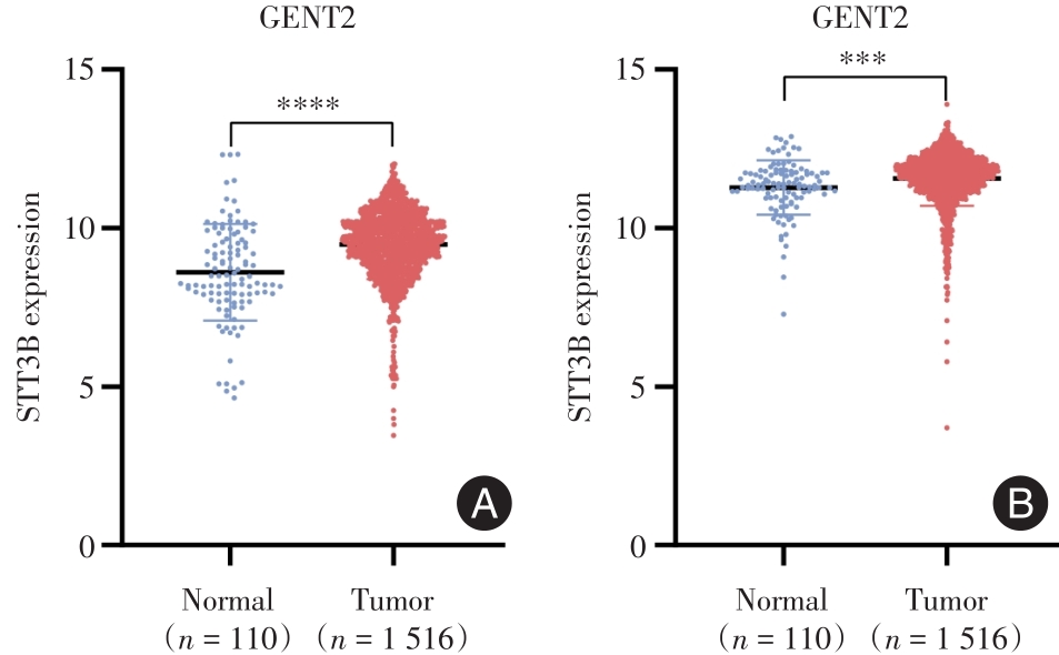
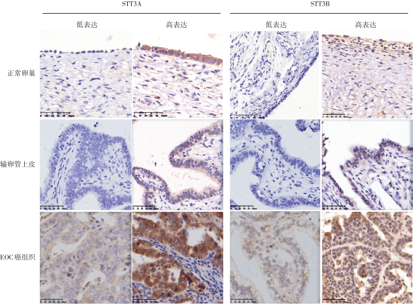
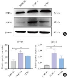
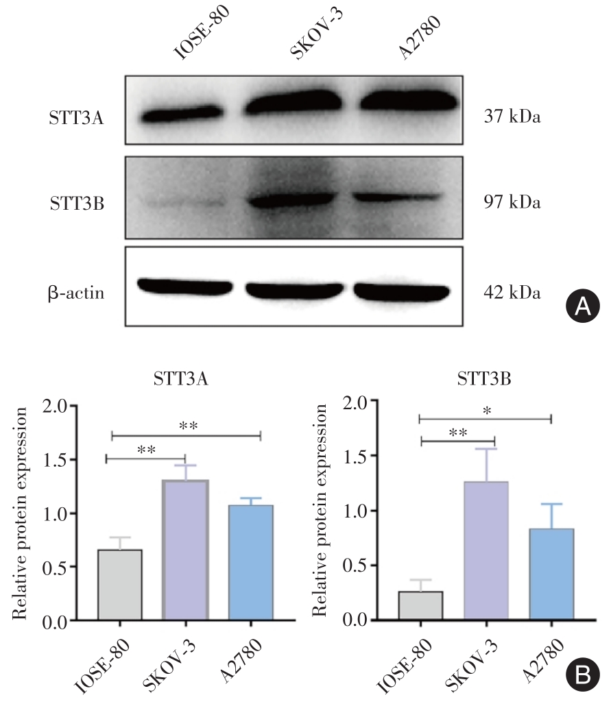
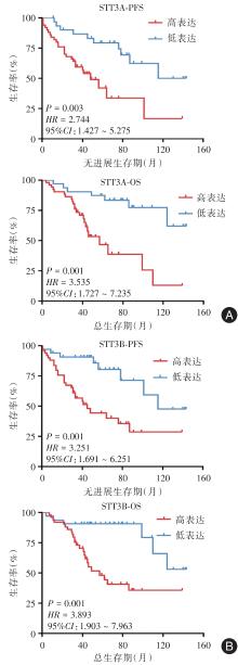
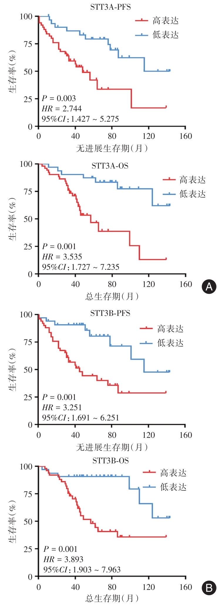
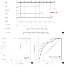
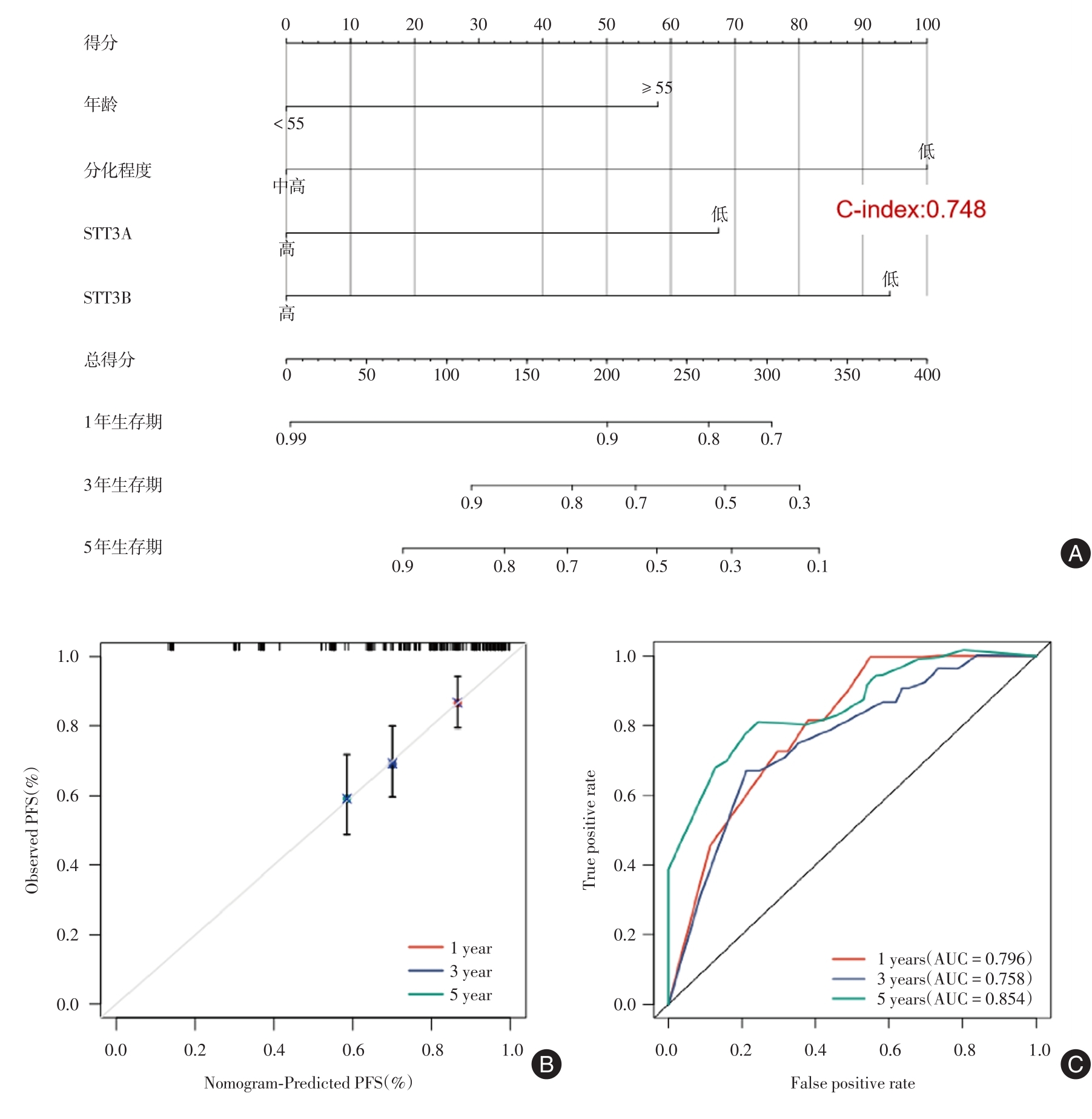
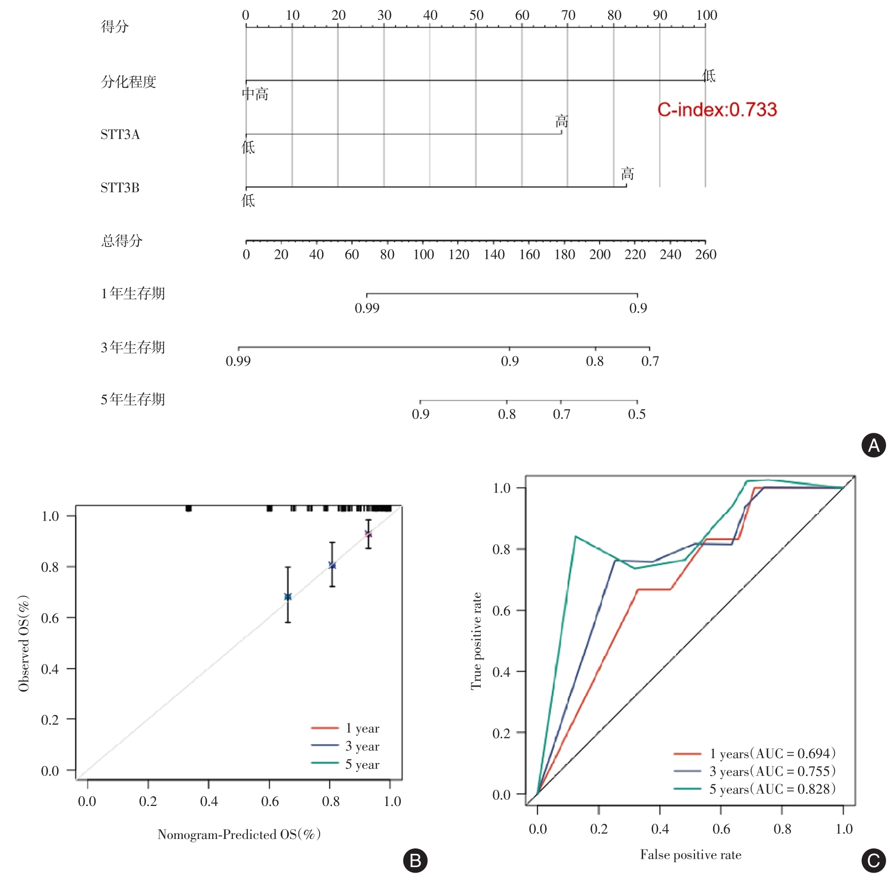
目的 探究上皮性卵巢癌(EOC)组织中STT3A和STT3B的表达水平与患者临床病理特征及预后的关系。 方法 选择接受手术治疗的88例EOC患者癌组织为实验组,22例非EOC患者(子宫或卵巢良性肿瘤)的正常输卵管上皮及其正常卵巢组织为对照组。收集患者的临床资料并进行随访。采用免疫组织化学染色方法检测STT3A和STT3B的表达,分析其表达水平与患者临床病理特征及预后的关系。 结果 STT3A和STT3B在EOC组织中的表达水平显著高于对照组(P < 0.05),且STT3A表达水平与肿瘤分化程度、淋巴结转移密切相关(P < 0.05)。STT3B的表达水平与FIGO分期、血清CA125水平、淋巴脉管间隙浸润、肿瘤分化程度、淋巴结转移具有显著的相关性(P < 0.05)。Kaplan-Meier生存分析表明,STT3A和STT3B高表达与EOC患者的不良预后相关(P < 0.05)。多因素Cox回归分析发现,STT3A和STT3B高表达是EOC患者预后的独立危险因素(P < 0.05)。Western blot结果显示STT3A和STT3B在卵巢癌细胞系中高表达(P < 0.05)。 结论 STT3A和STT3B在EOC癌组织中高表达,且其表达水平与患者不良预后有关,可能是判断EOC患者预后的潜在标志物。

关键词: 上皮性卵巢癌, STT3A, STT3B, 临床病理特征, 预后

Abstract:

Objective To explore the relationship between the expression levels of STT3A and STT3B in Epithelial Ovarian Cancer (EOC) and the clinicopathological features and prognosis of the patients. Methods The clinical information of 88 patients with EOC who underwent surgical treatment and 22 patients with normal tubal epithelium and normal ovarian tissue in our department from 2010 to 2019 were collected and followed up. The expressions of STT3A and STT3B in EOC and control groups were detected by immunohistochemistry, and their relationships with clinicopathological features and prognosis of patients were analyzed. Results The expression of STT3A and STT3B in EOC patients was significantly higher than that of control group (P < 0.05), and the expression of STT3A was related to tumor differentiation degree and lymph node metastasis (P < 0.05). The expression of STT3B was significantly correlated with FIGO staging, serum CA125 level, Lymph?vascular space invasion, degree of tumor differentiation and lymph node metastasis (P < 0.05). Kaplan?Meier survival analysis showed that the higher expressions of STT3A and STT3B were associated with poor prognosis (P < 0.05). Multivariate Cox regression analysis revealed that the high expressions of STT3A and STT3B were independent risk factors for the prognosis of EOC patients (P < 0.05). Western Blot results showed that STT3A and STT3B were significantly expressed in ovarian cancer (P < 0.05). Conclusion STT3A and STT3B are extremely expressed in EOC tissues and are associated with poor prognosis of EOC patients, and may be potential markers for the prognosis of EOC patients.

Key words: epithelial ovarian cancer, STT3A, STT3B, clinicopathological features, prognosis

中图分类号: