| [1] |
Weetman A P. An update on the pathogenesis of Hashimoto's thyroiditis[J]. J Endocrinol Invest, 2021, 44(5):883-890.doi: 10.1007/s40618-020-01477-1 .
|
| [2] |
|
| [3] |
|
| [4] |
Issa P P, Omar M, Buti Y, et al. Hashimoto's Thyroiditis Minimizes Lymph Node Metastasis in BRAF Mutant Papillary Thyroid Carcinomas[J]. Biomedicines, 2022, 10(8):2051.doi: 10.3390/biomedicines10082051 .
|
| [5] |
|
| [6] |
|
| [7] |
Siyu Duanyang, Qu Wenhao, Wang Yanfang, et al. Multimodal ultrasound and artificial intelligence for characterization of thyroid nodules in Hashimoto's thyroiditis: Current challenges and future perspectives[J]. Eur J Med Res, 2026,Online ahead of print.doi: 10.1186/s40001-026-04092-7 .
|
| [8] |
Jiang Wenchao, Zhou Guanjie, Bai Honghua, et al. MCFL: Multimodal collaborative fusion learning for Hashimoto's thyroiditis recognition[J]. IEEE Trans Image Process, 2026, 35: 402-417. doi: 10.1109/TIP.2025.3649346 .
|
| [9] |
Han Zhijiang, Xie Lesi, Wei Peiying, et al. Ultrasound gray scale ratio for differential diagnosis of papillary thyroid microcarcinoma from benign micronodule in patients with Hashimoto′s thyroiditis[J]. BMC Endocr Disord, 2022, 22(1): 187. doi: 10.1186/s12902-022-01028-0 .
|
| [10] |
Ragusa F, Fallahi P, Elia G, et al. Hashimotos' thyroiditis: Epidemiology, pathogenesis, clinic and therapy[J]. Best Pract Res Clin Endocrinol Metab, 2019, 33(6):101367.doi: 10.1016/j.beem.2019.101367 .
|
| [11] |
|
| [12] |
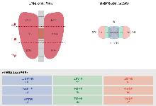
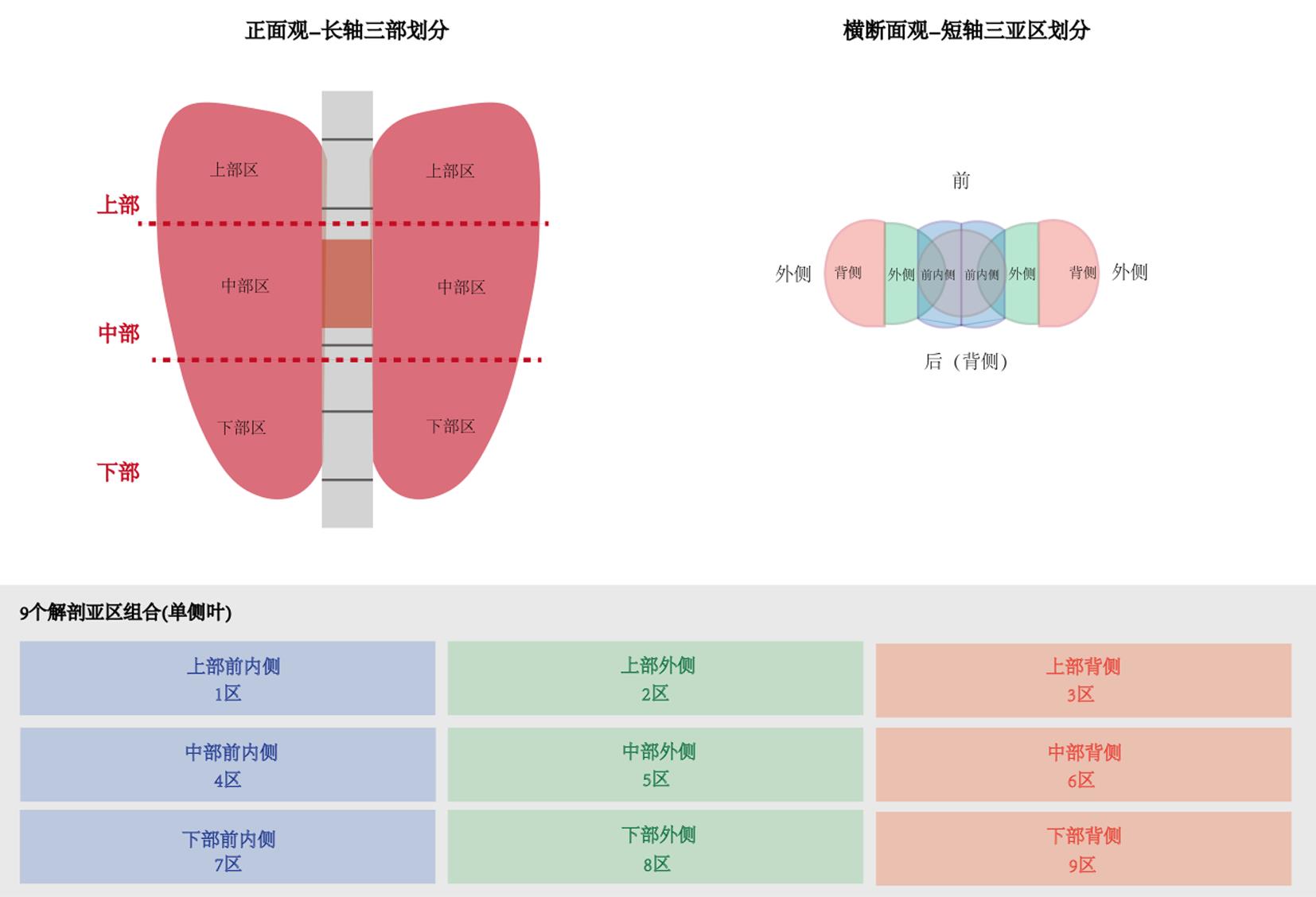
Luo Dingyuan. The Luo-Dingyuan thyroid vascular-lymphatic segmentation classification: A functional anatomy-based framework to facilitate precision management and thyroid function preservation[J]. Cancer Manag Res, 2026, 18: 607765. doi: 10.2147/CMAR.S607765 .
|
| [13] |
|
| [14] |
|
| [15] |
Orlando G, Scerrino G, Corigliano A, et al. Papillary Thyroid Microcarcinoma: Active Surveillance Against Surgery. Considerations of an Italian Working Group From a Systematic Review[J]. Front Oncol, 2022, 12:859461.doi: 10.3389/fonc.2022.859461 .
|
| [16] |
Cohen S M, Noel J E, Baroody M, et al. Prognostication of papillary thyroid microcarcinoma based on preoperative ultrasound[J]. Front Endocrinol (Lausanne), 2023, 14:1101705.doi: 10.3389/fendo.2023.1101705 .
|
| [17] |
廖博雅. 甲状腺乳头状癌合并桥本甲状腺炎的临床及病理学特征分析[D]. 衡阳:南华大学, 2019.
|
| [18] |
Teng Chao, Pang Kunkun, Zhang Lulu, et al. Impact of Hashimoto's thyroiditis on ultrasound diagnosis of papillary thyroid carcinoma: A retrospective study[J]. Front Oncol, 2025, 15: 1551114. doi: 10.3389/fonc.2025.1551114 .
|
| [19] |
Wang Zhiyuan, Gui Zhiqiang, Wang Zhihong, et al. Clinical and ultrasonic risk factors for high-volume central lymph node metastasis in cN0 papillary thyroid microcarcinoma: A retrospective study and meta-analysis[J]. Clin Endocrinol, 2023, 98(4): 609-621. doi: 10.1111/cen.14834 .
|
| [20] |
Cai Yifeng, Yang Ruixuan, Yang Shuhui, et al. Comparison of the C-TIRADS, ACR-TIRADS, and ATA guidelines in malignancy risk stratification of thyroid nodules[J]. Quant Imaging Med Surg, 2023, 13(7): 4514-4525. doi: 10.21037/qims-22-826 .
|
| [21] |
|
| [22] |
Feng Na, Wei Peiying, Kong Xiangkai, et al. The value of ultrasound grayscale ratio in the diagnosis of papillary thyroid microcarcinomas and benign micronodules in patients with Hashimoto's thyroiditis: A two-center controlled study[J]. Front Endocrinol, 2022, 13: 949847. doi: 10.3389/fendo.2022.949847 .
|
| [23] |
Gao Lei, Wen Xiuli, Yue Guanghui, et al. The predictive value of a nomogram based on ultrasound radiomics, clinical factors, and enhanced ultrasound features for central lymph node metastasis in papillary thyroid microcarcinoma[J]. Ultrason Imaging, 2025, 47(2): 93-103. doi: 10.1177/01617346251313982 .
|
| [24] |
|
| [25] |
Gong Jiantao, Zhu Baining, Liu Wenji, et al. Risk factors for lymph node metastasis in papillary thyroid carcinoma: A retrospective study[J]. Horm Metab Res, 2023, 55(5): 315-322. doi: 10.1055/a-2057-8358 .
|
| [26] |
Trimboli P, Castellana M, Piccardo A, et al. The ultrasound risk stratification systems for thyroid nodule have been evaluated against papillary carcinoma. A meta-analysis[J]. Rev Endocr Metab Disord, 2021, 22(2):453-460.doi: 10.1007/s11154-020-09592-3 .
|


 )