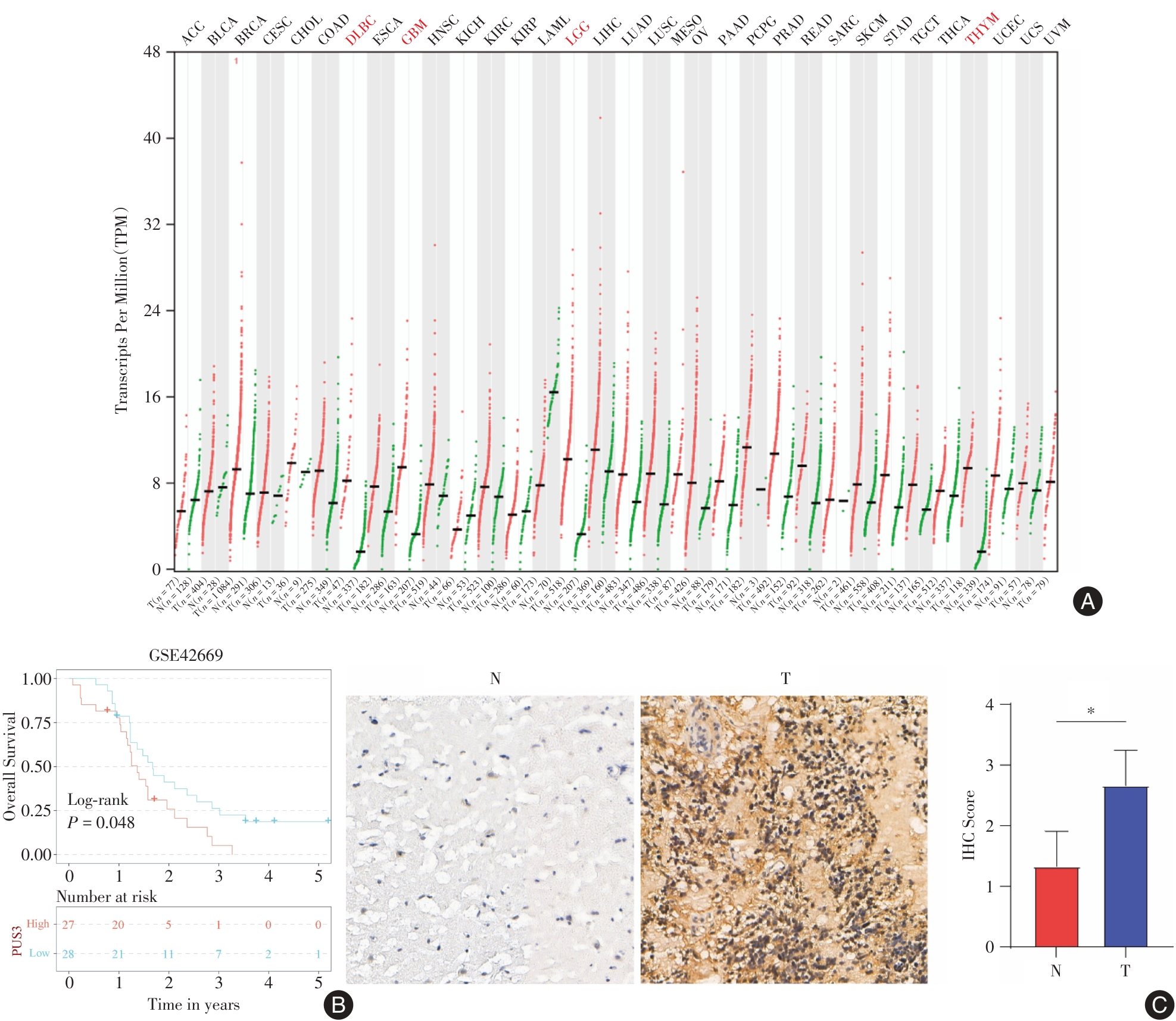
| 1 |
CUI Q, YIN K, ZHANG X, et al. Targeting PUS7 suppresses tRNA pseudouridylation and glioblastoma tumorigenesis[J]. Nat Cancer, 2021, 2(9): 932-949. doi:10.1038/s43018-021-00238-0
doi: 10.1038/s43018-021-00238-0
|
| 2 |
WELLER M, WEN P Y, CHANG S M, et al. Glioma[J].Nat Rev Dis Primers, 2024, 10(1): 33. doi:10.1038/s41572-024-00516-y
doi: 10.1038/s41572-024-00516-y
|
| 3 |
李梦欣, 惠磊, 汲乾坤, 等. ANXA1在脑胶质母细胞瘤中表达上调并促进胶质母细胞瘤的恶性进展. 实用医学杂志, 2023, 39(8): 928-935.
|
| 4 |
ZHANG D Y, MING G L, SONG H. PUS7: A targetable epitranscriptomic regulator of glioblastoma growth[J].Trends Pharmacol Sci, 2021, 42(12): 976-978. doi:10.1016/j.tips.2021.10.002
doi: 10.1016/j.tips.2021.10.002
|
| 5 |
GLADSON C L, PRAYSON R A, LIU W M. The pathobiology of glioma tumors[J].. Annu Rev Pathol, 2010, 5: 33-50. doi:10.1146/annurev-pathol-121808-102109
doi: 10.1146/annurev-pathol-121808-102109
|
| 6 |
LIU K, TSUNG K, ATTENELLO F J. Characterizing Cell Stress and GRP78 in Glioma to Enhance Tumor Treatment[J].Front Oncol, 2020, 10: 608911. doi:10.3389/fonc.2020.608911
doi: 10.3389/fonc.2020.608911
|
| 7 |
YASINJAN F, XING Y, GENG H, et al. Immunotherapy: a promising approach for glioma treatment[J].Front Immunol, 2023, 14: 1255611. doi:10.3389/fimmu.2023.1255611
doi: 10.3389/fimmu.2023.1255611
|
| 8 |
蔡嘉怡,陈思羽,蔡女略,等.微小RNA-378-5p对胶质瘤血管生成的影响[J].实用医学杂志,2025,41(2):186-194.
|
| 9 |
FU M, GU J, WANG M, et al. Emerging roles of tRNA-derived fragments in cancer[J]. Mol Cancer, 2023, 22(1): 30. doi:10.1186/s12943-023-01739-5
doi: 10.1186/s12943-023-01739-5
|
| 10 |
PENZO M, MONTANARO L. Turning Uridines around: Role of rRNA Pseudouridylation in Ribosome Biogenesis and Ribosomal Function[J]. Biomolecules, 2018, 8(2): 38. doi:10.3390/biom8020038
doi: 10.3390/biom8020038
|
| 11 |
ABDELRAHMAN H A, AL‐SHAMSI A M, ALI B R, et al. A null variant in PUS3 confirms its involvement in intellectual disability and further delineates the associated neurodevelopmental disease[J].Clinical Genetics, 2018, 94(6): 586-587. doi:10.1111/cge.13443
doi: 10.1111/cge.13443
|
| 12 |
Nøstvik M, KATETA S M, SchönEWOLF-GREULICH B, et al. Clinical and molecular delineation of PUS3-associated neurodevelopmental disorders[J]. Clin Genet, 2021, 100(5): 628-633. doi:10.1111/cge.14051
doi: 10.1111/cge.14051
|
| 13 |
SONG D, GUO M, XU S,et al.HSP90-dependent PUS7 overexpression facilitates the metastasis of colorectal cancer cells by regulating LASP1 abundance[J]. J Exp Clin Cancer Res,2021,40(1):170. doi:10.1186/s13046-021-01951-5
doi: 10.1186/s13046-021-01951-5
|
| 14 |
HU Y X, DIAO L T, HOU Y R, et al. Pseudouridine synthase 1 promotes hepatocellular carcinoma through mRNA pseudouridylation to enhance the translation of oncogenic mRNAs[J].Hepatology, 2024, 80(5): 1058-1073. doi:10.1097/hep.0000000000000702
doi: 10.1097/hep.0000000000000702
|
| 15 |
LUO W, XU Z, WANG H, et al. HIF1A-repressed PUS10 regulates NUDC/Cofilin1 dependent renal cell carcinoma migration by promoting the maturation of miR-194-5p[J].Cell Biosci, 2023, 13(1): 153. doi:10.1186/s13578-023-01094-4
doi: 10.1186/s13578-023-01094-4
|
| 16 |
LU S, WEI X, TAO L, et al. A novel tRNA-derived fragment tRF-3022b modulates cell apoptosis and M2 macrophage polarization via binding to cytokines in colorectal cancer[J]. J Hematol Oncol, 2022, 15(1): 176. doi:10.1186/s13045-022-01388-z
doi: 10.1186/s13045-022-01388-z
|
| 17 |
CHARETTE M, GRAY M W. Pseudouridine in RNA: What, where, how, and why[J]. IUBMB Life, 2000, 49(5): 341-351. doi:10.1080/152165400410182
doi: 10.1080/152165400410182
|
| 18 |
GUZZI N, CIEŚLA M, NGOC P C T,et al.Pseudouridylation of tRNA-Derived Fragments Steers Translational Control in Stem Cells[J]. Cell,2018,173(5):1204-1216.e26. doi:10.1016/j.cell.2018.03.008
doi: 10.1016/j.cell.2018.03.008
|
| 19 |
ZHANG D Y, MING G L, SONG H. PUS7: A targetable epitranscriptomic regulator of glioblastoma growth[J]. Trends Pharmacol Sci, 2021, 42(12): 976-978. doi:10.1016/j.tips.2021.10.002
doi: 10.1016/j.tips.2021.10.002
|
| 20 |
WU Y, PENG S, CHENG B, et al. FOXA1-dependent PUS1 regulates EIF3b stability in a non-enzymatic pathway mediating prostate cancer bone metastasis[J]. Int J Biol Sci, 2024, 20(11): 4566-4584. doi:10.7150/ijbs.100905
doi: 10.7150/ijbs.100905
|
| 21 |
HU Y X, DIAO L T, HOU Y R, et al. Pseudouridine synthase 1 promotes hepatocellular carcinoma through mRNA pseudouridylation to enhance the translation of oncogenic mRNAs[J].Hepatology, 2024,80(5):1058-1073. doi:10.1097/hep.0000000000000702
doi: 10.1097/hep.0000000000000702
|
| 22 |
BARBIERI I, KOUZARIDES T. Role of RNA modifications in cancer[J].Nat Rev Cancer, 2020, 20(6): 303-322. doi:10.1038/s41568-020-0253-2
doi: 10.1038/s41568-020-0253-2
|
| 23 |
SADRKHANLOO M, ENTEZARI M, OROUEI S, et al. STAT3-EMT axis in tumors: Modulation of cancer metastasis, stemness and therapy response[J]. Pharmacol Res, 2022, 182: 106311. doi:10.1016/j.phrs.2022.106311
doi: 10.1016/j.phrs.2022.106311
|
| 24 |
Barzegar Behrooz A, Talaie Z, Jusheghani F, et al. Wnt and PI3K/Akt/mTOR Survival Pathways as Therapeutic Targets in Glioblastoma[J]. Int J Mol Sci, 2022, 23(3): 1353. doi:10.3390/ijms23031353
doi: 10.3390/ijms23031353
|
| 25 |
WU F, YANG J, LIU J, et al. Signaling pathways in cancer-associated fibroblasts and targeted therapy for cancer[J]. Signal Transduct Target Ther, 2021, 6(1): 218. doi:10.1038/s41392-021-00641-0
doi: 10.1038/s41392-021-00641-0
|
| 26 |
MANNING B D, TOKER A.AKT/PKB Signaling: Navigating the Network[J].Cell. 2017,169(3):381-405. doi:10.1016/j.cell.2017.04.001
doi: 10.1016/j.cell.2017.04.001
|
| 27 |
HUANG L, CHEN W, TAN Z, et al. Mrc1+ macrophage-derived IGF1 mitigates crystal nephropathy by promoting renal tubule cell proliferation via the AKT/Rb signaling pathway[J]. Theranostics, 2024, 14(4): 1764-1780. doi:10.7150/thno.89174
doi: 10.7150/thno.89174
|
| 28 |
ZUO Y, ZHANG C Z, REN Q, et al. Activation of mitochondrial-associated apoptosis signaling pathway and inhibition of PI3K/Akt/mTOR signaling pathway by voacamine suppress breast cancer progression[J]. Phytomedicine, 2022, 99: 154015. doi:10.1016/j.phymed.2022.154015
doi: 10.1016/j.phymed.2022.154015
|


 )
)