实用医学杂志 ›› 2025, Vol. 41 ›› Issue (12): 1816-1824.doi: 10.3969/j.issn.1006-5725.2025.12.007

• 基础研究 • 上一篇    

线粒体自噬调控前列腺组织中NLRP3炎症小体的表达在实验性自身免疫性前列腺炎大鼠中的作用机制

陆良喜1,陆海旺2,王文杰3,史珺3,黄志敏2,宾彬2()   

  1. 1.广西中医药大学第一附属医院仁爱分院男科 (广西 南宁 530001 )
    2.广西中医药大学第一附属医院男科
    1.广西 南宁 530023),广西中医药大学基础医学院,(广西南宁 530001
  • 收稿日期:2025-03-19 出版日期:2025-06-25 发布日期:2025-07-02
  • 通讯作者: 宾彬 E-mail:billbinn@sina.com
  • 基金资助:
    国家自然科学基金青年科学基金项目(82205118)

To investigate the mechanism of mitochondrial autophagy regulating the expression of NLRP3 inflammasome in prostate tissue in rats with experimental autoimmune prostatitis

Liangxi LU1,Haiwang LU2,Wenjie WANG3,Jun SHI3,Zhimin HUANG2,Bin BIN2()   

  1. *.Department of Andrology,Ren'ai Branch of the First Affiliated Hospital of Guangxi University of Chinese Medicine,Nanning 530001,Guangxi,China
  • Received:2025-03-19 Online:2025-06-25 Published:2025-07-02
  • Contact: Bin BIN E-mail:billbinn@sina.com

摘要:

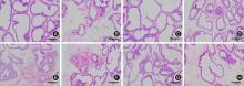
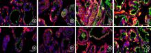
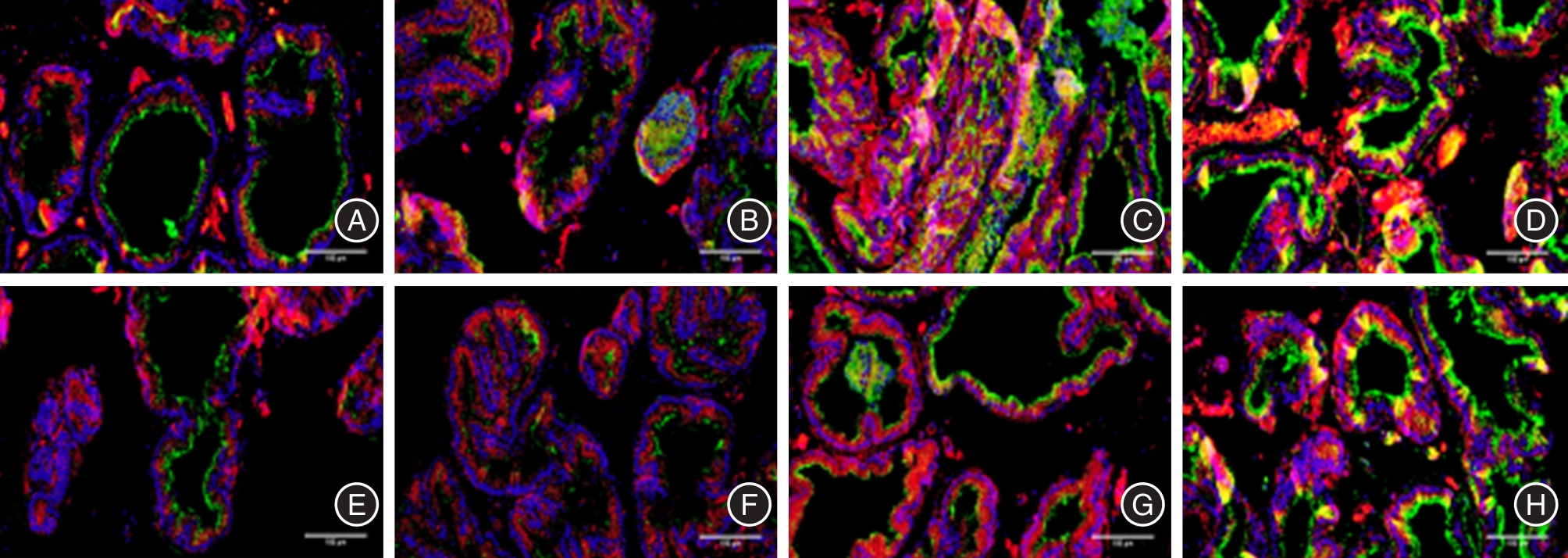
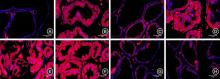
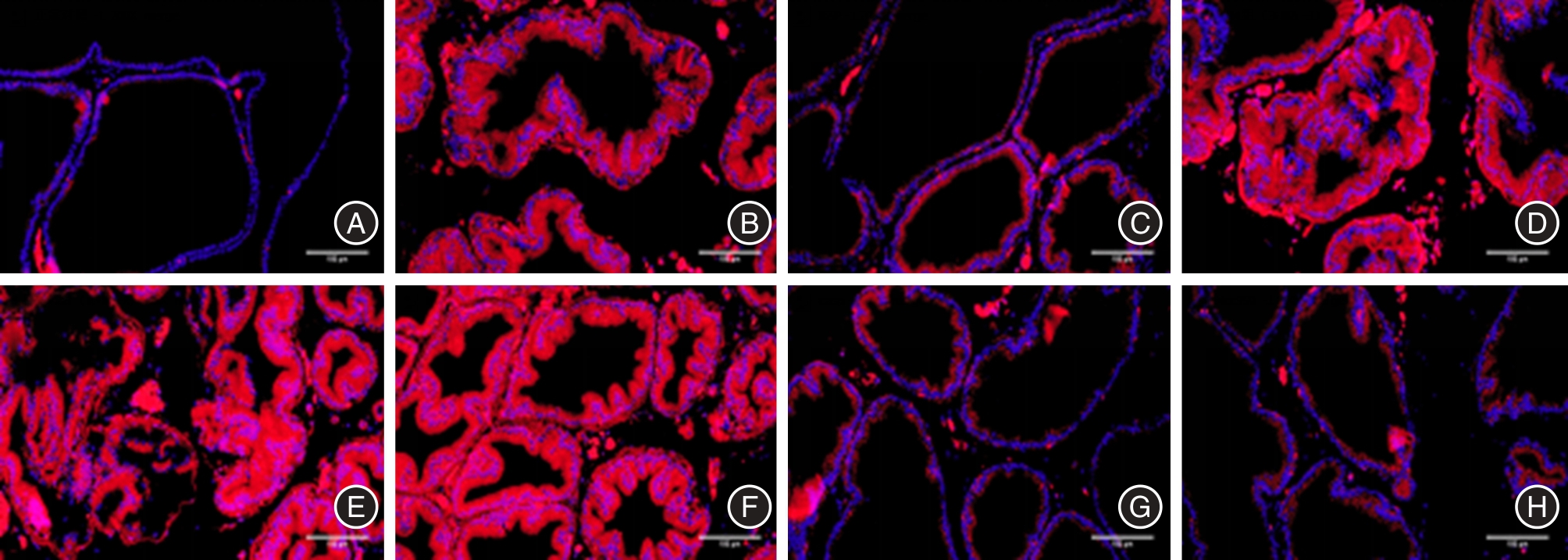
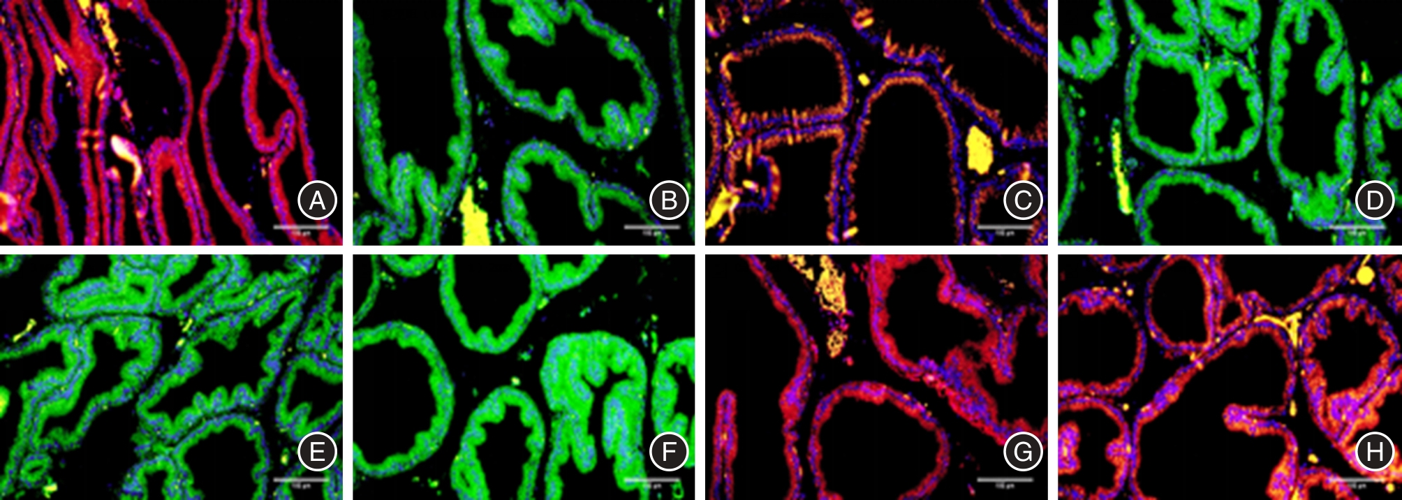
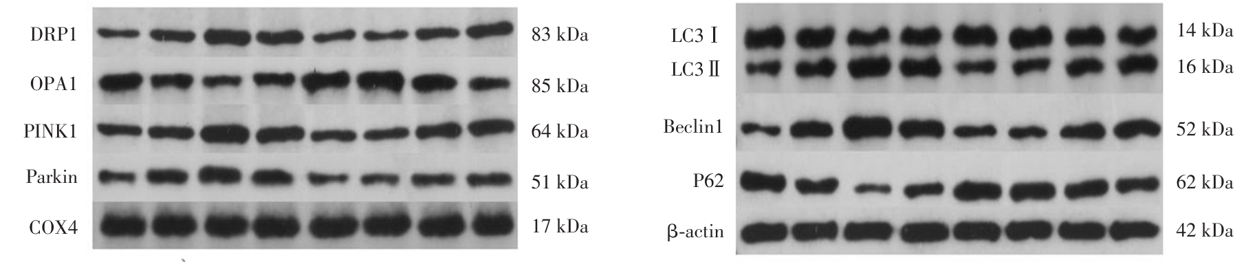
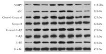
目的 探讨在实验性自身免疫性前列腺炎(experimental autoimmune prostatitis,EAP)大鼠前列腺组织中线粒体自噬调控NLRP3炎症小体的机制,以期为新药物研发提供理论支持。 方法 将40只SD雄性大鼠数字表随机分为8组,即正常组(N)、模型组(M)、雷帕霉素组(RAP)、雷帕霉素+线粒体自噬抑制剂组(RAP + Mdivi-1)、自噬抑制剂组(3MA)、Caspase1抑制剂组(Caspase1)、线粒体自噬抑制剂组(Mdivi-1)、NLRP3 抑制剂组(NLRP3),每组5只。药物干预后,采用HE染色、免疫荧光、比色法、WB法等观察相关指标。 结果 与N组比较,M组大鼠前列腺腺体结构损伤明显;与M组比较,RAP组、Caspase-1组、NLRP3组前列腺腺体结构有改善;而3-MA组、Mdivi-1组大鼠前列腺组织结构无改善,甚至破坏更明显。与N组比较,M组大鼠前列腺组织LC3-II和LAMP-1共表达增强,线粒体膜电位下降,ROS释放水平明显增加。与M组比较,RAP组、NLRP3组上述指标明显改善;但是,3-MA组、Mdivi-1组上述指标变得更差。与N组比较,M组大鼠前列腺线粒体DRP1、PINK1、Parkin蛋白表达升高,OPA1蛋白表达降低。与M组比较,RAP组、NLRP3组DRP1、PINK1、Parkin蛋白表达显著升高,RAP组OPA1蛋白表达显著降低;3-MA组、Mdivi-1组DRP1、PINK1、Parkin蛋白表达显著下降;Caspase-1组Parkin蛋白表达降低,但是DRP1、OPA1、PINK1蛋白表达差异无统计学意义。与N组比较,M组大鼠前列腺组织自噬蛋白LC3II/LC3I、Beclin1和炎症小体相关蛋白NLRP3、ASC、Cleaced-Caspase1、Cleaced-IL-1β、IL-18表达升高,P62蛋白表达下降;与M组比较,RAP组、NLRP3组LC3II/LC3I、Beclin1蛋白表达显著升高,P62、NLRP3、ASC、Cleaced-Caspase1、Cleaced-IL-1β、IL-18蛋白表达显著降低;3-MA 组、Mdivi-1组LC3II/LC3I、Beclin1蛋白表达显著下降,P62、NLRP3、ASC、Cleaced-Caspase1、IL-18蛋白表达升高;与M组比较,Caspase-1组NLRP3、ASC、Cleaced-Caspase1、Cleaced-IL-1β、IL-18蛋白表达显著降低,LC3II/LC3I、Beclin1、P62蛋白表达差异无统计学意义。 结论 NLRP3炎症小体参与EAP大鼠前列腺炎症进程,线粒体自噬通过调控前列腺组织中NLRP3炎症小体的活化介导EAP大鼠前列腺炎的发生发展。

关键词: 慢性前列腺炎, 线粒体自噬, Pink1/Parkin通路, NLRP3炎症小体, 炎症

Abstract:

Objective To investigate the mechanism of mitochondrial autophagy regulating the expression of NLRP3 inflammasome in prostate tissue in experimental autoimmune prostatitis(EAP)rats and to provide a theoretical basis for the study of new drug development. Methods A numerical table of 40 SD male rats was randomly divided into 8 groups. Namely, normal group (N), model group (M), rapamycin group (RAP), rapamycin + mitochondrial autophagy inhibitor group (RAP+Mdivi-1), autophagy inhibitor group (3MA), mitochondrial autophagy inhibitor group (Mdivi-1), Caspase1 inhibitor group (Caspase1), NLRP3 inhibitor group (NLRP3), 5 animals per group. After drug intervention, HE staining, immunofluorescence, colorimetry, and WB method were used to observe the relevant indexes. Results Compared with group N, the structural damage of prostate gland was obvious in group M. Compared with the M group, the prostate gland structure in RAP group, Caspase-1 group and NLRP3 group were improved. However, that in 3-MA group and Mdivi-1 group was not improved, and even destroyed more obviously. Compared with group N, the co-expression of LC3-II and LAMP-1 was enhanced, mitochondrial membrane potential was decreased, ROS release level was significantly increased in prostate tissue of rats in group M. Compared with the M group, the above indexes in RAP group and NLRP3 group were significantly improved. However, the above indexes in 3-MA group and Mdivi-1 group became worse. Compared with group N, the protein expressions of DRP1, PINK1 and Parkin in prostate mitochondria of rats in group M were increased, and the protein expressions of OPA1 was decreased. Compared with group M, the protein expressions of DRP1, PINK1 and Parkin in RAP group and NLRP3 group were significantly increased, while those in 3-MA group and Mdivi-1 group were significantly decreased. OPA1 protein expression was significantly decreased in the RAP group. The protein expression of Parkin in Caspase-1 group was decreased, but the protein expression of DRP1, OPA1 and PINK1 had no significant difference. Compared with group N, the protein expressions of LC3II/LC3I, Beclin1, NLRP3, ASC, Cleaced-Caspase1, Cleaced-IL-1β, and IL-18 in prostate tissue of rats in group M were increased, while the protein expressions of P62 was decreased. Compared with M group, LC3II/LC3I and Beclin1 protein expressions in RAP group and NLRP3 group were significantly increased, while those in 3-MA group and Mdivi-1 group were significantly decreased. Compared with M group, P62, NLRP3, ASC, Cleaced-Caspase1, Cleaced-IL-1β and IL-18 protein expressions in RAP group and NLRP3 group were significantly decreased, while those in 3-MA group and Mdivi-1 group were significantly increased. Compared with M group, the protein expressions of NLRP3, ASC, Cleaced-Caspase1, Cleaced-IL-1β, and IL-18 in Caspase-1 group were significantly reduced, but the protein expressions of LC3Ⅱ/LC3Ⅰ, Beclin1, and P62 were not statistically significant. Conclusions NLRP3 inflammatosome is involved in the progression of chronic prostatitis in EAP rats. Mitochondrial autophagy mediates the occurrence and development of prostatitis in EAP by regulating the activation of NLRP3 inflammasome in prostate tissue.

Key words: chronic prostatitis, mitochondrial autophagy, Pink1/Parkin pathway, NLRP3 inflammasome, inflammation

中图分类号: