实用医学杂志 ›› 2024, Vol. 40 ›› Issue (18): 2618-2622.doi: 10.3969/j.issn.1006-5725.2024.18.019

• 医学检查与临床诊断 • 上一篇    下一篇

超声生理盐水造影对新生儿环状胰腺的定量诊断价值

杨博洋1,方倩1,郑懿2,王红英1()   

  1. 1.广州医科大学附属妇女儿童医疗中心,超声科,(广州 510623 )
    2.广州医科大学附属妇女儿童医疗中心,新生儿外科,(广州 510623 )
  • 收稿日期:2024-06-05 出版日期:2024-09-25 发布日期:2024-09-30
  • 通讯作者: 王红英 E-mail:gzfe1122@163.com
  • 基金资助:
    国家自然科学基金青年项目(82101807)

Quantitative diagnosis of annular pancreas in neonates with saline⁃contrast ultrasound

Boyang YANG1,Qian FANG1,Yi ZHENG2,Hongying. WANG1()   

  1. Department of Ultrasound,Women and Children's Medical Center Affiliated to Guangzhou Medical University,Guangzhou 510623,China
  • Received:2024-06-05 Online:2024-09-25 Published:2024-09-30
  • Contact: Hongying. WANG E-mail:gzfe1122@163.com

摘要:

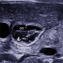
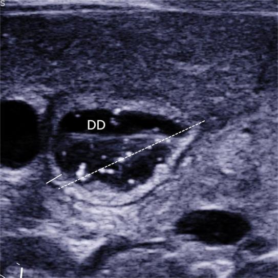
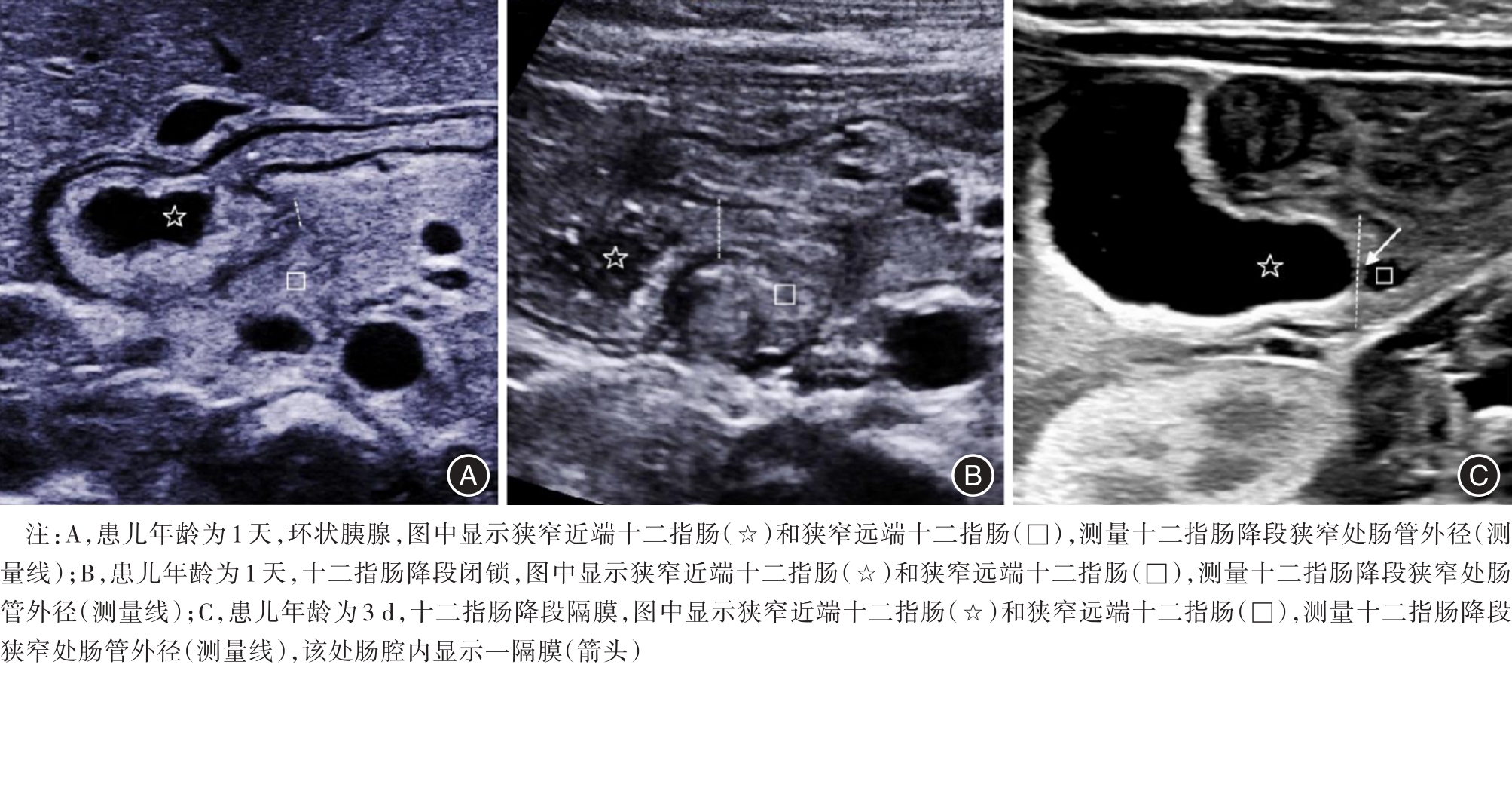
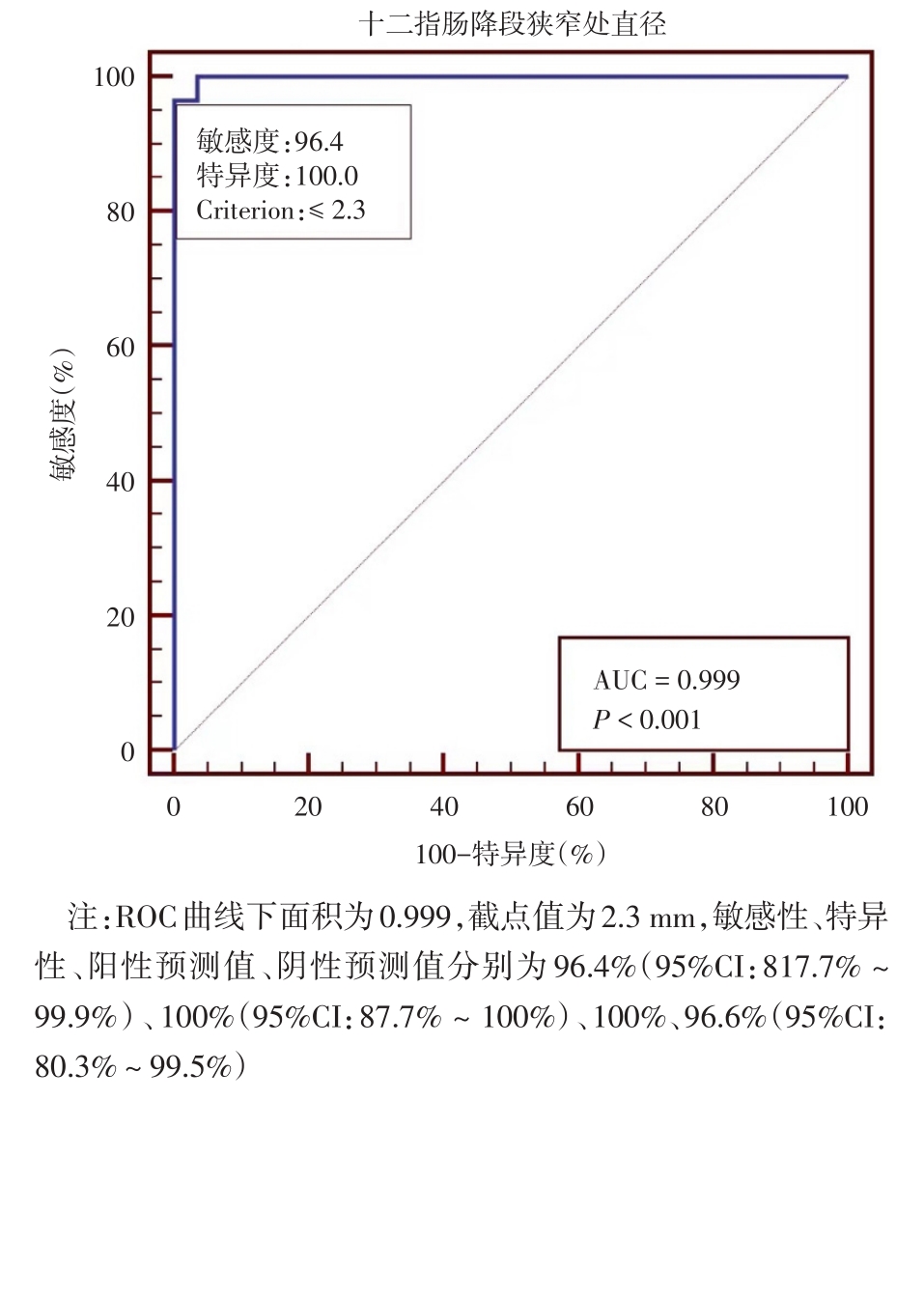
目的 探讨超声参数在新生儿环状胰腺诊断中的应用价值。 方法 收集2016年3月至2018年6月间,于本单位就诊的十二指肠降段梗阻的新生儿56例,均经上消化道超声造影检查。测量超声参数包括十二指肠降段扩张段外径(dilated duodenum diameter, DDD)、十二指肠降段扩张段肠壁厚度(dilated duodenum thickness, DDT)、十二指肠降段狭窄处外径(stenotic duodenum diameter, SDD)。据术中诊断情况分为环状胰腺组(A组)28例、十二指肠降段闭锁组(B组)3例、十二指肠降段隔膜组(C组)25例。采用单因素方差分析比较三组间的三种超声参数差异有无统计学意义,两两比较行LSD-t检验。如两两比较差异均有统计学意义,采用受试者工作特征曲线计算通过超声参数来诊断环状胰腺的曲线下面积及最佳截点值。 结果 三组中的DDD分别为(23.2 ± 2.4)、(25 ± 1.0)、(19.4 ± 2.6)mm,三组比较显示差异有统计学意义(F = 19.406,P < 0.001),两两比较显示A组、B组与C组相比,差异有统计学意义(t = 5.7,P < 0.05;t = 3.793,P < 0.05);A组与B组相比差异无统计学意义(t = 1.232,P > 0.05)。三组DDT分别为(3.0 ± 0.6)、(3.2 ± 0.4)、(2.4 ± 0.3)mm,三组比较显示差异有统计学意义(F = 12.487,P < 0.001),两两比较显示A组、B组与C组相比,差异有统计学意义(t = 4.695,P < 0.05;t = 2.778,P < 0.05),A组与B组相比,差异无统计学意义(t = 0.666,P > 0.05);三组中的SDD分别为(1.9 ± 0.3)、(3.6 ± 0.8)、(5.5 ± 0.7)mm,三组比较显示差异有统计学意义(F = 333.556,P < 0.001),两两比较,显示差异均有统计学意义(t = 5.521,P < 0.05;t = 6.142,P < 0.05;t = 25.828,P < 0.05)。采用SDD ≤ 2.3 mm来诊断环状胰腺的敏感性、特异性、阳性预测值、阴性预测值分别为96.4%、100%、100%、96.6%,曲线下面积为0.999。 结论 SDD可用于诊断新生儿环状胰腺。

关键词: 新生儿, 环状胰腺, 超声检查, 十二指肠降段扩张段外径, 十二指肠降段扩张段肠壁厚度, 十二指肠降段狭窄处外径

Abstract:

Objective To investigate the clinical utility of ultrasound parameters in diagnosing neonatal annular pancreas. Methods A total of 56 neonates with descending duodenal obstruction underwent saline?contrast ultrasound examination. Ultrasonic parameters, including dilated duodenum diameter (DDD), dilated duodenum thickness (DDT), and stenosis duodenum diameter (SDD), were measured. The cases were categorized into three groups: annular pancreas (group A, n = 28), descending duodenal atresia (group B, n = 3), and descending duodenal septum (group C, n = 25). One?way ANOVA was employed to compare statistical differences among the three ultrasonic parameters across the groups, followed by LSD?t test for pairwise comparisons between two groups showing significant differences. Receiver operating characteristics analysis was conducted to calculate the area under the curve and determine the cut?off value if there were statistically significant differences observed. Results In group A, group B, group C, the diameters of DDD were measured as(23.2 ± 2.4)mm, (25 ± 1.0)mm, (19.4 ± 2.6)mm, respectively. When comparing these three groups, there was a statistically significant difference between group A or group B and group C in terms of DDD diameter measurements (t = 5.7, P < 0.05; t = 3.793, P < 0.05). However, no statistically significant difference was observed between group A and group B for DDD diameter measurements (t = 1.232, P > 0.05). The corresponding values for DDT were measured as (3.0 ± 0.6)mm, (3.2 ± 0.4) mm,and(2.4 ± 0.3) mm,respectively. When comparing these three groups,the results showed a statistically significant difference between either Group A or Group B compared to Group C in terms of DDT measurement (t = 4.695, P < 0.05; t = 2.778, P < 0.05). However,no statistically significant difference was found between Group A and Group B regarding the measurement of DDT (t = 0.666, P > 0.05). The SDD measurements in the three groups were (1.9 ± 0.3) mm, (3.6 ± 0.8)mm, (5.5 ± 0.7)mm, respectively. Statistical analysis using LSD?t test revealed significant differences between the three groups (t = 5.521, P < 0.05; t = 6.142, P < 0.05; t = 25.828, P < 0.05).The sensitivity, specificity, positive predictive value, and negative predictive value for detecting annular pancreas at a cutoff of 2.3 mm were determined to be 96.4%, 100%, 100%, and 96.6%, respectively, with an area under the receiver operating characteristic curve of 0.999. Conclusion SDD could be employed for the diagnosis of neonatal annular pancreas.

Key words: neonate, annular pancreas, ultrasound, dilated duodenum diameter, dilated duodenum thickness, stenotic duodenum diameter

中图分类号: