实用医学杂志 ›› 2023, Vol. 39 ›› Issue (20): 2572-2578.doi: 10.3969/j.issn.1006-5725.2023.20.003

• 基础研究 • 上一篇    下一篇

连接蛋白43在氧化低密度脂蛋白诱导RAW264.7细胞自噬中的作用

熊思渊1,2,4,王璐1,2,3,田俊杰1,2,4,谷扬5,马雅静5,马克涛1,2,3,张莹莹1,3,5,李新芝1,2,4()   

  1. 1.新疆地方与民族高发病教育部重点实验室 (新疆 石河子 832000 )
    2.国家卫健委中亚高发病防治重点实验室 (新疆 石河子 832000 )
    3.石河子大学医学院 生理学教研室,(新疆 石河子 832000 )
    4.石河子大学医学院 病理生理学教研室 (新疆 石河子 832000 )
    5.石河子大学医学院第一附属医院检验科 (新疆 石河子 832000 )
  • 收稿日期:2023-04-16 出版日期:2023-10-25 发布日期:2023-11-15
  • 通讯作者: 李新芝 E-mail:Lixinzhi@shzu.edu.cn
  • 基金资助:
    国家自然科学基金项目(81860286);中国医学科学院中央级公益性科研院所基本科研业务费专项资金资助(2020-PT330-003);兵团国际合作项目(2020BC004);“天山英才培养”计划、石河子大学青年创新培育人才计划项目(KX01860301)

Role of Cx43 in ox⁃LDL induced RAW264.7 autophagy

Siyuan XIONG1,2,4,Lu WANG1,2,3,Junjie TIAN1,2,4,Yang GU5,Yajing MA5,Ketao MA1,2,3,Yingying ZHANG1,3,5,Xinzhi. LI1,2,4()   

  1. 1.Key Laboratory of Xinjiang Regional and Ethnic High Incidence,Ministry of Education,Shihezi 832000,China
    2.Key Laboratory of Prevention and Control of High Morbidity in Central Asia,National Health Commission,Shihezi 832000,China
    4.Department of Pathophysiology,College of Medicine,Shihezi University,Shihezi 832000,China
  • Received:2023-04-16 Online:2023-10-25 Published:2023-11-15
  • Contact: Xinzhi. LI E-mail:Lixinzhi@shzu.edu.cn

摘要:

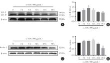
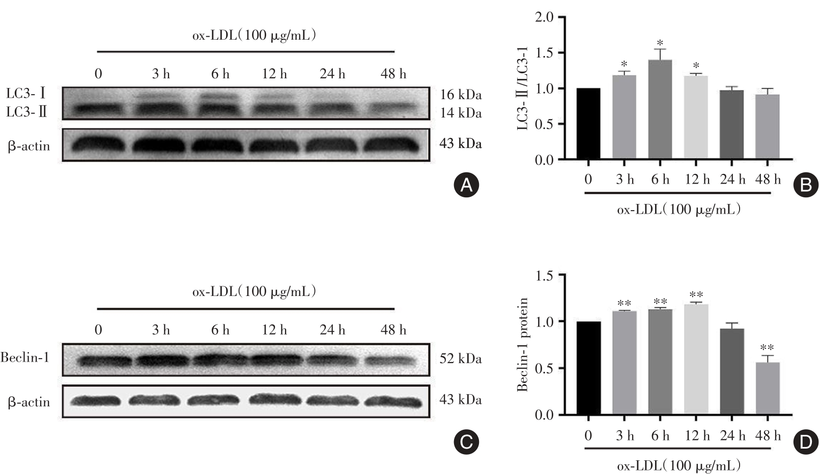
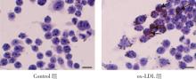
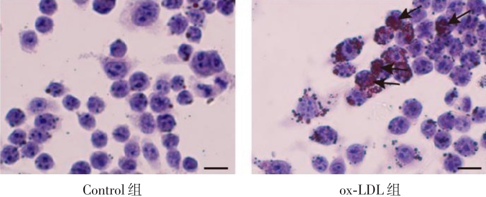
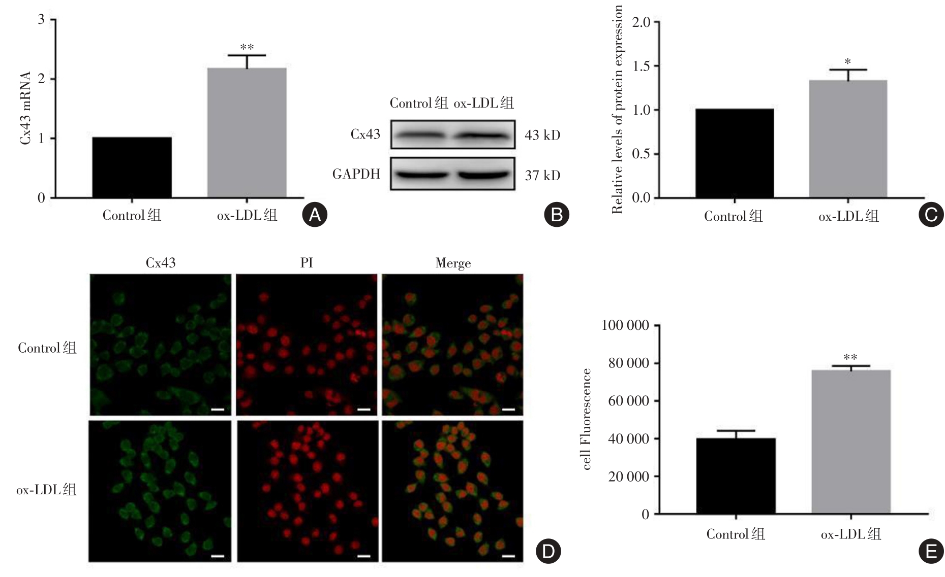
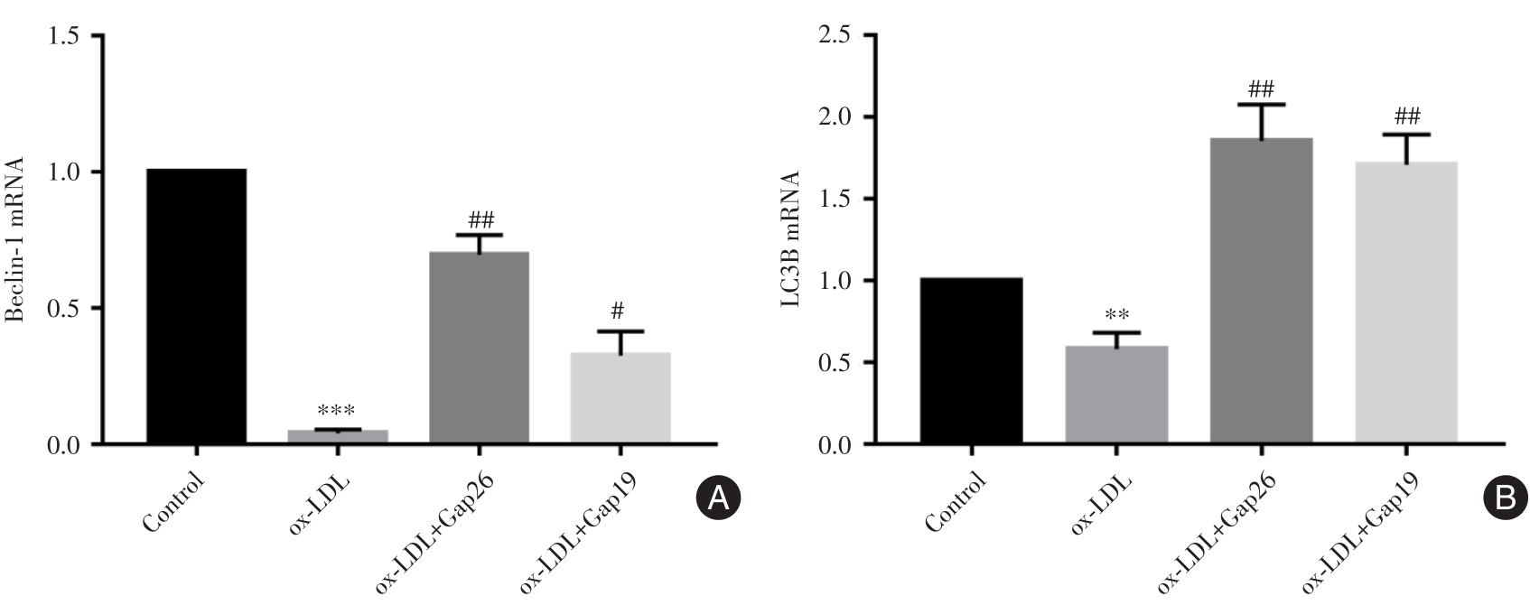
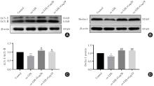
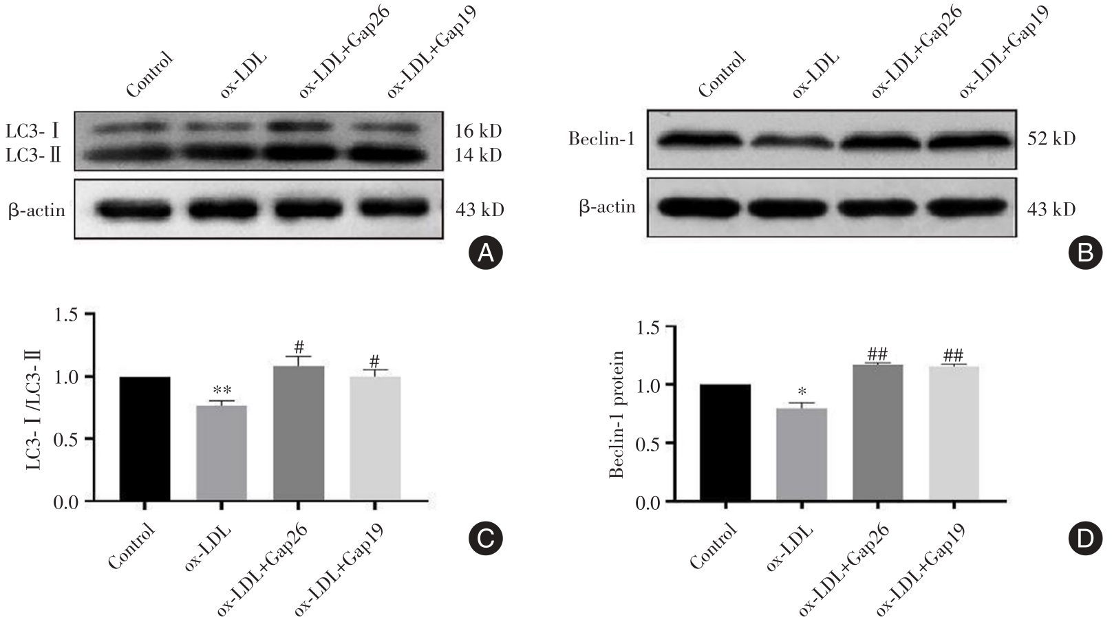
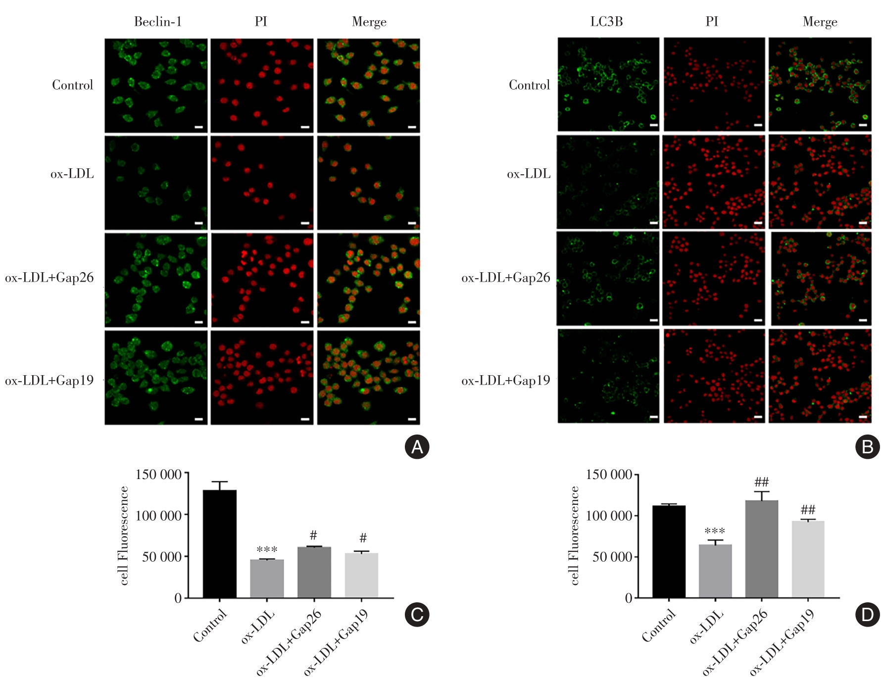
目的 探究连接蛋白43(Connexins43,Cx43)在氧化低密度脂蛋白(oxidized low-density lipoprotein,ox-LDL)诱导小鼠单核巨噬细胞系(RAW264.7细胞)自噬中的作用。 方法 利用ox-LDL(100 μg/mL,24 h)构建RAW264.7自噬及泡沫化模型。实验分为Control组、ox-LDL组、ox-LDL+Gap26组、ox-LDL+Gap19组。qRT-PCR检测各组Cx43、自噬蛋白Beclin-1及LC3B mRNA水平。Western blot检测Cx43、Beclin-1及LC3B蛋白水平。细胞免疫荧光技术检测Cx43、Beclin-1 及LC3B 在RAW264.7细胞上的定位和表达。 结果 油红O染色结果显示,ox-LDL干预RAW264.7细胞24 h后,ox-LDL组细胞浆内出现红染脂滴,即细胞发生泡沫化。Western blot、qRT-PCR及免疫荧光结果显示,ox-LDL干预RAW264.7细胞24 h后,ox-LDL组细胞中Cx43的表达水平升高。此外,Western blot、qRT-PCR及免疫荧光结果表明,ox-LDL干预RAW264.7细胞24 h后,与Control组相比,ox-LDL组细胞的Beclin-1、LC3BmRNA及蛋白表达水平降低;与ox-LDL组相比,ox-LDL+Gap26组及ox-LDL+Gap19组的自噬相关蛋白表达水平升高。 结论 阻断Cx43能促进ox-LDL诱导RAW264.7细胞自噬。

关键词: 连接蛋白43, 巨噬细胞, 自噬

Abstract:

Objective This study aims to explore the role of connexin 43 (Cx43) in the autophagy of RAW264.7 cell induced by oxidized low-density lipoprotein (ox-LDL). Methods The ox-LDL (100 μg/mL, 24 h) was used to establish the model of autophagy and foam cell formation. RAW264.7 mouse mononuclear macrophage lines were randomly divided into control group, ox-LDL group, ox-LDL+Gap26 group and ox-LDL+Gap19 group. The mRNA levels of Cx43, autophagy protein Beclin-1 and LC3B were detected by qRT-PCR. The protein levels of Cx43, Beclin-1 and LC3B were detected by Western blot. The expression and localization of Cx43, Beclin-1 and LC3B were detected by cellular immunofluorescence. Results The results of oil red O staining showed that red lipid droplets appeared in the cell plasma of the ox-LDL group and cells were foamy 24 h after ox-LDL intervention in RAW264.7 cells. By Western blot, qRT-PCR and immunofluorescence 24 h after ox-LDL intervention in RAW264.7 cells, the expression level of Cx43 in the cells of the ox-LDL group was increased, the expression levels of Beclin-1, LC3B mRNA and protein were decreased in the cells of the ox-LDL group compared with the control group, the expression levels of Beclin-1 and LC3B were decreased in the ox-LDL+Gap26 group compared with the ox-LDL group, and the expression levels of autophagy-related proteins in the ox LDL+Gap19 group were increased compared with the ox-LDL group. Conclusion Cx43 blocking can significantly promote RAW264.7 autophagy induced by ox-LDL.

Key words: Cx43, macrophage, autophagy

中图分类号: