

The Journal of Practical Medicine ›› 2026, Vol. 42 ›› Issue (13): 2444-2453.doi: 10.3969/j.issn.1006-5725.2026.13.021
• Treatise:Mechanism and Practice • Previous Articles
Yuxiu LI,Zhe YANG,Zhenfen ZHANG,Binwei XU,Yan HONG( )
)
Received:2026-04-29
Online:2026-07-10
Published:2026-07-14
Contact:
Yan HONG
E-mail:hongyanb@163.com
CLC Number:
Yuxiu LI,Zhe YANG,Zhenfen ZHANG,Binwei XU,Yan HONG. Ferulic acid improves delayed wound healing caused by arsenic poisoning by inhibiting NF-κB signaling[J]. The Journal of Practical Medicine, 2026, 42(13): 2444-2453.
Tab.1
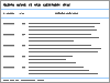
Primer sequences"
| 基因 | 上游引物(5′→3′) | 下游引物(5′→3′) |
|---|---|---|
| GAPDH | GCAAGTTCAACGGCACAG | GCCAGTAGACTCCACGACAT |
| IL-1β | GAGCCTGTGTTTCCTCCTTG | CAAGTGCAAGGCTATGACCA |
| IL-6 | CTCCCAACAGACCTGTCTATAC | CCATTGCACAACTCTTTTCTCA |
| TNF-α | CCAGTGTGGGAAGCTGTCTT | AAGCAAAAGAGGAGGCAACA |
| IFN-γ | CAGCAACAGCAAGGCGAAAAAGG | TTTCCGCTTCCTGAGGCTGGAT |
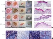
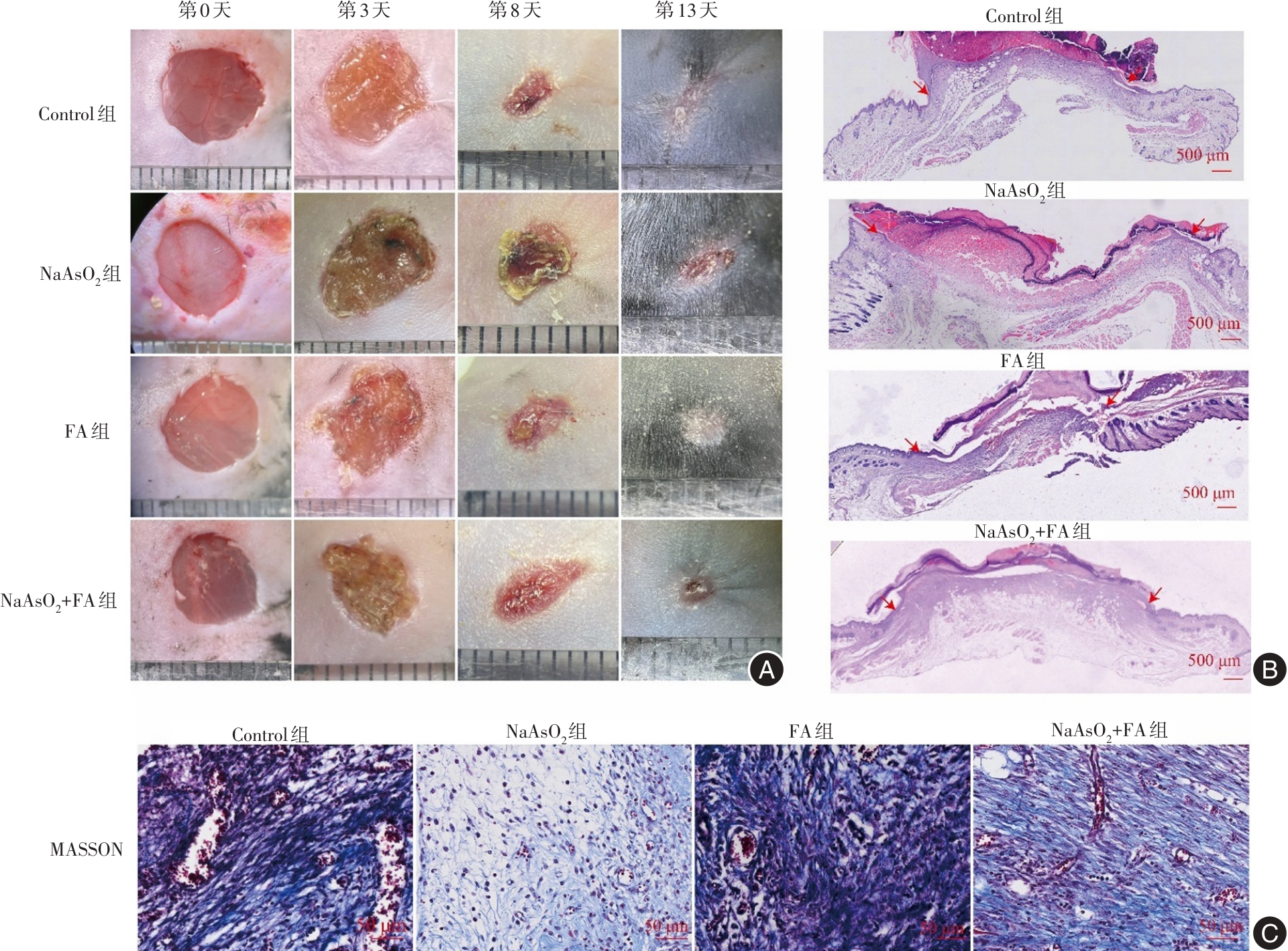
Fig.1
FA ameliorates arsenic poisoning-induced delayed wound healing in mice"
Tab.2
Wound healing rates of each group of mice (n = 5)"
| 时间 | Control组 | NaAsO2组 | FA组 | NaAsO2+FA组 | F值 | P值 |
|---|---|---|---|---|---|---|
| 第3天 | 31.15 ± 5.18 | 2.80 ± 2.01* | 28.85 ± 2.99 | 19.96 ± 4.75# | 63.73 | 0.000 1 |
| 第8天 | 76.16 ± 3.90 | 35.24 ± 4.78* | 71.38 ± 4.77 | 51.91 ± 6.82# | 79.24 | 0.000 1 |
| 第13天 | 100.00 ± 0.00 | 80.34 ± 6.38* | 100.00 ± 0.00 | 91.79 ± 3.35# | 40.02 | 0.0001 |
Tab.3
The indicators of wound healing degree for each group of mice(n = 5)"
| 指标 | Control组 | NaAsO2组 | FA组 | NaAsO2+FA组 | F值 | P值 |
|---|---|---|---|---|---|---|
| 未愈合伤口长度/cm | 0.55 ± 0.15 | 1.19 ± 0.13* | 0.61 ± 0.12 | 0.90 ± 0.20# | 22.10 | 0.000 1 |
| 伤口组织胶原蛋白占比/% | 72.24 ± 6.12 | 39.69 ± 5.43* | 75.57 ± 5.35 | 57.92 ± 5.60# | 50.56 | 0.000 1 |
Tab.4
The expression of inflammatory factors in the wound tissues of each group of mice (n = 5)"
| 炎性因子 | Control组 | NaAsO2组 | FA组 | NaAsO2+FA组 | F值 | P值 |
|---|---|---|---|---|---|---|
| IL-1β | 1.01 ± 0.15 | 3.47 ± 0.34* | 0.92 ± 0.22 | 2.14 ± 0.23# | 156.2 | 0.000 1 |
| IL-6 | 1.19 ± 0.17 | 2.97 ± 0.21* | 0.99 ± 0.14 | 1.85 ± 0.21# | 150.3 | 0.000 1 |
| TNF-α | 1.01 ± 0.16 | 2.80 ± 0.18* | 1.09 ± 0.16 | 1.76 ± 0.14# | 163.5 | 0.000 1 |
| IFN-γ | 1.06 ± 0.13 | 3.90 ± 0.23* | 1.10 ± 0.13 | 2.72 ± 0.28# | 279.3 | 0.000 1 |
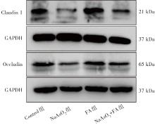
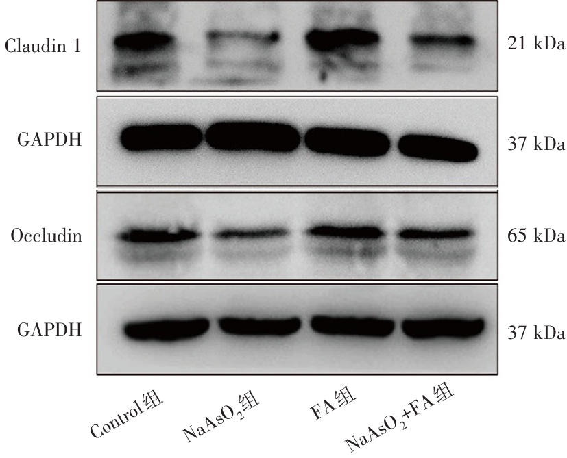
Fig.2
FA ameliorates arsenic poisoning-induced reduction of Claudin-1 and Occludin expression in wound tissue of mice"
Tab.5
The expression of tight junction proteins in the wound tissues of each group of mice (n = 5)"
| 紧密连接蛋白 | Control组 | NaAsO2组 | FA组 | NaAsO2+FA组 | F值 | P值 |
|---|---|---|---|---|---|---|
| Claudin-1 | 1.21 ± 0.16 | 0.22 ± 0.035* | 1.09 ± 0.26 | 0.66 ± 0.07# | 60.70 | 0.000 1 |
| Occludin | 1.15 ± 0.15 | 0.31 ± 0.04* | 0.97 ± 0.19 | 0.69 ± 0.08# | 49.57 | 0.000 1 |
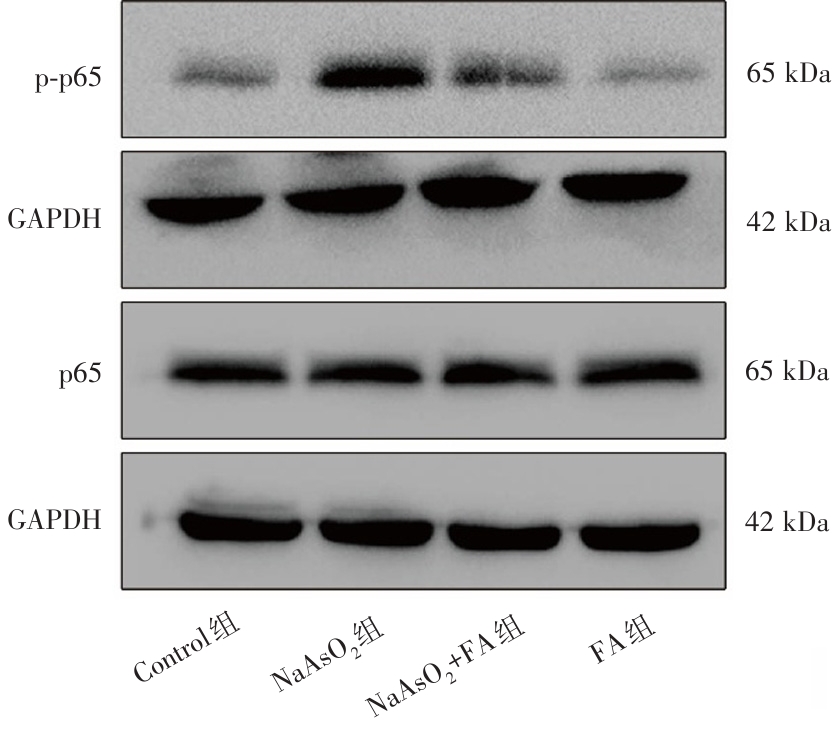
Fig.3
FA attenuates the activation of the NF-κB signaling pathway in arsenic-induced wound tissue in mice"
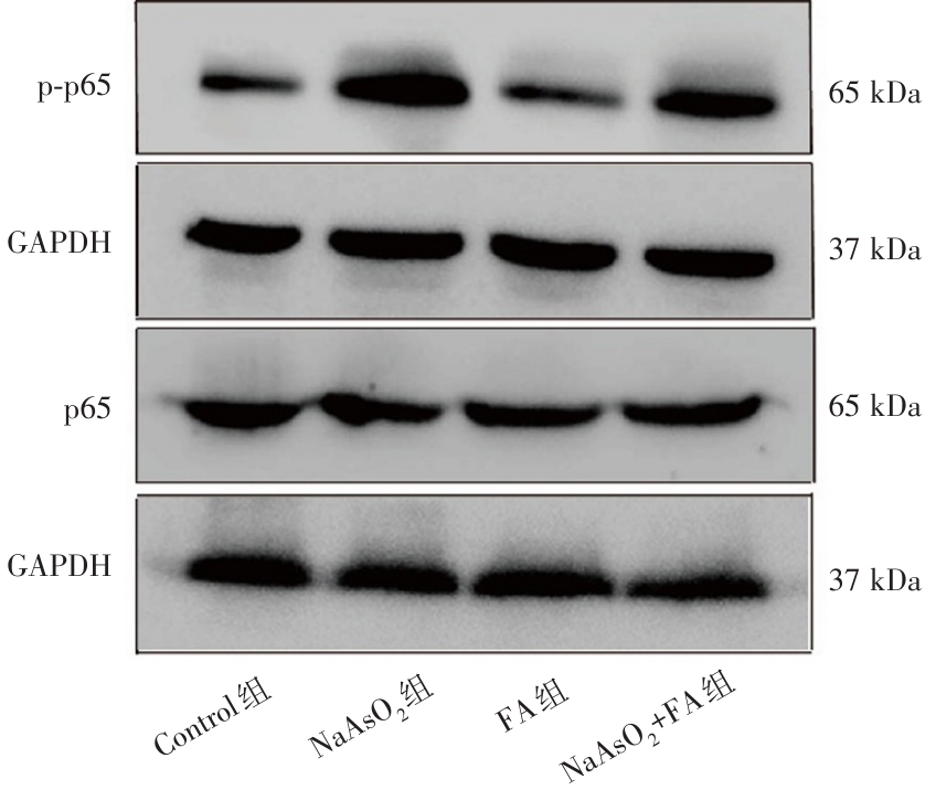
Fig 4
FA improves the reduced proliferation and migration capacity of HUVEC cells induced by arsenic"
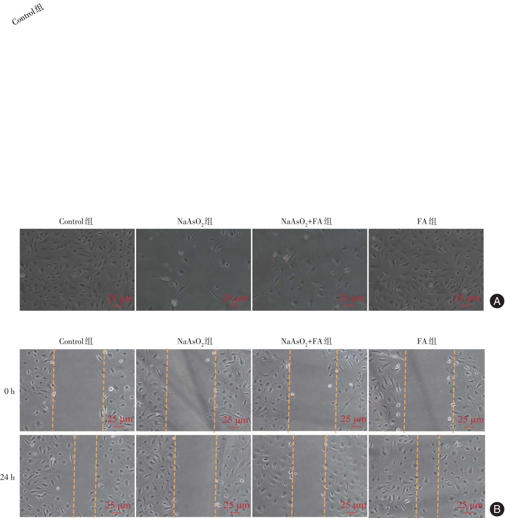
Tab.6
Proliferation activity of HUVEC cells in each group (n = 5)"
| 时间 | Control组 | NaAsO2组 | NaAsO2+FA组 | FA组 | F值 | P值 |
|---|---|---|---|---|---|---|
| 24 h | 1.01 ± 0.12 | 0.72 ± 0.04* | 0.86 ± 0.08 | 1.07 ± 0.23 | 13.35 | 0.000 1 |
| 48 h | 1.29 ± 0.07 | 0.73 ± 0.03* | 0.97 ± 0.05# | 1.28 ± 0.06 | 118.80 | 0.000 1 |
| 72 h | 1.50 ± 0.10 | 0.84 ± 0.03* | 1.18 ± 0.04# | 1.47 ± 0.04 | 102.10 | 0.000 1 |
Tab.7
Expression of inflammatory factors in HUVEC cells of each group (n = 5)"
| 炎性因子 | Control组 | NaAsO2组 | NaAsO2+FA组 | FA组 | F值 | P值 |
|---|---|---|---|---|---|---|
| IL-1β | 0.99 ± 0.13 | 3.36 ± 0.31* | 2.08 ± 0.24# | 0.93 ± 0.14 | 138.8 | 0.000 1 |
| IL-6 | 0.98 ± 0.12 | 2.91 ± 0.28* | 1.99 ± 0.17# | 0.91 ± 0.09 | 142.6 | 0.000 1 |
| TNF-α | 1.03 ± 0.16 | 2.83 ± 0.19* | 1.92 ± 0.18# | 1.00 ± 0.17 | 123.2 | 0.000 1 |
| IFN-γ | 0.97 ± 0.09 | 4.01 ± 0.33* | 2.76 ± 0.29# | 1.06 ± 0.17 | 183.7 | 0.000 1 |
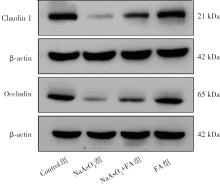
Fig.5
FA improves claudin-1 and occludin expression induced by arsenic in HUVECs"
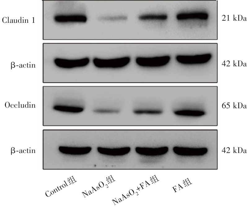
Tab.8
Expression of tight junction proteins in HUVEC cells of each group (n = 5)"
| 紧密连接蛋白 | Control组 | NaAsO2组 | NaAsO2+FA组 | FA组 | F值 | P值 |
|---|---|---|---|---|---|---|
| Claudin-1 | 1.03 ± 0.15 | 0.24 ± 0.07* | 0.52 ± 0.10# | 1.12 ± 0.13 | 56.35 | 0.000 1 |
| Occludin | 0.95 ± 0.14 | 0.20 ± 0.05* | 0.43 ± 0.04# | 0.99 ± 0.17 | 91.03 | 0.000 1 |
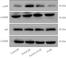
Fig.6
FA attenuates arsenic-induced activation of the NF-κB signaling pathway in HUVECs"
| [1] |
XU Y, TOKAR E J, PI J. Arsenic as an environmental toxicant and a therapeutic agent: Foe and friend[J]. Toxicol Appl Pharmacol, 2021, 415: 115438. doi:10.1016/j.taap.2021.115438 .
doi: 10.1016/j.taap.2021.115438 |
| [2] |
THAKUR M, RACHAMALLA M, NIYOGI S, et al. Molecular mechanism of arsenic-induced neurotoxicity including neuronal dysfunctions[J]. Int J Mol Sci, 2021, 22(18): 10077. doi:10.3390/ijms221810077 .
doi: 10.3390/ijms221810077 |
| [3] |
RAHAMAN M S, RAHMAN M M, MISE N, et al. Environmental arsenic exposure and its contribution to human diseases, toxicity mechanism and management[J]. Environ Pollut, 2021, 289: 117940. doi:10.1016/j.envpol.2021.117940 .
doi: 10.1016/j.envpol.2021.117940 |
| [4] |
SU Q, HE Y, PAN H, et al. Toxicity of inorganic arsenic to animals and its treatment strategies[J]. Comp Biochem Physiol Part C Toxicol Pharmacol, 2023, 271: 109654. doi:10.1016/j.cbpc. 2023.109654 .
doi: 10.1016/j.cbpc. 2023.109654 |
| [5] |
DRESLER S R, PINTO B I, SALANGA M C, et al. Arsenic impairs wound healing processes in dermal fibroblasts and mice[J]. Int J Mol Sci, 2024, 25(4): 2161. doi:10.3390/ijms25042161 .
doi: 10.3390/ijms25042161 |
| [6] |
GUO S, DIPIETRO L A. Factors affecting wound healing[J]. J Dent Res, 2010, 89(3): 219-229. doi:10.1177/0022034509359125 .
doi: 10.1177/0022034509359125 |
| [7] |
BARRIENTOS S, BREM H, STOJADINOVIC O, et al. Clinical application of growth factors and cytokines in wound healing[J]. Wound Repair Regen, 2014, 22(5): 569-578. doi:10.1111/wrr.12205 .
doi: 10.1111/wrr.12205 |
| [8] |
VOLKSDORF T, HEILMANN J, EMING S A, et al. Tight junction proteins claudin-1 and occludin are important for cutaneous wound healing[J]. Am J Pathol, 2017, 187(6): 1301-1312. doi:10.1016/j.ajpath.2017.02.006 .
doi: 10.1016/j.ajpath.2017.02.006 |
| [9] |
GENTRY P R, MCDONALD T B, SULLIVAN D E, et al. Analysis of genomic dose-response information on arsenic to inform key events in a mode of action for carcinogenicity[J]. Environ And Mol Mutagen, 2010, 51(1): 1-14. doi:10.1002/em.20505 .
doi: 10.1002/em.20505 |
| [10] |
SHI Y, WEI Y, QU S, et al. Arsenic induces apoptosis of human umbilical vein endothelial cells through mitochondrial pathways[J]. Cardiovasc Toxicol, 2010, 10(3): 153-160. doi:10.1007/s12012-010-9073-z .
doi: 10.1007/s12012-010-9073-z |
| [11] |
VERRET W J, CHEN Y, AHMED A, et al. A randomized, double-blind placebo-controlled trial evaluating the effects of vitamin E and selenium on arsenic-induced skin lesions in Bangladesh[J]. J Occup Environ Med, 2005, 47(10): 1026-1035. doi:10.1097/01.jom.0000183095.45050.97 .
doi: 10.1097/01.jom.0000183095.45050.97 |
| [12] |
PURUSHOTHAMAN J R, RIZWANULLAH M. Ferulic acid: A comprehensive review[J]. Cureus, 2024, 16(8): e68063. doi:10.7759/cureus.68063 .
doi: 10.7759/cureus.68063 |
| [13] |
KIM K H, JUNG J H, CHUNG W S, et al. Ferulic acid induces keratin 6α via inhibition of nuclear β-catenin accumulation and activation of Nrf2 in wound-induced inflammation[J]. Biomedicines, 2021, 9(5): 459. doi:10.3390/biomedicines9050459 .
doi: 10.3390/biomedicines9050459 |
| [14] | 邵帅, 高月, 刘建香, 等. 阿魏酸调节Thbd信号通路减轻血管内皮细胞辐射损伤的作用机制研究 [C] .重庆:中国毒理学会第七次全国会员代表大会暨中国毒理学会第六次中青年学者科技论坛, 2018. |
| [15] |
GHASEMI-DEHNOO M, AMINI-KHOEI H, LORIGOOINI Z, et al. Ferulic acid ameliorates ulcerative colitis in a rat model via the inhibition of two LPS-TLR4-NF-κB and NF-κB-INOS-NO signaling pathways and thus alleviating the inflammatory, oxidative and apoptotic conditions in the colon tissue[J]. Inflammopharmacology, 2023, 31(5): 2587-2597. doi:10.1007/s10787-023-01277-y .
doi: 10.1007/s10787-023-01277-y |
| [16] |
ZHANG D, JING B, CHEN Z N, et al. Ferulic acid alleviates sciatica by inhibiting neuroinflammation and promoting nerve repair via the TLR4/NF-κB pathway[J]. CNS Neurosci Ther, 2023, 29(4): 1000-1011. doi:10.1111/cns.14060 .
doi: 10.1111/cns.14060 |
| [17] |
AMBROZOVA N, ULRICHOVA J, GALANDAKOVA A. Models for the study of skin wound healing. The role of Nrf2 and NF-κB[J]. Biomed Pap Med Fac Univ Palacky Olomouc Czech Repub, 2017, 161(1): 1-13. doi:10.5507/bp.2016.063 .
doi: 10.5507/bp.2016.063 |
| [18] |
ZULKEFLI N, CHE ZAHARI C N M, SAYUTI N H, et al. Flavonoids as potential wound-healing molecules: Emphasis on pathways perspective[J]. Int J Mol Sci, 2023, 24(5): 4607. doi:10.3390/ijms24054607 .
doi: 10.3390/ijms24054607 |
| [19] |
GHAISAS M M, KSHIRSAGAR S B, SAHANE R S. Evaluation of wound healing activity of ferulic acid in diabetic rats[J]. Int Wound J, 2014, 11(5): 523-532. doi:10.1111/j.1742-481X. 2012.01119.x .
doi: 10.1111/j.1742-481X. 2012.01119.x |
| [20] |
LI S, MA J, LI J, et al. Sprayable self-assembly multifunctional bioactive poly(ferulic acid) hydrogel for rapid MRSA infected wound repair[J]. J Biomed Mater Res A, 2024, 112(3): 390-401. doi:10.1002/jbm.a.37636 .
doi: 10.1002/jbm.a.37636 |
| [21] |
ZENG Q, ZHANG A. Assessing potential mechanisms of arsenic-induced skin lesions and cancers: Human and in vitro evidence[J]. Environ Pollut, 2020, 260: 113919. doi:10.1016/j.envpol. 2020.113919 .
doi: 10.1016/j.envpol. 2020.113919 |
| [22] |
GUO Z, HU Q, TIAN J, et al. Proteomic profiling reveals candidate markers for arsenic-induced skin keratosis[J]. Environ Pollut, 2016, 218: 34-38. doi:10.1016/j.envpol.2016.08.035 .
doi: 10.1016/j.envpol.2016.08.035 |
| [23] |
PINTO B I, LUJAN O R, RAMOS S A, et al. Estrogen mitigates the negative effects of arsenic contamination in anIn VitroWound model[J]. Appl Vitro Toxicol, 2018, 4(1): 24-29. doi:10.1089/aivt.2017.0020 .
doi: 10.1089/aivt.2017.0020 |
| [24] |
SUZUKI A, NOMURA T, JOKURA H, et al. Beneficial effects of oral supplementation with ferulic acid, a plant phenolic compound, on the human skin barrier in healthy men[J]. Int J Vitam Nutr Res, 2023, 93(1): 54-60. doi:10.1024/0300-9831/a000699 .
doi: 10.1024/0300-9831/a000699 |
| [25] |
KIM J, KIM J, LEE Y I, et al. Effect of a topical antioxidant serum containing vitamin C, vitamin E, and ferulic acid after Q-switched 1064-nm Nd: YAG laser for treatment of environment-induced skin pigmentation[J]. J Cosmet Dermatol, 2020, 19(10): 2576-2582. doi:10.1111/jocd.13323 .
doi: 10.1111/jocd.13323 |
| [26] |
PEÑA O A, MARTIN P. Cellular and molecular mechanisms of skin wound healing[J]. Nat Rev Mol Cell Biol, 2024, 25(8): 599-616. doi:10.1038/s41580-024-00715-1 .
doi: 10.1038/s41580-024-00715-1 |
| [27] | 方兴艳, 田侦丽, 赵哲仪, 等. 亚砷酸钠对人脐静脉内皮细胞损伤及鞘氨醇激酶1/1-磷酸鞘氨醇信号轴的影响[J]. 中国组织工程研究, 2023, 27(26): 4161-4167. |
| [28] |
ZHU X, HUANG B, ZHAO F, et al. p38-mediated FOXN3 phosphorylation modulates lung inflammation and injury through the NF-κB signaling pathway[J]. Nucleic Acids Res, 2023, 51(5): 2195-2214. doi:10.1093/nar/gkad057 .
doi: 10.1093/nar/gkad057 |
| [29] |
WANG P H, HUANG B S, HORNG H C, et al. Wound healing[J]. J Chin Med Assoc, 2018, 81(2): 94-101. doi:10.1016/j.jcma.2017.11.002 .
doi: 10.1016/j.jcma.2017.11.002 |
| [30] |
GAN R, LIU H, WU S, et al. Curcumin alleviates arsenic trioxide-induced inflammation and pyroptosis via the NF-κB/NLRP3 signaling pathway in the hypothalamus of ducks[J]. Biol Trace Elem Res, 2023, 201(5): 2503-2511. doi:10.1007/s12011-022-03321-4 .
doi: 10.1007/s12011-022-03321-4 |
| [31] |
ZHAO D, JIAO S, YI H. Arsenic exposure induces small intestinal toxicity in mice by barrier damage and inflammation response via activating RhoA/ROCK and TLR4/Myd88/NF-κB signaling pathways[J]. Toxicol Lett, 2023, 384: 44-51. doi:10.1016/j.toxlet.2023.07.007 .
doi: 10.1016/j.toxlet.2023.07.007 |
| [32] |
WEI Z, XUE Y, XUE Y, et al. Ferulic acid attenuates non-alcoholic steatohepatitis by reducing oxidative stress and inflammation through inhibition of the ROCK/NF-κB signaling pathways[J]. J Pharmacol Sci, 2021, 147(1): 72-80. doi:10.1016/j.jphs. 2021.05.006 .
doi: 10.1016/j.jphs. 2021.05.006 |
| [33] |
HUANG C, HUANGFU C, BAI Z, et al. Multifunctional carbomer based ferulic acid hydrogel promotes wound healing in radiation-induced skin injury by inactivating NLRP3 inflammasome[J]. J Nanobiotechnology, 2024, 22(1): 576. doi:10.1186/s12951-024-02789-7 .
doi: 10.1186/s12951-024-02789-7 |
| Viewed | ||||||
|
Full text |
|
|||||
|
Abstract |
|
|||||
