

The Journal of Practical Medicine ›› 2024, Vol. 40 ›› Issue (23): 3306-3316.doi: 10.3969/j.issn.1006-5725.2024.23.005
• Basic Research • Previous Articles
Xiang JIA1,2,Tianjie XU1,2,Jiaxin FAN1,2,Xiaoling GUO1,2,Kainan LIU3,Hui ZHANG4,Yongsheng WANG1,2,Qian. WANG1,2( )
)
Received:2024-08-23
Online:2024-12-10
Published:2024-12-16
Contact:
Qian. WANG
E-mail:tswqxx008@126.com
CLC Number:
Xiang JIA,Tianjie XU,Jiaxin FAN,Xiaoling GUO,Kainan LIU,Hui ZHANG,Yongsheng WANG,Qian. WANG. Metformin exerts a protective effect on articular cartilage in osteoarthritis rats by activating the SIRT1/p53 signaling pathway[J]. The Journal of Practical Medicine, 2024, 40(23): 3306-3316.
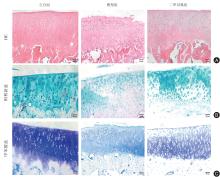
Fig.1
Observation of knee cartilage in rats of each group (× 200)"
Tab.1
OARSI score of rats"
| 组别 | 例数 | 分数 |
|---|---|---|
| 空白组 | 3 | 0.000 ± 0.000 |
| 模型组 | 3 | 9.667 ± 2.082? |
| 二甲双胍组 | 3 | 3.333 ± 1.528# |
| F值 | 33.650 | |
| P值 | 0.001 |
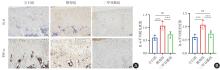
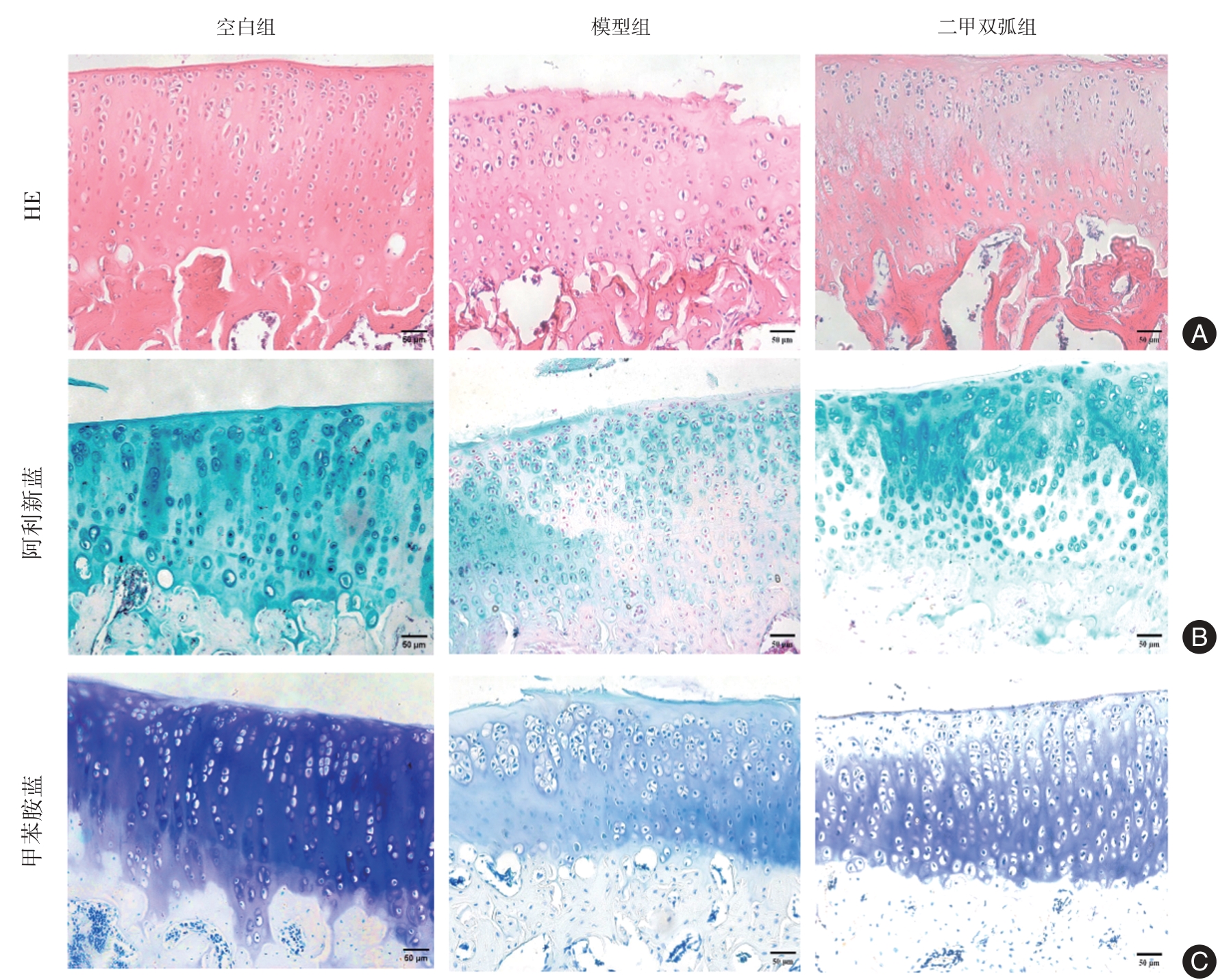
Fig.2
Immunohistochemical staining results of IL-6 and TNF-α in rat cartilage tissue (× 200)"

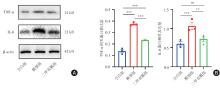
Fig.3
Western blot results of IL-6 and TNF-α in rat cartilage tissue"
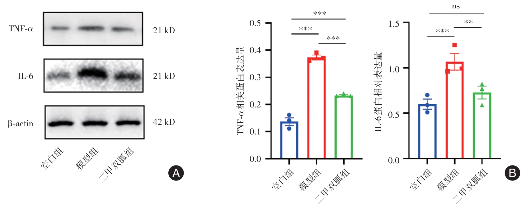
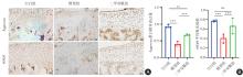
Fig.4
Immunohistochemical staining results of Aggrecan and SOX9 in rat cartilage tissue (× 200)"

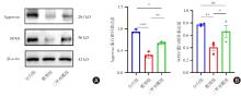
Fig.5
Western blot results of Aggrecan and SOX9 in rat cartilage"
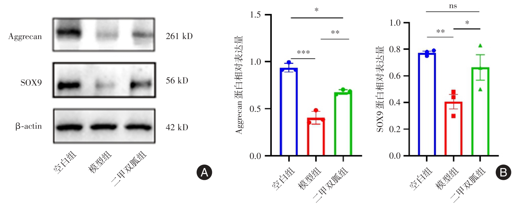
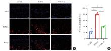
Fig.6
TUNEL fluorescence staining results in rat cartilage tissue (× 200)"

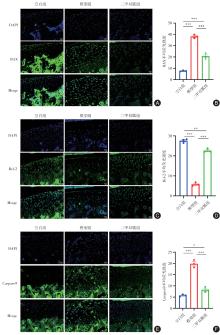
Fig. 7
Immunofluorescence staining results of BAX, Bcl-2 and Caspase-9 in rat cartilage tissue (× 200)"
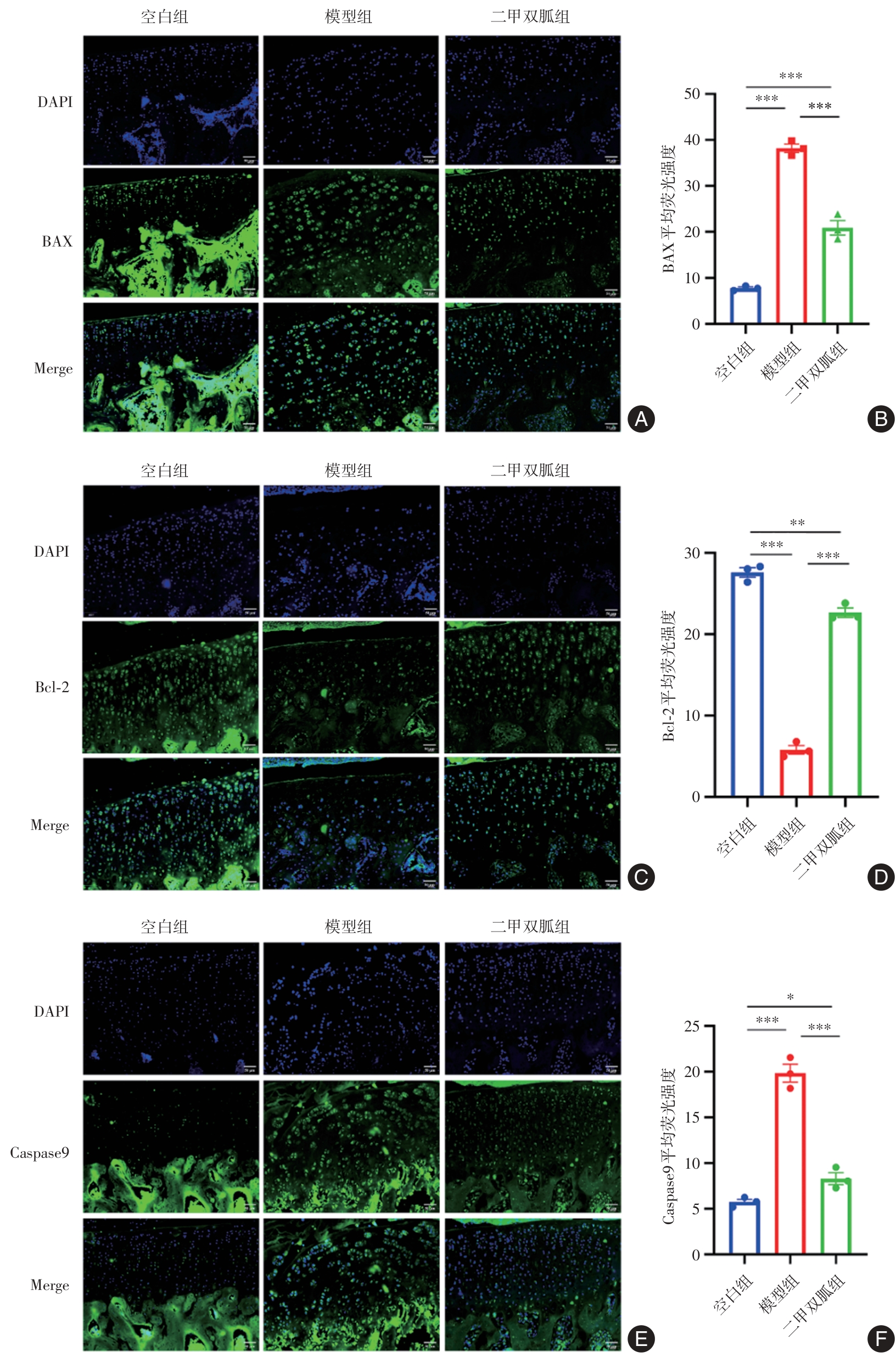
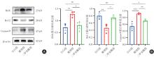
Fig.8
Western blot results of BAX, Bcl-2 and Caspase-9 in rat cartilage tissue"
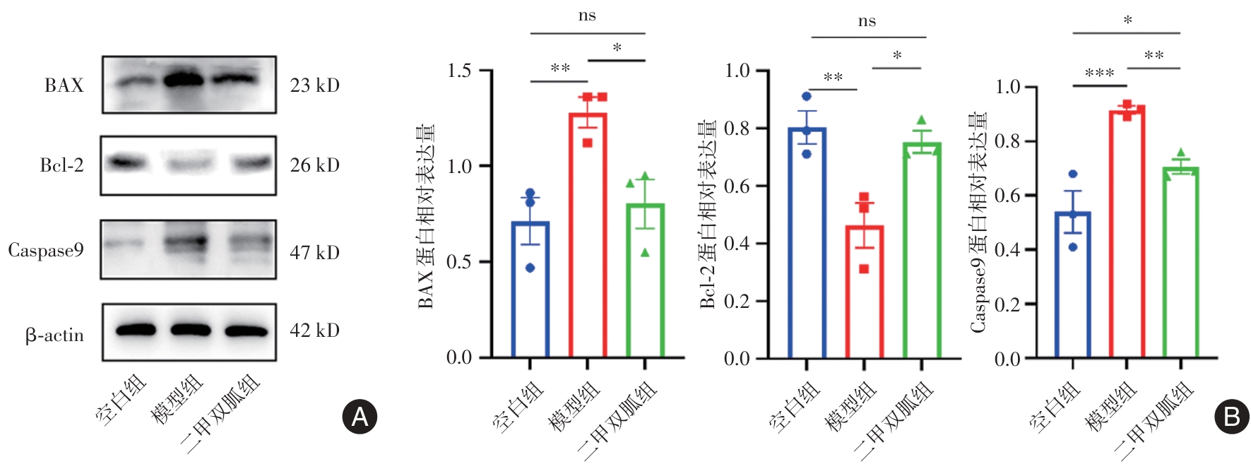
Fig.9
Immunohistochemical staining results of SIRT1 and p53 in rat cartilage tissue (× 200)"
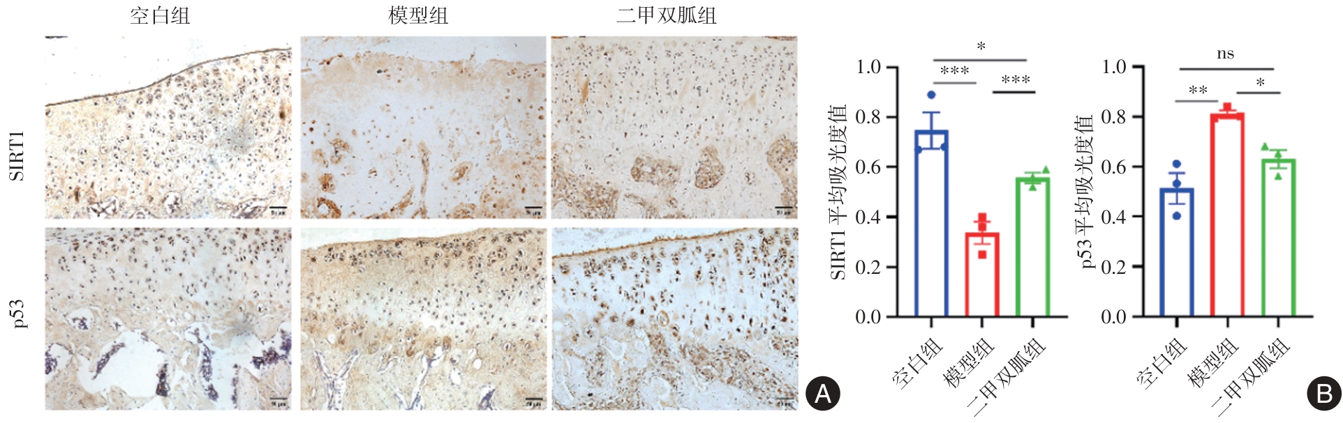
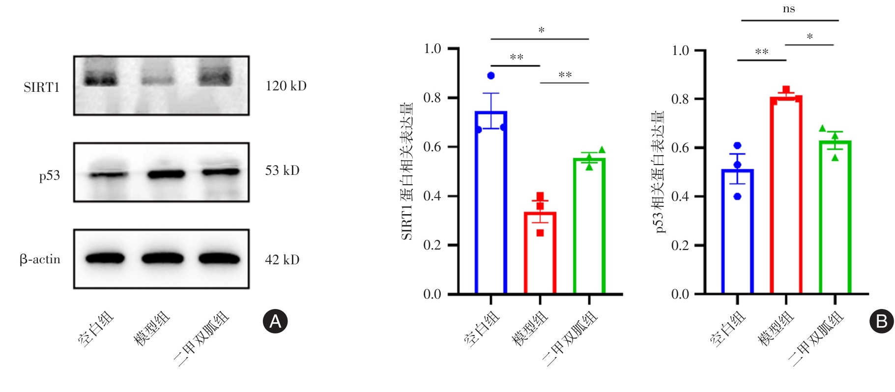
Fig.10
Western blot results of SIRT1 and p53 in rat cartilage tissue"
| 1 |
DIAMOND L E, GRANT T, UHLRICH S D. Osteoarthritis year in review 2023: Biomechanics[J]. Osteoarthritis Cartilage, 2024, 32(2): 138-147. doi:10.1016/j.joca.2023.11.015
doi: 10.1016/j.joca.2023.11.015 |
| 2 |
MINNIG M C C, GOLIGHTLY Y M, NELSON A E. Epidemiology of osteoarthritis: Literature update 2022-2023[J]. Curr Opin Rheumatol, 2024, 36(2): 108-112. doi:10.1097/bor.0000000000000985
doi: 10.1097/bor.0000000000000985 |
| 3 | PERRUCCIO A V, YOUNG J J, WILFONG J M, 等. Osteoarthritis year in review 2023: Epidemiology & therapy[J]. Osteoarthritis Cartilage, 2024, 32(2): 159-165. |
| 4 |
XU X, SUN Y, CEN X, et al. Metformin activates chaperone-mediated autophagy and improves disease pathologies in an Alzheimer disease mouse model[J]. Protein Cell, 2021, 12(10): 769-787. doi:10.1007/s13238-021-00858-3
doi: 10.1007/s13238-021-00858-3 |
| 5 |
FORETZ M, GUIGAS B, VIOLLET B. Metformin: Update on mechanisms of action and repurposing potential[J]. Nat Rev Endocrinol, 2023, 19(8): 460-476. doi:10.1038/s41574-023-00833-4
doi: 10.1038/s41574-023-00833-4 |
| 6 | 田珂, 冷秋枫, 吕晶, 等. 二甲双胍通过NLRP3炎症小体通路对皮肤角质形成细胞增殖和凋亡的双向调节研究[J]. 中国全科医学, 2025,28(6):742-750. |
| 7 |
KUSWANTO W, BAKER M C. Repurposing drugs for the treatment of osteoarthritis[J]. Osteoarthritis Cartilage, 2024, 32(8): 886-895. doi:10.1016/j.joca.2024.05.008
doi: 10.1016/j.joca.2024.05.008 |
| 8 |
HE M, LU B, OPOKU M, et al. Metformin Prevents or Delays the Development and Progression of Osteoarthritis: New Insight and Mechanism of Action[J]. Cells, 2022, 11(19): 3012. doi:10.3390/cells11193012
doi: 10.3390/cells11193012 |
| 9 |
CHEN C, ZHOU M, GE Y, et al. SIRT1 and aging related signaling pathways[J]. Mech Ageing Dev, 2020, 187: 111215. doi:10.1016/j.mad.2020.111215
doi: 10.1016/j.mad.2020.111215 |
| 10 |
CHEN L, HUADONG Z, YANQING L, et al. Novel Role of the SIRT1 in Endocrine and Metabolic Diseases[J]. Int J Biol Sci, 2023, 19(2):484-501. doi:10.7150/ijbs.78654
doi: 10.7150/ijbs.78654 |
| 11 | 贲莹, 张天雅, 田佳鑫, 等. 基于SIRT1/p53介导的细胞凋亡途径探讨补阳还五汤对糖尿病周围神经病变的治疗作用及方中黄芪用量[J]. 中国实验方剂学杂志, 2022, 28(2): 1-10. |
| 12 |
ZHOU M, LIU B, YE H M, et al. ROS-induced imbalance of the miR-34a-5p/SIRT1/p53 axis triggers chronic chondrocyte injury and inflammation[J]. Heliyon, 2024, 10(11): e31654. doi:10.1016/j.heliyon.2024.e31654
doi: 10.1016/j.heliyon.2024.e31654 |
| 13 | 冯晓峰, 张荣凯, 祁伟仲, 等. 二甲双胍干预骨关节炎模型小鼠早期骨关节炎软骨及软骨下骨变化[J]. 中国组织工程研究, 2019, 23(19): 3031-3036. |
| 14 |
LI J, ZHANG B, LIU W X, et al. Metformin limits osteoarthritis development and progression through activation of AMPK signalling[J]. Ann Rheum Dis, 2020, 79(5): 635-645. doi:10.1136/annrheumdis-2019-216713corr1
doi: 10.1136/annrheumdis-2019-216713corr1 |
| 15 | 徐田杰, 樊佳欣, 郭小玲, 等. 二甲双胍抑制PI3K/AKT/mTOR信号通路保护骨关节炎模型大鼠关节软骨[J]. 中国组织工程研究, 2025, 29(5): 1003-1012. |
| 16 |
LI D, RUAN G, ZHANG Y, et al. Metformin attenuates osteoarthritis by targeting chondrocytes, synovial macrophages and adipocytes[J]. Rheumatology(Oxford), 2023, 62(4): 1652-1661. doi:10.1093/rheumatology/keac467
doi: 10.1093/rheumatology/keac467 |
| 17 | 许学猛, 刘文刚, 许树柴, 等. 膝骨关节炎(膝痹)中西医结合临床实践指南[J]. 实用医学杂志, 2021, 37(22): 2827-2833. |
| 18 |
YAO Q, WU X, TAO C, et al. Osteoarthritis: Pathogenic signaling pathways and therapeutic targets[J]. Signal Transduct Target Ther, 2023, 8(1):56. doi:10.1038/s41392-023-01330-w
doi: 10.1038/s41392-023-01330-w |
| 19 |
TONG L, YU H, HUANG X, et al. Current understanding of osteoarthritis pathogenesis and relevant new approaches[J]. Bone Res, 2022, 10(1):60. doi:10.1038/s41413-022-00226-9
doi: 10.1038/s41413-022-00226-9 |
| 20 |
JONES I A, TOGASHI R, WILSON M L, et al. Intra-articular treatment options for knee osteoarthritis[J]. Nat Rev Rheumatol, 2019, 15(2): 77-90. doi:10.1038/s41584-018-0123-4
doi: 10.1038/s41584-018-0123-4 |
| 21 |
LI D, RUAN G, ZHANG Y, et al. Metformin attenuates osteoarthritis by targeting chondrocytes, synovial macrophages and adipocytes[J]. Rheumatology (Oxford), 2023, 62(4): 1652-1661. doi:10.1093/rheumatology/keac467
doi: 10.1093/rheumatology/keac467 |
| 22 |
ZAKI S, BLAKER C L, LITTLE C B. OA foundations-Exper-imental models of osteoarthritis[J]. Osteoarthritis Cartilage, 2022, 30(3): 357-380. doi:10.1016/j.joca.2021.03.024
doi: 10.1016/j.joca.2021.03.024 |
| 23 |
SZYMCZAK-PAJOR I, WENCLEWSKA S, ŚLIWIŃSKA A. Metabolic Action of Metformin[J]. Pharmaceuticals(Basel), 2022, 15(7): 810. doi:10.3390/ph15070810
doi: 10.3390/ph15070810 |
| 24 |
FENG X, PAN J, LI J, et al. Metformin attenuates cartilage degeneration in an experimental osteoarthritis model by regulating AMPK/mTOR[J]. Aging(Alany NY), 2020, 12(2): 1087-1103. doi:10.18632/aging.102635
doi: 10.18632/aging.102635 |
| 25 | 黄夏荣, 周君, 孙光华, 等. 电针对老年大鼠关节软骨及软骨下骨极化相关蛋白表达的影响[J]. 实用医学杂志, 2023, 39(12): 1473-1479. |
| 26 |
FUJII Y, LIU L, YAGASAKI L, et al. Cartilage Homeostasis and Osteoarthritis[J]. In J Mol Scis, 2022, 23(11): 6316. doi:10.3390/ijms23116316
doi: 10.3390/ijms23116316 |
| 27 |
CHEN Y, QIU F, YU B, et al. Metformin, an AMPK Activator, Inhibits Activation of FLSs but Promotes HAPLN1 Secretion[J]. Mol Ther Methods Clin Dev, 2020, 17: 1202-1214. doi:10.1016/j.omtm.2020.05.008
doi: 10.1016/j.omtm.2020.05.008 |
| 28 |
TYLUTKA A, WALAS Ł, ZEMBRON-LACNY A. Level of IL-6, TNF, and IL-1β and age-related diseases: A systematic review and meta-analysis[J]. Front Immunol, 2024, 15: 1330386. doi:10.3389/fimmu.2024.1330386
doi: 10.3389/fimmu.2024.1330386 |
| 29 |
NEGISHI Y, ADILI A, DE VEGA S, et al. IL-6 Reduces Spheroid Sizes of Osteophytic Cells Derived from Osteoarthritis Knee Joint via Induction of Apoptosis[J]. Am J Pathol, 2024, 194(1): 135-149. doi:10.1016/j.ajpath.2023.10.005
doi: 10.1016/j.ajpath.2023.10.005 |
| 30 |
WANG L, HE C. Nrf2-mediated anti-inflammatory polarization of macrophages as therapeutic targets for osteoarthritis[J]. Front Immunol, 2022, 13:967193. doi:10.3389/fimmu.2022.967193
doi: 10.3389/fimmu.2022.967193 |
| 31 |
NILSSON N, ALIM M D A, DIETRICH-ZAGONEL F, et al. The Delayed Presentation of Achilles Tendon Ruptures Is Associated With Marked Alterations in the Gene Expression of COL1A1, MMPs, TIMPs, and IL-6[J]. Am J Sports Med, 2024, 52(1): 164-173. doi:10.1177/03635465231212669
doi: 10.1177/03635465231212669 |
| 32 | 刘子歌, 陈德胜. 破骨细胞因子和抗破骨细胞因子在骨代谢调控网络中作用的研究进展[J]. 医学研究杂志, 2024, 53(6): 175-178. |
| 33 |
TANG H, GONG X, DAI J, et al. The IRF1/GBP5 axis promotes osteoarthritis progression by activating chondrocyte pyroptosis[J]. J Orthop Translat, 2024, 44: 47-59. doi:10.1016/j.jot.2023.11.005
doi: 10.1016/j.jot.2023.11.005 |
| 34 |
ZHOU Z, LV C, WANG Y, et al. BuShen JianGu Fang alleviates cartilage degeneration via regulating multiple genes and signaling pathways to activate NF-κB/Sox9 axis[J]. Phytomedicine, 2023, 113: 154742. doi:10.1016/j.phymed.2023.154742
doi: 10.1016/j.phymed.2023.154742 |
| 35 | 谭清梅, 杨诚, 廖坚文, 等. 二甲双胍通过AMPK信号通路对小鼠骨关节炎保护作用的研究[J]. 中国临床解剖学杂志, 2017, 35(4): 413-418. |
| 36 | 丁丽宏, 耿世佳, 王玉杰. 蟛蜞菊内酯对肺炎链球菌感染的肺泡上皮细胞凋亡及炎症因子分泌的调节作用[J]. 实用医学杂志, 2024, 40(3): 316-320. |
| 37 | 尹路, 蒋川锋, 陈俊杰, 等. 沙苑子苷A对关节软骨细胞凋亡的影响[J]. 中国组织工程研究, 2025, 29(8): 1541-1547. |
| 38 |
DADSENA S, CUEVAS ARENAS R, VIEIRA G, et al. Lipid unsaturation promotes BAX and BAK pore activity during apoptosis[J]. Nat Commun, 2024, 15(1): 4700. doi:10.1038/s41467-024-49067-6
doi: 10.1038/s41467-024-49067-6 |
| 39 |
VANDENABEELE P, BULTYNCK G, SAVVIDES S N. Pore-forming proteins as drivers of membrane permeabilization in cell death pathways[J]. Nat Rev Mol Cell Biol, 2023, 24(5): 312-333. doi:10.1038/s41580-022-00564-w
doi: 10.1038/s41580-022-00564-w |
| 40 | 李田洋, 高小凤, 王宝娟, 等. 骨炎消巴布剂对膝骨关节炎家兔软骨细胞凋亡及Bcl-2、Bax表达的影响[J]. 中国免疫学杂志, 2023, 39(8): 1647-1652. |
| 41 |
AI Y, MENG Y, YAN B, et al. The biochemical pathways of apoptotic, necroptotic, pyroptotic, and ferroptotic cell death[J]. Mol Cell, 2024, 84(1): 170-179. doi:10.1016/j.molcel.2023.11.040
doi: 10.1016/j.molcel.2023.11.040 |
| 42 |
NOGALES C, MAMDOUH Z M, LIST M, et al. Network pharmacology: Curing causal mechanisms instead of treating symptoms[J]. Trends Pharmacol Sci, 2022, 43(2): 136-150. doi:10.1016/j.tips.2021.11.004
doi: 10.1016/j.tips.2021.11.004 |
| 43 | 曾红玉, 叶贵珊, 武琦, 等. OXSR1活性对p53依赖和非依赖途径介导的犬肾细胞凋亡的影响[J]. 中国畜牧兽医, 2024,51(10): 4222-4234. |
| 44 |
YANG Y, LIU Y, WANG Y, et al. Regulation of SIRT1 and Its Roles in Inflammation[J]. Front Immunol, 2022, 13: 831168. doi:10.3389/fimmu.2022.831168
doi: 10.3389/fimmu.2022.831168 |
| 45 |
XU Y, WAN W. Acetylation in the regulation of autophagy[J]. Autophagy, 2023, 19(2): 379-387. doi:10.1080/15548627.2022.2062112
doi: 10.1080/15548627.2022.2062112 |
| 46 |
LI M, HU J, ZHOU J, et al. Grass carp (Ctenopharyngodon idella) deacetylase SIRT1 targets p53 to suppress apoptosis in a KAT8 dependent or independent manner[J]. Fish Shellfish Immunol, 2024, 144: 109264. doi:10.1016/j.fsi.2023.109264
doi: 10.1016/j.fsi.2023.109264 |
| [1] | Shan LUO,Ying FENG,Dandan FAN,Wenxin ZHENG,Xingrong GUO,Xuzhi. RUAN. ANGPTL8 knockout reduces lipopolysaccharide⁃induced hepatic lipid deposition [J]. The Journal of Practical Medicine, 2024, 40(9): 1197-1203. |
| [2] | Wei HE,Liping LIU,Jingwei ZHUO,Xiaodong ZHANG,Tong YANG,Jubin. FENG. CCR5 blockade reduces tumor growth by inducing apoptosis and impairing immunosuppression of tumor microenvironment [J]. The Journal of Practical Medicine, 2024, 40(9): 1204-1210. |
| [3] | Shangzeng WANG,Bei ZHANG,Zhen WANG,Shang MA,Deyang RUANGZHANG,Zhiying YIN,Yunqi ZHU,Kunpeng HU,Shao CHENG. Comparison of the effects of CR and PS prostheses in the treatment of knee osteoarthritis [J]. The Journal of Practical Medicine, 2024, 40(9): 1251-1256. |
| [4] | Wenxin LI,Minjun LU,Li LIN,Yueqin LIU,Xiaolan. ZHU. circRAF1 regulates the proliferation and apoptosis of human ovarian granulosa cells [J]. The Journal of Practical Medicine, 2024, 40(7): 910-917. |
| [5] | Zhen YANG,Shaoru JIANG,Xiaoyan CHEN,Xiaolin CHEN,Weimin DENG,Xinyu. GUO. Effect of Jinghou Zengzhi Granules on ovarian GDF9 secretion and granulosa cells apoptosis in controlled ovarian hyperstimulation rats [J]. The Journal of Practical Medicine, 2024, 40(7): 918-923. |
| [6] | Ying ZHOU,Dajun JIANG,Yong TIAN,Yongxiang GU,Guohui. YANG. Inhibition of TRAF6 ameliorates myocardial inflammatory injury and cardiac dysfunction via regulating cardiomyocyte inflammation in sepsis mice [J]. The Journal of Practical Medicine, 2024, 40(5): 608-614. |
| [7] | Xiaoyan WANG,Xiaoyi ZOU,Xiang ZHU,Ting WANG,Yetao QIANG,Siyuan ZHOU,Peng ZHANG,Ping ZHANG. Iron overload regulates atherosclerotic activity of foam cells induced by oxLDL [J]. The Journal of Practical Medicine, 2024, 40(3): 295-301. |
| [8] | Runwei MA,Chunjie MU,Wenting GUI,Yao DENG,Minzhang ZHAO,Min LIU,Yi SONG. LncRNA SENCR targeted miR⁃206 regulates proliferation and apoptosis of human vascular smooth muscle cells of aortic dissection tissues [J]. The Journal of Practical Medicine, 2024, 40(3): 302-308. |
| [9] | Lihong DING,Shijia GENG,Yujie WANG. Effects of wedelobata on apoptosis and secretion of inflammatory cytokines in the alveolar epithelium infected by Streptococcus pneumonia [J]. The Journal of Practical Medicine, 2024, 40(3): 316-320. |
| [10] | Chunfang KONG,Anna LI,Bo KE,Weirong DING,Tingting LIU,Huan FU,Tingting ZHANG,Chenghao JIN,Mei. WU. The inhibitory effect and molecular mechanism of 6-gingerol on human multiple myeloma cells [J]. The Journal of Practical Medicine, 2024, 40(23): 3291-3297. |
| [11] | Bing′er WU,Qing LI,Kerong YANG,Jian ZHANG,Yi YU,Lei LEI,Bo. HU. Impact of spermidine on proliferation and apoptosis in diffuse large B⁃cell lymphoma cell lines [J]. The Journal of Practical Medicine, 2024, 40(22): 3130-3137. |
| [12] | Zhe SHI,Xialin ZUO,Linhui PENG,Zhiwei LU,Kongping. LI. Effect of M1 microglial polarization on secondary damage in the thalamus after cerebral cortical infarction [J]. The Journal of Practical Medicine, 2024, 40(22): 3138-3145. |
| [13] | Shengnan JIANG,Wenbing ZHI,Jing CHEN,Tingting SUN,Zongren XU,Shuai LIU,Hong ZHANG,Ye LI,Yang LIU. Sophora davidii Hance leaves total alkaloids Attenuate Lipopolysaccharide-induced inflammatory response in RAW264.7 cell by Inhibiting the MAPK/NF-κB signaling pathway [J]. The Journal of Practical Medicine, 2024, 40(20): 2835-2840. |
| [14] | Guojiang YIN,Bixi LI,Pengxiao WEI,Yuqin YAN,Xiaoyang SONG,Kun LI. Effect of anterior quadratus lumborum block at the lateral supra⁃arcuate ligament on postoperative analgesia and inflammatory response in elderly patients undergoing robot⁃assisted radical prostatectomy [J]. The Journal of Practical Medicine, 2024, 40(2): 202-206. |
| [15] | Chunmei ZHANG,Xinhui HUANG,Jinqiu HU,Xiaoyan BI,Fuli. YA. Sulforaphane protects human platelets from high glucose⁃induced cellular apoptosis through down-regulating PI3K/Akt signaling pathway [J]. The Journal of Practical Medicine, 2024, 40(18): 2530-2536. |
| Viewed | ||||||
|
Full text |
|
|||||
|
Abstract |
|
|||||