

The Journal of Practical Medicine ›› 2026, Vol. 42 ›› Issue (8): 1301-1311.doi: 10.3969/j.issn.1006-5725.2026.08.001
• Chronic Disease Control • Next Articles
Xun ZHANG1,Juan XIAO1,Chaochao YU2,Zhaoxie YU1,Zhipeng FENG1,Yankun PAN1,Shuo YANG1,Feng SHEN1,3,4,5( )
)
Received:2025-11-21
Online:2026-04-25
Published:2026-04-28
Contact:
Feng SHEN
E-mail:40052958@qq.com
CLC Number:
Xun ZHANG,Juan XIAO,Chaochao YU,Zhaoxie YU,Zhipeng FENG,Yankun PAN,Shuo YANG,Feng SHEN. The effect of electroacupuncture at the Heart-Pericardium Meridian points on the brain functional network connectivity in APP/PS1 mice[J]. The Journal of Practical Medicine, 2026, 42(8): 1301-1311.
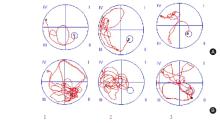
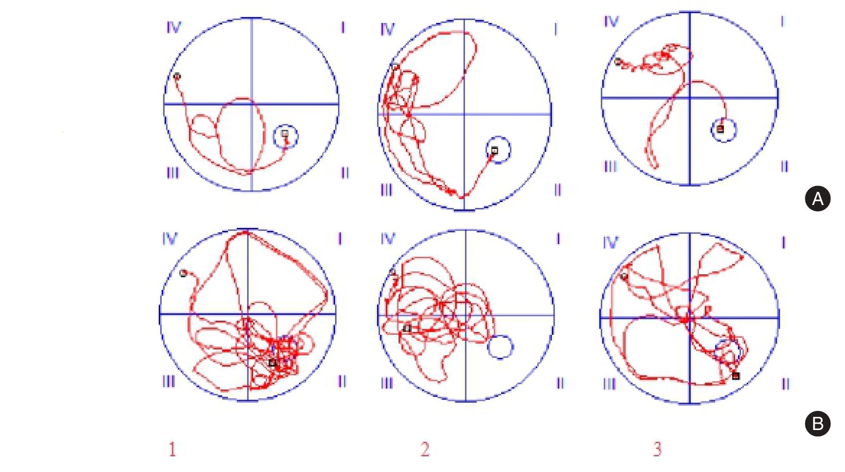
Fig.1
Morris water maze trajectory diagrams of mice in each group"
Tab.1
Comparison of latency in each group of mice(n = 6)"
| 组别 | 第1天 | 第2天 | 第3天 | 第4天 | 第5天 |
|---|---|---|---|---|---|
| 对照组 | 55.90 ± 4.12 | 38.83 ± 4.84 | 29.89 ± 3.39 | 18.79 ± 4.26 | 6.62 ± 2.78 |
| 模型组 | 58.22 ± 2.71 | 52.70 ± 5.61* | 46.88 ± 4.23* | 39.64 ± 3.74* | 28.41 ± 4.26* |
| 电针组 | 57.77 ± 3.88 | 39.75 ± 5.22# | 31.11 ± 2.64# | 19.65 ± 4.59# | 6.97 ± 3.14# |
| F值 | 0.836 | 7.972 | 44.375 | 30.566 | 41.336 |
| P值 | 0.455 | < 0.01 | < 0.01 | < 0.01 | < 0.01 |
Tab.2
Comparison of platform crossing times and target quadrant exploration time (n = 6)"
| 8.00 ± 1.41 | 36.25 ± 5.17 | |
| 2.00 ± 0.89* | 10.62 ± 3.08* | |
| 6.50 ± 2.07# | 35.14 ± 7.20# | |
| 24.718 | 42.884 | |
| < 0.01 | < 0.01 |

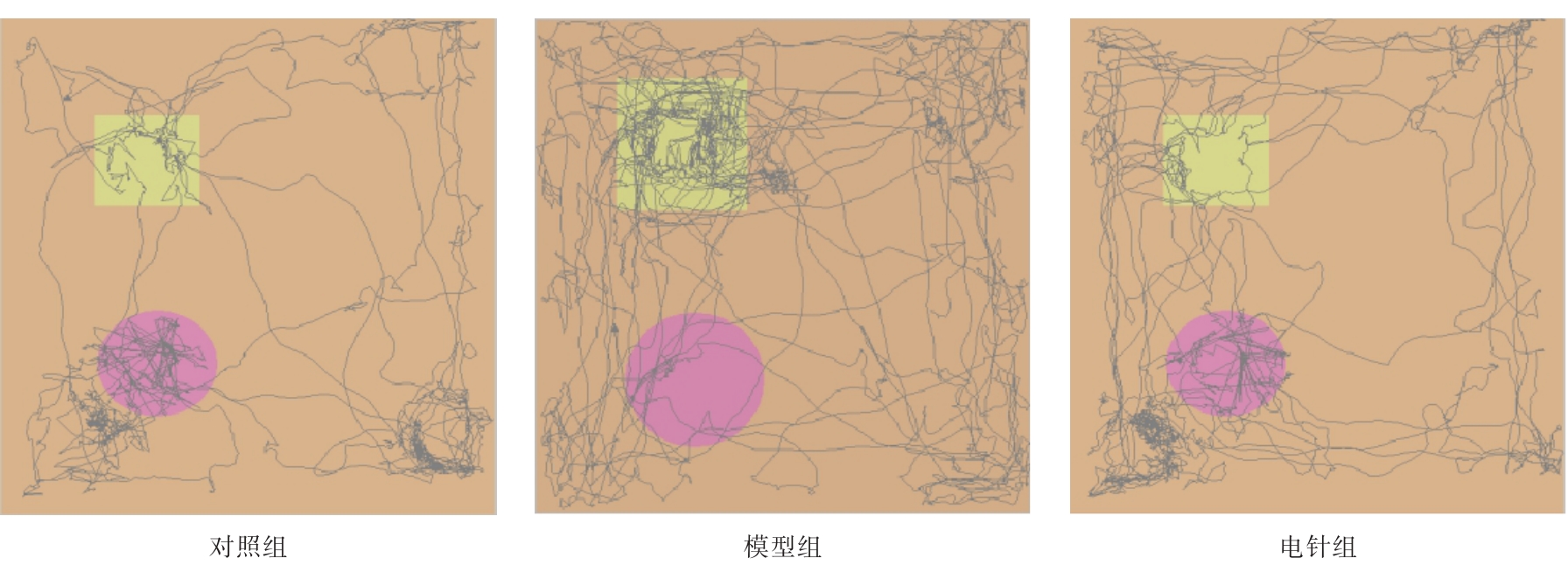
Fig.2
Shows the experimental trajectories of the new object in each group of mice"
Tab.3
Comparison of cognitive index of mice in each group (n = 6)"
| 组别 | 认知指数 |
|---|---|
| 对照组 | 68.58 ± 9.38 |
| 模型组 | 33.39 ± 4.74* |
| 电针组 | 66.59 ± 9.38# |
| F值 | 35.43 |
| P值 | < 0.01 |
Fig.3
The comparison results of functional connectivity between the model group and the control group(with the right hippocampus as the ROI)"
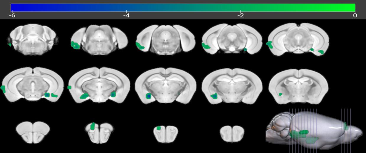
Fig.4
The comparison results of functional connectivity between the model group and the control group(with the left hippocampus as the ROI)"


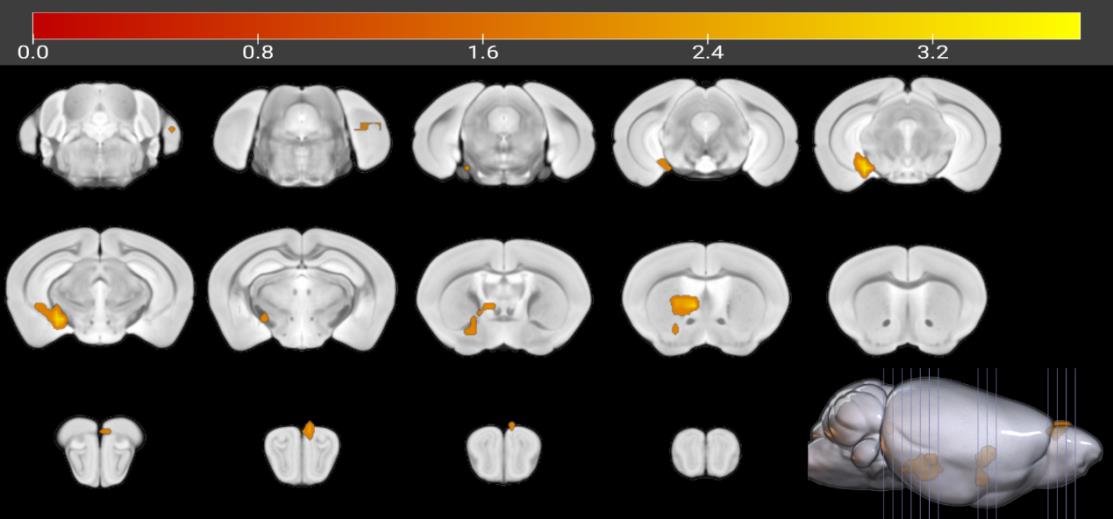
Fig.5
The comparison results of functional connectivity between the electroacupuncture group and the model group(with the right hippocampus as the ROI)"
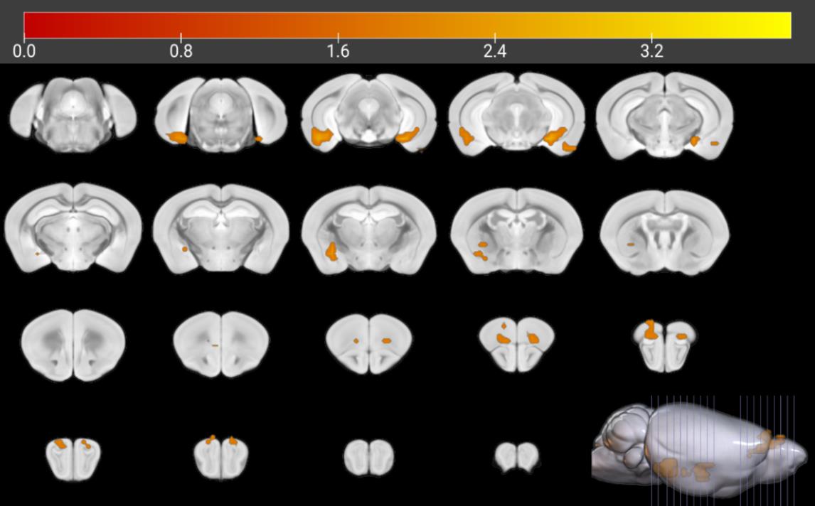
Fig.6
Shows the comparison results of functional connectivity between the electroacupuncture group and the model group(with the left hippocampus as the ROI)"
Tab.4
Comparison of functional connectivity between the model group and the control group (with the right hippocampus as ROI, n = 6)"
| 脑区 | T值 | 体素 | MNI峰值坐标 | ||
|---|---|---|---|---|---|
| X | Y | Z | |||
| 左侧后梨状区过渡区域 | -3.766 5 | 13 | -33 | -18 | -15 |
| 右侧中央杏仁核 | -6.223 3 | 30 | 24 | -6 | -15 |
| 左侧齿状回 | -5.342 5 | 17 | -18 | -24 | -12 |
| 右侧内嗅区 | -4.019 0 | 59 | 45 | -21 | -3 |
| 右侧主嗅球 | -3.044 1 | 11 | 9 | 42 | 12 |
Tab.5
Comparison of functional connectivity between the model group and the control group (with the left hippocampus as ROI, n = 6)"
| 脑区 | T值 | 体素 | MNI峰值坐标 | ||
|---|---|---|---|---|---|
| X | Y | Z | |||
| 右侧后梨状区过渡区域 | -4.230 9 | 22 | 33 | -18 | -21 |
| 左侧主嗅球 | -4.456 0 | 43 | -3 | 30 | 9 |
| 左侧内嗅区 | -3.256 9 | 12 | -27 | -33 | 9 |
Tab.6
Comparison of functional connectivity between the electroacupuncture group and the model group (with the right hippocampus as ROI, n = 6)"
| 脑区 | T值 | 体素 | MNI峰值坐标 | ||
|---|---|---|---|---|---|
| X | Y | Z | |||
| 左侧后梨状区过渡区域 | 3.286 6 | 10 | -33 | -18 | -18 |
| 右侧中央杏仁核 | 3.532 5 | 25 | 24 | 0 | -12 |
| 右侧后梨状区过渡区域 | 3.752 6 | 46 | 33 | -24 | -12 |
| 左侧齿状回 | 3.914 3 | 34 | -21 | -21 | -12 |
| 右侧主嗅球 | 3.391 3 | 31 | 9 | 42 | 12 |
| 左侧主嗅球 | 3.504 3 | 20 | -9 | 39 | 3 |
Tab.7
Comparison of Functional Connectivity between the electroacupuncture group and the model group (with the left hippocampus as ROI, n = 6)"
| 脑区 | T值 | 体素 | MNI峰值坐标 | ||
|---|---|---|---|---|---|
| X | Y | Z | |||
| 右侧外侧隔核 | 3.784 6 | 29 | 6 | 12 | -6 |
| 右侧海马亚CA3 | 3.752 3 | 33 | 21 | -18 | -12 |
| 左侧主嗅球 | 3.575 5 | 9 | -6 | 45 | 6 |
| 左侧内嗅区 | 3.081 6 | 9 | -27 | -33 | 6 |
Tab.8
Contents of DA and NE in the hippocampus of mice in each group (n = 6)"
| 组别 | DA含量/(ng/mL) | NE含量/(pg/mL) |
|---|---|---|
| 对照组 | 145.17 ± 2.66 | 1 305.08 ± 60.47 |
| 模型组 | 91.63 ± 5.35* | 1 036.91 ± 27.10* |
| 电针组 | 136.20 ± 21.00# | 1 263.94 ± 96.63# |
| F值 | 10.35 | 9.117 |
| P值 | < 0.05 | < 0.05 |
Tab.9
Contents of DA and NE in the prefrontal cortex of mice in each group (n = 6)"
| 组别 | DA含量/(ng/mL) | NE含量/(pg/mL) |
|---|---|---|
| 对照组 | 170.89 ± 6.62 | 961.09 ± 58.40 |
| 模型组 | 131.65 ± 6.16* | 794.31 ± 20.23* |
| 电针组 | 160.96 ± 12.41# | 906.94 ± 38.50# |
| F值 | 15.877 | 12.288 |
| P值 | < 0.01 | < 0.01 |
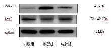
Fig.7
Western blotting of GSK-3β and SynI proteins in the hippocampal tissues of mice in each group"
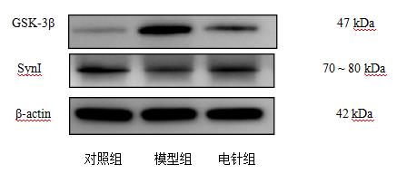
Tab.10
Expression Levels of GSK-3β and SynI in the hippocampus of mice in each group (n =3)"
| 组别 | ||
|---|---|---|
| 对照组 | ||
| 模型组 | ||
| 电针组 | ||
| < 0.01 | < 0.01 |
| [1] |
SCHELTENS P, DE STROOPER B, KIVIPELTO M, et al. Alzheimer's disease[J]. Lancet,2021,397(10284):1577-1590. doi: 10.1016/S0140-6736(20)32205-4 .
doi: 10.1016/S0140-6736(20)32205-4 |
| [2] |
JIA L F, DU Y F, CHU L, et al. Prevalence, risk factors, and management of dementia and mild cognitive impairment in adults aged 60 years or older in China: A cross-sectional study[J]. Lancet Public Health, 2020, 5(12): e661-e671.doi: 10.1016/S2468-2667(20)30185-7 .
doi: 10.1016/S2468-2667(20)30185-7 |
| [3] |
朱丹迪,潘伟刚,刘超猛,等.阿尔茨海默病药物联合治疗研究进展[J].神经疾病与精神卫生,2021,21(12):898-902.doi:10.3969/j.issn.1009-6574.2021.12.012 .
doi: 10.3969/j.issn.1009-6574.2021.12.012 |
| [4] |
陈生弟,方嵘.应重视早期阿尔茨海默病的非药物治疗[J].中国现代神经疾病杂志,2021,21(11):913-917.doi:10.3969/j.issn.1672-6731.2021.11.001 .
doi: 10.3969/j.issn.1672-6731.2021.11.001 |
| [5] |
YU X, LU F, CHEN J, et al. A bibliometric analysis of acupuncture treatment and cognitive impairment[J]. Front Neurol,2025,16:1495191. doi: 10.3389/fneur.2025.1495191 .
doi: 10.3389/fneur.2025.1495191 |
| [6] | 王昊,惠鑫,赵百孝.针灸治疗阿尔茨海默病临床研究进展[J]. 中华中医药杂志,2020,35(4):1945-1948. |
| [7] |
XU M, LIU J, LIU Q, et al. Preliminary study on early diagnosis of Alzheimer's disease in APP/PS1 transgenic mice using multimodal magnetic resonance imaging[J]. Front Aging Neurosci,2024,16:1326394.doi:10.3389/fnagi.2024. 1326394 .
doi: 10.3389/fnagi.2024. 1326394 |
| [8] |
PINI L, GEROLDI C, GALLUZZI S, et al. Age at onset reveals different functional connectivity abnormalities in prodromal Alzheimer's disease[J]. Brain Imaging Behav, 2020, 14(6): 2594-2605. doi: 10.1007/s11682-019-00212-6 .
doi: 10.1007/s11682-019-00212-6 |
| [9] |
WEARN A, TARDIF C L, LEPPERT I R, et al. Quantitative MRI of the hippocampus reveals microstructural trajectories of aging and Alzheimer's disease pathology[J]. Proc Natl Acad Sci U S A, 2025, 122(44): e2502674122. doi: 10.1073/pnas.2502674122 .
doi: 10.1073/pnas.2502674122 |
| [10] |
SERRA L, BOZZALI M, FADDA L, et al. The role of hippocampus in the retrieval of autobiographical memories in patients with amnestic mild cognitive impairment due to Alzheimer's disease[J]. J Neuropsychol,2020,14(1): 46-68. doi: 10.1111/jnp.12174 .
doi: 10.1111/jnp.12174 |
| [11] |
韩英妹, 李一杰, 张衡, 等. 基于MRI分析阿尔茨海默病大尺度脑网络研究进展[J].实用医学杂志, 2024, 40(4): 575-579. doi: 10.3969/j.issn.1006-5725.2024.04. 024 .
doi: 10.3969/j.issn.1006-5725.2024.04. 024 |
| [12] |
FAGIANI F, LANNI C, RACCHI M, et al. (Dys)regulation of Synaptic Activity and Neurotransmitter Release by β-Amyloid: A Look Beyond Alzheimer's Disease Pathogenesis[J]. Front Mol Neurosci, 2021, 14: 635880. doi: 10.3389 /fnmol.2021.635880 .
doi: 10.3389 /fnmol.2021.635880 |
| [13] |
WANG L, BI L, QIU Y, et al. Effectiveness of electro-acupuncture for cognitive improvement on Alzheimer's disease quantified via PET imaging of sphingosine-1-phosphate receptor 1[J]. Alzheimers Dement, 2024, 20(12): 8331-8345. doi: 10.1002/alz.14260 .
doi: 10.1002/alz.14260 |
| [14] |
ZHANG R Z, ZHANG X, ZHU T T, et al. Study on the Effects of Acupuncture with the "Yizhi Tiaoshen" Acupoint Formula on Blood Oxygen Metabolism and Neural Function in Key Brain Regions of AD Rats[J]. Degener Neurol Neuromuscul Dis, 2024, 14: 103-114. doi: 10.2147/DNND.S468770 .
doi: 10.2147/DNND.S468770 |
| [15] |
LI J, YANG M, DAI Y,et al. Electroacupuncture regulates Rab5a-mediating NGF transduction to improve learning and memory ability in the early stage of AD mice[J]. CNS Neurosci Ther,2024,30(5):e14743. doi: 10.1111/cns.14743 .
doi: 10.1111/cns.14743 |
| [16] | 中国针灸学会.实验动物常用穴位名称与定位第3部分:小鼠[J].针刺研究,2021,46(5):445-446. |
| [17] |
何川,王丽,潘小丽,等.基于β2AR/β-arrestin2/NF-κB通路探讨预电针“内关”“间使”对阿尔茨海默病样大鼠学习记忆及蓝斑核-海马神经环路的作用机制[J].北京中医药大学学报,2024,47(11):1612-1622.doi: 10. 3969 / j. issn. 1006-2157. 2024. 11. 017 .
doi: 10. 3969 / j. issn. 1006-2157. 2024. 11. 017 |
| [18] |
刘孔, 娄必丹, 俞赟丰, 等. 针刺心包经穴治疗阿尔茨海默病的理论探讨[J]. 云南中医中药杂志, 2022, 43(7): 14-16.doi:10.16254/j.cnki.53-1120/ r.2022.07.020 .
doi: 10.16254/j.cnki.53-1120/ r.2022.07.020 |
| [19] |
张丽颖,王洪峰. 基于数据挖掘技术针灸治疗阿尔茨海默病的选穴规律分析[J]. 长春中医药大学学报, 2018,34(5):911-914.doi:10.13463/j.cnki.cczyy. 2018.05.028 .
doi: 10.13463/j.cnki.cczyy. 2018.05.028 |
| [20] |
NGUYEN-DUC J, DE RIEDMATTEN I, SPENCER A P C, et al. Mapping Activity and Functional Organisation of the Motor and Visual Pathways Using ADC-fMRI in the Human Brain[J]. Hum Brain Mapp,2025,46(2):e70110. doi: 10.1002/hbm.70110 .
doi: 10.1002/hbm.70110 |
| [21] |
魏玉婷,苏明莉,朱田田,等.针刺“益智调神”穴方对阿尔茨海默病患者海马与全脑功能连接的影响[J].中国针灸,2023,43(12):1351-1357.doi:10.13703/ j.0255-2930.20230405-0002 .
doi: 10.13703/ j.0255-2930.20230405-0002 |
| [22] |
何肖君,李珑,梁胜祥,等.电针“百会”“神庭”对阿尔茨海默病小鼠早期空间识别功能与脑区局部一致性的影响[J].福建中医药,2021,52(7):30-33. doi:10. 13260/j.cnki.jfjtcm.012308 .
doi: 10. 13260/j.cnki.jfjtcm.012308 |
| [23] |
PAN F F, HUANG Q, HUANG C C, et al. Associations of hippocampal volumes, brain hypometabolism, and plasma NfL with amyloid, tau, and cognitive decline[J]. Alzheimers Dement, 2025, 21(2): e70005. doi: 10.1002/alz.70005 .
doi: 10.1002/alz.70005 |
| [24] |
陈佳悦,黄雪燕,姚倩倩,等. β-淀粉样蛋白耐受在阿尔茨海默病中的研究进展[J]. 中华神经医学杂志, 2024, 23(8): 837-841. doi:10.3760/cma.j.cn 115354-20240617-00353 .
doi: 10.3760/cma.j.cn 115354-20240617-00353 |
| [25] |
RIBARIČ S. Detecting Early Cognitive Decline in Alzheimer's Disease with Brain Synaptic Structural and Functional Evaluation[J]. Biomedicines,2023,11(2):355. doi: 10.3390/biomedicines11020355 .
doi: 10.3390/biomedicines11020355 |
| [26] |
胡杨,周怡民,申美伦,等.早期阿尔茨海默病患者嗅觉功能障碍及其检测[J].空军军医大学学报,2024,45(4):461-464,471.doi:10.13276/j.issn. 2097-1656.2024.04.019 .
doi: 10.13276/j.issn. 2097-1656.2024.04.019 |
| [27] |
CARNEMOLLA S E, KUMFOR F, LIANG C T, et al.Olfactory Bulb Integrity in Frontotemporal Dementia and Alzheimer's Disease[J]. J Alzheimers Dis,2022,89(1):51-66. doi: 10.3233/JAD-220080 .
doi: 10.3233/JAD-220080 |
| [28] |
杨斯闵,李璐迪,曹月,等.基于3D脑结构MRI定量分析对轻度认知障碍及阿尔茨海默病患者嗅觉皮层的研究[J].磁共振成像,2023,14(1):20-24,31. doi:10.12015/issn.1674-8034.2023.01.004 .
doi: 10.12015/issn.1674-8034.2023.01.004 |
| [29] |
BOUHABEN J, DELGADO-LIMA A H, DELGADO-LOSADA M L. The role of olfactory dysfunction in mild cognitive impairment and Alzheimer's disease: A meta-analysis[J]. Arch Gerontol Geriatr,2024,123:105425. doi: 10.1016/j.archger.2024.105425 .
doi: 10.1016/j.archger.2024.105425 |
| [30] |
XU M, LIU J, LIU Q, et al. Preliminary study on early diagnosis of Alzheimer's disease in APP/PS1 transgenic mice using multimodal magnetic resonance imaging[J]. Front Aging Neurosci,2024,16:1326394.doi: 10.3389/fnagi.2024.1326394 .
doi: 10.3389/fnagi.2024.1326394 |
| [31] |
JEFFS Q L, PRATHER J F, TODD W D. Potential neural substrates underlying circadian and olfactory disruptions in preclinical Alzheimer's disease[J]. Front Neurosci,2023,17:1295998. doi: 10.3389/fnins.2023.1295998 .
doi: 10.3389/fnins.2023.1295998 |
| [32] |
PŁOSKA A, CIEŚLIK P, SIEKIERZYCKA A, et al. Neurochemical changes underlying cognitive impairment in olfactory bulbectomized rats and the impact of the mGlu5-positive allosteric modulator CDPPB[J]. Brain Res,2021,1768:147577. doi: 10.1016/j.brainres.2021.147577 .
doi: 10.1016/j.brainres.2021.147577 |
| [33] |
MOEIN TAGHAVI H, KARIMPOOR M, VAN STAALDUINEN E K, et al. Elevated tau in the piriform cortex in Alzheimer's but not Parkinson's disease using PET-MR[J]. Alzheimers Dement (Amst),2024,16(4):e70040. doi: 10.1002/dad2.70040 .
doi: 10.1002/dad2.70040 |
| [34] |
AUDRONYTE E, PAKULAITE-KAZLIENE G, SUTNIKIENE V, et al. Odor Discrimination as a Marker of Early Alzheimer's Disease[J]. J Alzheimers Dis,2023,94(3):1169-1178. doi: 10.3233/JAD-230077 .
doi: 10.3233/JAD-230077 |
| [35] |
KIM S, NAM Y, KIM H S, et al. Alteration of Neural Pathways and Its Implications in Alzheimer's Disease[J]. Biomedicines,2022,10(4):845.doi: 10.3390/biomedicines10040845 .
doi: 10.3390/biomedicines10040845 |
| [36] |
SHI Y, LI Y, CI R, et al. Dynamic functional connectivity and transcriptomic signatures reveal stage-dependent brain network dysfunction in Alzheimer's disease spectrum[J]. Alzheimers Res Ther,2025,17(1):247. doi: 10.1186/s13195-025-01898-1 .
doi: 10.1186/s13195-025-01898-1 |
| [37] |
AKYUZ E, ARULSAMY A, ASLAN F S, et al. An Expanded Narrative Review of Neurotransmitters on Alzheimer's Disease: The Role of Therapeutic Interventions on Neurotransmission[J]. Mol Neurobiol,2025,62(2):1631-1674. doi: 10.1007/s12035-024-04333-y .
doi: 10.1007/s12035-024-04333-y |
| [38] |
MUÑOZ DE LEÓN-LÓPEZ C A, NAVARRO-LOBATO I, KHAN Z U. The Role of Astrocytes in Synaptic Dysfunction and Memory Deficits in Alzheimer's Disease[J]. Biomolecules, 2025, 15(7): 910. doi: 10.3390/biom15070910 .
doi: 10.3390/biom15070910 |
| [39] |
XING B, LI Y C, GAO W J. Norepinephrine versus dopamine and their interaction in modulating synaptic function in the prefrontal cortex[J]. Brain Res,2016,1641(Pt B):217-233.doi: 10.1016/j.brainres.2016.01.005 .
doi: 10.1016/j.brainres.2016.01.005 |
| [40] |
TAN X, LIANG Z, LI Y,et al. Isoorientin, a GSK-3β inhibitor, rescues synaptic dysfunction, spatial memory deficits and attenuates pathological progression in APP/PS1 model mice[J]. Behav Brain Res, 2021, 398: 112968. doi: 10.1016/j.bbr.2020.112968 .
doi: 10.1016/j.bbr.2020.112968 |
| [41] |
MU L, XIA D, CAI J, et al. Treadmill Exercise Reduces Neuroinflammation, Glial Cell Activation and Improves Synaptic Transmission in the Prefrontal Cortex in 3 × Tg-AD Mice[J]. Int J Mol Sci,2022,23(20):12655. doi: 10.3390/ijms232012655 .
doi: 10.3390/ijms232012655 |
| [42] |
MOSCHETTA M, RAVASENGA T, DE FUSCO A, et al. Ca binding to synapsin I regulates resting Ca and recovery from synaptic depression in nerve terminals[J]. Cell Mol Life Sci, 2022,79(12):600.doi: 10.1007/s00018-022-04631-5 .
doi: 10.1007/s00018-022-04631-5 |
| [43] |
LEI L, LUO Y, KANG D,et al. Gypenoside IX restores Akt/GSK-3β pathway and alleviates Alzheimer's disease-like neuropathology and cognitive deficits[J]. Aging (Albany NY),2023,15(23):14172-14191. doi: 10.18632/aging.205295 .
doi: 10.18632/aging.205295 |
| [44] |
肖娟, 王佳, 鲁镇坤, 等. 电针调控前额叶皮层GLU/NMDAR2B通路改善APP/PS1小鼠突触可塑性损伤的作用机制[J].湖北中医药大学学报,2025,27(1):8-12.doi:10. 3969/j. issn. 1008 987x.2025.01.02 .
doi: 10. 3969/j. issn. 1008 987x.2025.01.02 |
| [45] |
MIJALKOV M, VERÉB D, CANAL-GARCIA A, et al. Directed Functional Brain Connectivity is Altered in Sub-threshold Amyloid-β Accumulation in Cognitively Normal Individuals[J]. Neurosci Insights,2023,18: 26331055231161625. doi: 10.1177/26331055231161625 .
doi: 10.1177/26331055231161625 |
| [1] | Rongxin LI,Li HUANG,Yueyang ZENG,Shuhui ZHANG,Yiran CHEN,Yuli LIU,Tieming. MA. Exploring the mechanism of electroacupuncture to improve cognitiveimpairment in alzheimer′s disease model rats based on NF⁃κB/NLRP3/Caspase⁃1 signaling pathway [J]. The Journal of Practical Medicine, 2025, 41(3): 322-329. |
| [2] | Ling QI,Chuan HE,Yao WANG,Xiaolei ZHANG,Jun. MA. The effect of electroacupuncture on the SIRT3/FOXO3/SOD2 signaling pathway in the substantia nigra of mice with Parkinson′s disease [J]. The Journal of Practical Medicine, 2025, 41(24): 3815-3823. |
| [3] | Bao WANG,Shulin MA,Xinhua YAO,Fan YANG,Kai WEN,Sijing LUO,Ying GAN,Yi. LU. Effect of intraoperative electroacupuncture analgesia on stress response to tracheal intubation in patients undergoing thyroid surgery under general anesthesia [J]. The Journal of Practical Medicine, 2024, 40(8): 1132-1136. |
| [4] | Yingmei HAN,Yijie LI,Heng ZHANG,Jing LV,Yi ZHANG,Yingbo QIAO,Nan LIN,Huiyong XU,Feng. WANG. Research progress of large-scale brain network of Alzheimer’s disease based on MRI analysis [J]. The Journal of Practical Medicine, 2024, 40(4): 575-579. |
| [5] | Ying GAN,Bao WANG,Jiayin YAO,Xinhua YAO,Dong LIU,Rong HUANG,Yi. LU. Analgesic effect of perioperative electroacupuncture stimulation on laparoscopic surgery [J]. The Journal of Practical Medicine, 2023, 39(20): 2612-2617. |
| [6] |
ZHONG Peirui, HE Xiaoyan, LIAO Ying, SUN Guanghua, LIU Jing, ZHOU Jun, LI Shuzhi, LIU Yuan, QU Mengjian..
Study on the mechanism of P53/P21 pathway in electroacupuncture inhibiting osteoblast aging in osteopo⁃ rotic rats [J]. The Journal of Practical Medicine, 2023, 39(2): 192-197. |
| [7] |
HUANG Xiarong, ZHOU Jun, SUN Guanghua, PENG Xinke, LIAO Yuan, LIU Jing, LUO Fu, ZHONG Peirui, PENG Ting, HU Lizhi..
Effect of electroacupuncture on articular cartilage and subchondral polarization⁃related protein expression in aged rats [J]. The Journal of Practical Medicine, 2023, 39(12): 1473-1479. |
| [8] |
LI Hanzhang, QI Ling, ZHANG Xiaolei, CHEN Xianglin, GUO Lei, GUO Shuqin, MA Jun..
Effect of electroacupuncture on GLP⁃1R⁃mediated PI3K/AKT/GSK⁃3β pathway in mice with Parkinson′s disease [J]. The Journal of Practical Medicine, 2021, 37(21): 2712-2716. |
| Viewed | ||||||
|
Full text |
|
|||||
|
Abstract |
|
|||||