

The Journal of Practical Medicine ›› 2026, Vol. 42 ›› Issue (8): 1312-1321.doi: 10.3969/j.issn.1006-5725.2026.08.002
• Chronic Disease Control • Previous Articles
Yameng LIU1,Chao WANG1,Mengxin WANG1,Shanning GAN1,Yanping ZHU2( )
)
Received:2025-11-20
Online:2026-04-25
Published:2026-04-28
Contact:
Yanping ZHU
E-mail:1119788145@qq.com
CLC Number:
Yameng LIU,Chao WANG,Mengxin WANG,Shanning GAN,Yanping ZHU. Effects of human umbilical cord mesenchymal stem cells on aquaporin-4 after white matter injury in neonatal rats[J]. The Journal of Practical Medicine, 2026, 42(8): 1312-1321.
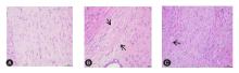
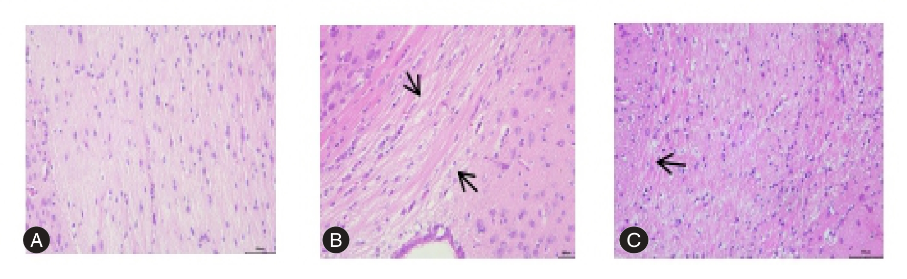
Fig.1
Pathological changes of brain histopathology(HE,×400)"
Tab.1
Brain water content in brain tissue of rats in each group at different time points after modeling(n=6)"
| 组别 | 脑含水量 | |||
|---|---|---|---|---|
| 7 d | 14 d | t值 | P值 | |
| 假手术组 | 85.74 ± 0.18 | 82.81 ± 0.18 | 28.339 | < 0.001 |
| WMI组 | 86.40 ± 0.25* | 83.94 ± 0.27* | 16.172 | < 0.001 |
| HUC-MSCs组 | 85.76 ± 0.50# | 82.91 ± 0.13# | 13.492 | < 0.001 |
| F值 | 7.235 | 56.961 | ||
| P值 | 0.006 | < 0.001 | ||
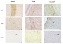
Fig.2
Expression of MBP, GFAP and AQP4 in brain tissue of neonatal rats(immunohistochemical staining,×200)"
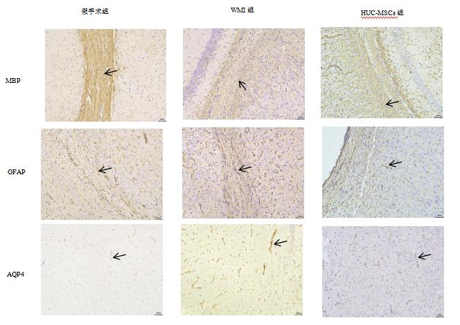
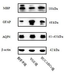
Fig.3
Expression levels of MBP, GFAP and AQP4 protein in neonatal rat brain tissue"
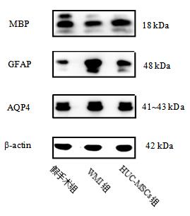
Tab.2
Expression levels of MBP,GFAP and AQP4 in rats in each group(n = 6)"
| 组别 | MBP | GFAP | AQP4 | |||||
|---|---|---|---|---|---|---|---|---|
| 平均光密度值 | 相对蛋白表达量 | 平均光密度值 | 相对蛋白表达量 | 平均光密度值 | 相对蛋白表达量 | |||
| 假手术组 | 0.22 ± 0.01 | 1.27 ± 0.11 | 0.17 ± 0.01 | 0.82 ± 0.08 | 0.16 ± 0.02 | 0.82 ± 0.09 | ||
| WMI组 | 0.16 ± 0.00* | 0.76 ± 0.07* | 0.26 ± 0.02* | 1.19 ± 0.09* | 0.24 ± 0.17* | 1.18 ± 0.10* | ||
| HUC-MSCs组 | 0.20 ± 0.01*# | 1.01 ± 0.08*# | 0.23 ± 0.02*# | 1.01 ± 0.04*# | 0.20 ± 0.01*# | 0.99 ± 0.04*# | ||
| F值 | 106.690 | 46.550 | 42.125 | 37.641 | 48.215 | 29.828 | ||
| P值 | < 0.001 | < 0.001 | < 0.001 | < 0.001 | < 0.001 | < 0.001 | ||
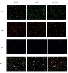
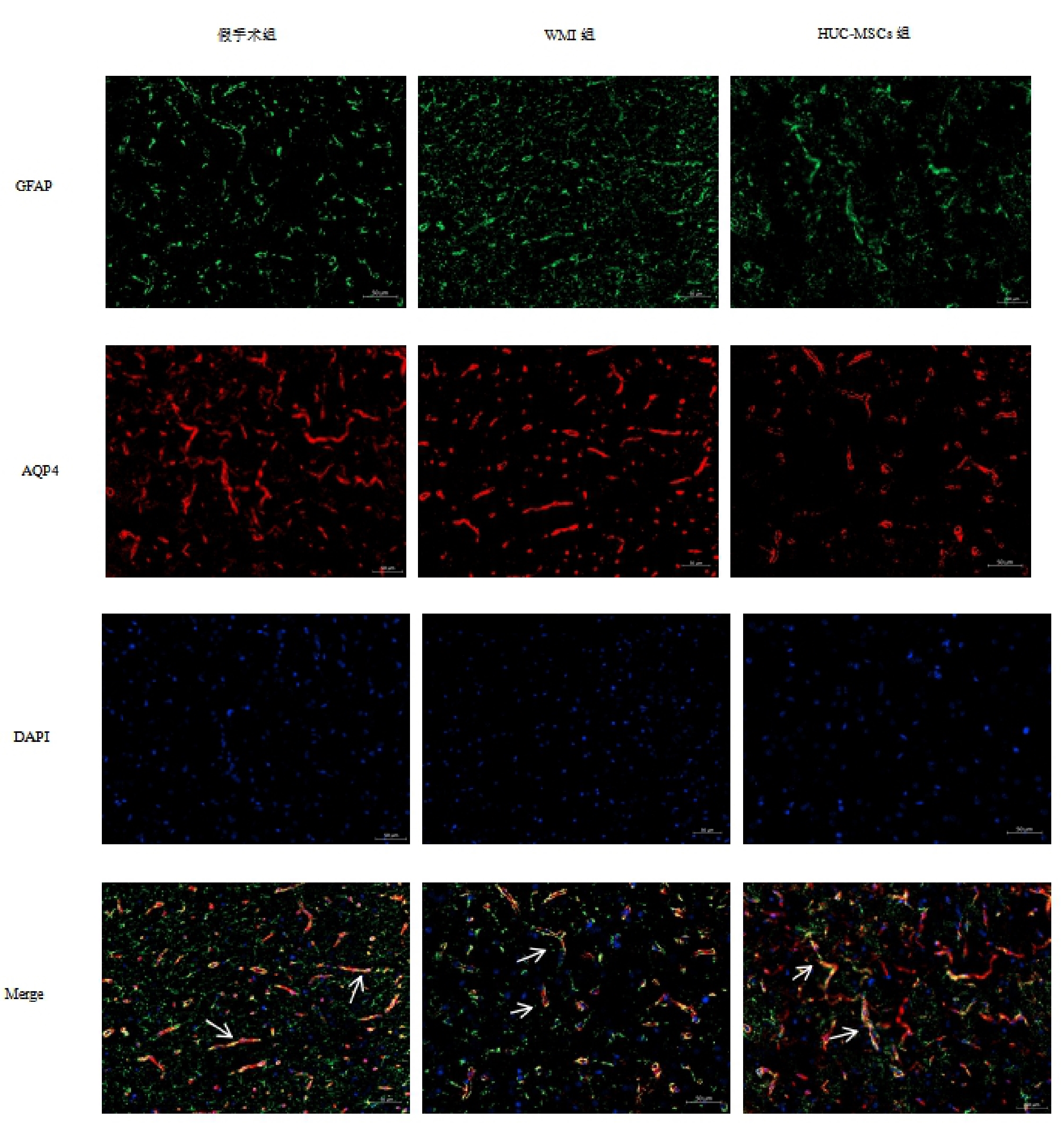
Fig.4
GFAP co-expressed with AQP4"
Tab.3
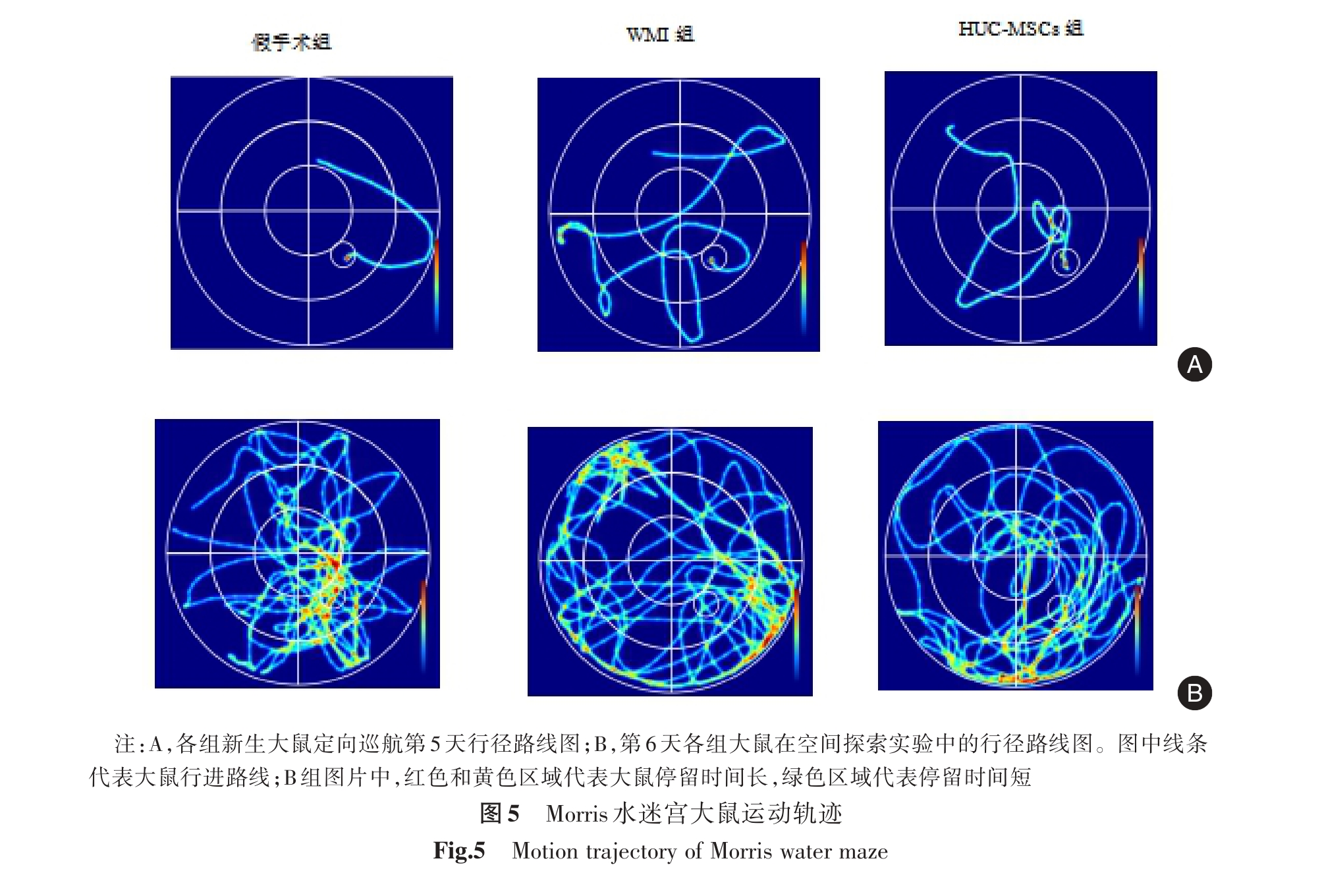
Escape incubation period,times of platform crossing and time of target quadrant residence(n = 6)"
| 组别 | 逃逸潜伏期/s | 穿越平台次数 | 目标象限停留时间/s | ||||
|---|---|---|---|---|---|---|---|
| DAY1 | DAY2 | DAY3 | DAY4 | DAY5 | |||
| 假手术组 | 53.82 ± 3.34 | 43.11 ± 2.06 | 36.87 ± 3.10 | 23.53 ± 2.11 | 9.06 ± 2.36 | 8.33 ± 1.75 | 48.60 ± 3.63 |
| WMI组 | 51.39 ± 6.47 | 52.44 ± 4.54* | 46.13 ± 5.05* | 35.89 ± 1.82* | 21.47 ± 5.80* | 4.83 ± 0.75* | 35.32 ± 3.03* |
| HUC-MSCs组 | 54.58 ± 3.98 | 47.12 ± 2.18*# | 41.46 ± 2.07*# | 28.96 ± 1.26*# | 14.85 ± 2.13*# | 6.67 ± 0.82*# | 42.08 ± 5.90*# |
| F值 | 0.723 | 13.292 | 9.789 | 85.813 | 15.876 | 12.830 | 13.880 |
| P值 | 0.501 | < 0.001 | 0.002 | < 0.001 | < 0.001 | 0.001 | < 0.001 |
"
| [1] |
AGUT T, ALARCON A, CABAÑAS F,et al. Preterm white matter injury: Ultrasound diagnosis and classification[J]. Pediatr Res, 2020, 87(): 37-49. doi: 10.1038/s41390-020-0781-1 .
doi: 10.1038/s41390-020-0781-1 |
| [2] |
REES P, CALLAN C, CHADDA K R,et al. Preterm Brain Injury and Neurodevelopmental Outcomes: A Meta-analysis[J]. Pediatrics, 2022, 150(6):e2022057442. doi: 10.1542/peds.2022-057442 .
doi: 10.1542/peds.2022-057442 |
| [3] |
RENZ P, STEINFORT M, HAESLER V,et al. Neuroinflammatory reactive astrocyte formation correlates with adverse outcomes in perinatal white matter injury[J]. Glia, 2024, 72(9): 1663-1673. doi: 10.1002/glia.24575 .
doi: 10.1002/glia.24575 |
| [4] |
SOBRAL A F, COSTA I, TEIXEIRA V,et al. Molecular Motors in Blood-Brain Barrier Maintenance by Astrocytes[J]. Brain Sci, 2025, 15(3): 279. doi: 10.3390/brainsci15030279 .
doi: 10.3390/brainsci15030279 |
| [5] |
CHEN T, BOSCO D B, YING Y,et al. The Emerging Role of Microglia in Neuromyelitis Optica[J]. Front Immunol, 2021, 12: 616301. doi: 10.3389/fimmu.2021.616301 .
doi: 10.3389/fimmu.2021.616301 |
| [6] |
HUANG Y, CHEN S, LUO Y,et al. Crosstalk between Inflammation and the BBB in Stroke[J]. Curr Neuropharmacol, 2020, 18(12): 1227-1236. doi: 10.2174/1570159X18666200620230321 .
doi: 10.2174/1570159X18666200620230321 |
| [7] |
BRANDEBURA A N, PAUMIER A, ONUR T S,et al. Astrocyte contribution to dysfunction, risk and progression in neurodegenerative disorders[J]. Nat Rev Neurosci, 2023, 24(1): 23-39. doi: 10.1038/s41583-022-00641-1 .
doi: 10.1038/s41583-022-00641-1 |
| [8] |
VANDEBROEK A, YASUI M. Regulation of AQP4 in the Central Nervous System[J]. Int J Mol Sci, 2020, 21(5):1603. doi: 10.3390/ijms21051603 .
doi: 10.3390/ijms21051603 |
| [9] |
AI L, USMAN M, LU H. Experimental study of cerebral edema and expression of VEGF and AQP4 in the penumbra area of rat brain trauma[J]. Sci Rep, 2025, 15(1): 17040. doi: 10.1038/s41598-025-02071-2 .
doi: 10.1038/s41598-025-02071-2 |
| [10] |
HAN F, BI J, QIAO L,et al. Stem Cell Therapy for Alzheimer's Disease[M]. Singapore:Springer, 2020: 39-55. doi:10.1007/978-981-15-4370-8_4
doi: 10.1007/978-981-15-4370-8_4 |
| [11] |
HOANG D M, PHAM P T, BACH T Q,et al. Stem cell-based therapy for human diseases[J]. Signal Transduct Target Ther, 2022, 7(1): 272. doi: 10.1038/s41392-022-01134-4 .
doi: 10.1038/s41392-022-01134-4 |
| [12] |
JIAO Y, SUN Y T, CHEN N F,et al. Human umbilical cord-derived mesenchymal stem cells promote repair of neonatal brain injury caused by hypoxia/ischemia in rats[J]. Neural Regen Res, 2022, 17(11): 2518-2525. doi: 10.4103/1673-5374.339002 .
doi: 10.4103/1673-5374.339002 |
| [13] |
WANG G, WU H L, LIU Y P,et al. Pre-clinical study of human umbilical cord mesenchymal stem cell transplantation for the treatment of traumatic brain injury: Safety evaluation from immunogenic and oncogenic perspectives[J]. Neural Regen Res, 2022, 17(2): 354-361. doi: 10.4103/1673-5374.317985 .
doi: 10.4103/1673-5374.317985 |
| [14] |
徐倩倩, 张书绢, 王超, 等. 人脐带间充质干细胞对新生大鼠脑白质损伤的修复作用[J]. 中华新生儿科杂志, 2025, 40(5): 297-305. doi: 10.3760/cma.j.cn101451-20241006-00348 .
doi: 10.3760/cma.j.cn101451-20241006-00348 |
| [15] |
LIU Y, MA Y, WANG Y, et al. Mesenchymal Stem Cells Attenuated Blood-Brain Barrier Disruption via Downregulation of Aquaporin-4 Expression in EAE Mice[J]. Mol Neurobiol, 2020, 57(9): 3891-3901. doi: 10.1007/s12035-020-01998-z .
doi: 10.1007/s12035-020-01998-z |
| [16] |
王超, 徐倩倩, 张书绢, 等. 人脐带间充质干细胞衍生的外泌体对新生大鼠 脑白质损伤小胶质细胞极化的影响[J]. 实用医学杂志, 2025, 41(16): 2447-2454. doi: 10.3969/j.issn.1006-5725.2025.16.002 .
doi: 10.3969/j.issn.1006-5725.2025.16.002 |
| [17] |
张军, 李明霞, 王超, 等. 不同剂量人脐带间充质干细胞移植对新生大鼠脑白质损伤的修复作用[J]. 中国当代儿科杂志, 2024, 26(4): 394-402. doi: 10.7499/j.issn.1008-8830.2310081 .
doi: 10.7499/j.issn.1008-8830.2310081 |
| [18] |
SUN C, ZOU N, ZHANG H C. The effect of magnetic guiding BMSCs on hypoxic-ischemic brain damage via magnetic resonance imaging evaluation[J]. Magn Reson Imaging, 2021, 79: 59-65. doi: 10.1016/j.mri.2021.03.008 .
doi: 10.1016/j.mri.2021.03.008 |
| [19] |
XIE Y, YANG Y, YUAN T. Brain Damage in the Preterm Infant: Clinical Aspects and Recent Progress in the Prevention and Treatment[J]. CNS Neurol Disord Drug Targets, 2023, 22(1): 27-40. doi: 10.2174/1871527321666220223092905 .
doi: 10.2174/1871527321666220223092905 |
| [20] |
GALINSKY R, DEAN J M, LINGAM I,et al. A Systematic Review of Magnesium Sulfate for Perinatal Neuroprotection: What Have We Learnt From the Past Decade [J]. Front Neurol, 2020, 11: 449. doi: 10.3389/fneur.2020.00449 .
doi: 10.3389/fneur.2020.00449 |
| [21] |
ZHANG M, WANG H, HE Y, et al. Effects and mechanisms of breastmilk stem cells in the treatment of white matter injury in newborn rats[J]. Stem Cell Res Ther, 2025, 16(1): 124. doi: 10.1186/s13287-025-04257-x .
doi: 10.1186/s13287-025-04257-x |
| [22] |
TSCHERRIG V, COTTAGNOUD S, HAESLER V, et al. MicroRNA Cargo in Wharton's Jelly MSC Small Extracellular Vesicles: Key Functionality to In Vitro Prevention and Treatment of Premature White Matter Injury[J]. Stem Cell Rev Rep, 2023, 19(7): 2447-2464. doi: 10.1007/s12015-023-10595-1 .
doi: 10.1007/s12015-023-10595-1 |
| [23] |
ELBAZ B, POPKO B. Molecular Control of Oligodendrocyte Development[J]. Trends Neurosci, 2019, 42(4): 263-277. doi: 10.1016/j.tins.2019.01.002 .
doi: 10.1016/j.tins.2019.01.002 |
| [24] |
GO V, SARIKAYA D, ZHOU Y, et al. Extracellular vesicles derived from bone marrow mesenchymal stem cells enhance myelin maintenance after cortical injury in aged rhesus monkeys[J]. Exp Neurol, 2021, 337: 113540. doi: 10.1016/j.expneurol.2020.113540 .
doi: 10.1016/j.expneurol.2020.113540 |
| [25] |
TAPIA-BUSTOS A, LESPAY-REBOLLEDO C, VÍO V, et al. Neonatal Mesenchymal Stem Cell Treatment Improves Myelination Impaired by Global Perinatal Asphyxia in Rats[J]. Int J Mol Sci, 2021, 22(6): 3275. doi: 10.3390/ijms22063275 .
doi: 10.3390/ijms22063275 |
| [26] |
GHANBARI A, RAD F, SHAHRAKI M H, et al. Human mesenchymal stem cells-derived microvesicles increase oligodendrogenesis and neurogenesis of cultured adult neural stem cells[J]. Neurosci Lett, 2024, 841: 137951. doi: 10.1016/j.neulet.2024.137951 .
doi: 10.1016/j.neulet.2024.137951 |
| [27] |
ZHANG J, BULLER B A, ZHANG Z G, et al. Exosomes derived from bone marrow mesenchymal stromal cells promote remyelination and reduce neuroinflammation in the demyelinating central nervous system[J]. Exp Neurol, 2022, 347: 113895. doi: 10.1016/j.expneurol.2021.113895 .
doi: 10.1016/j.expneurol.2021.113895 |
| [28] |
KOOHSARI S A, ABSALAN A, AZADI D. Human umbilical cord mesenchymal stem cell-derived extracellular vesicles attenuate experimental autoimmune encephalomyelitis via regulating pro and anti-inflammatory cytokines[J]. Sci Rep, 2021, 11(1): 11658. doi: 10.1038/s41598-021-91291-3 .
doi: 10.1038/s41598-021-91291-3 |
| [29] |
FAHIM I, ISHAQUE A, RAMZAN F, et al. Overexpression of OLIG2 and MYT1L Transcription Factors Enhance the Differentiation Potential of Human Mesenchymal Stem Cells into Oligodendrocytes[J]. Curr Issues Mol Biol, 2023, 45(5): 4100-4123. doi: 10.3390/cimb45050261 .
doi: 10.3390/cimb45050261 |
| [30] |
CARTA S, CHIODEGA V, PASQUALI R T. GFAP as a marker of astrocytic damage correlated with medication overuse in migraine[J]. Immunol Res, 2025, 73(1): 119. doi: 10.1007/s12026-025-09674-x .
doi: 10.1007/s12026-025-09674-x |
| [31] |
PIERRE W C, LONDONO I, QUINIOU C, et al. Modulatory effect of IL-1 inhibition following lipopolysaccharide-induced neuroinflammation in neonatal microglia and astrocytes[J]. Int J Dev Neurosci, 2022, 82(3): 243-260. doi: 10.1002/jdn.10179 . Epub 2022 Mar 27.
doi: 10.1002/jdn.10179 |
| [32] |
HEITHOFF B P, GEORGE K K, PHARES A N, et al. Astrocytes are necessary for blood-brain barrier maintenance in the adult mouse brain[J]. Glia, 2021, 69(2): 436-472. doi: 10.1002/glia.23908 .
doi: 10.1002/glia.23908 |
| [33] |
SUN C, LIN L, YIN L, et al. Acutely Inhibiting AQP4 With TGN-020 Improves Functional Outcome by Attenuating Edema and Peri-Infarct Astrogliosis After Cerebral Ischemia[J]. Front Immunol, 2022, 13:870029.doi: 10.3389/fimmu.2022.870029 .
doi: 10.3389/fimmu.2022.870029 |
| [34] |
DATTA A, SARMAH D, KAUR H, et al. Post-stroke Impairment of the Blood-Brain Barrier and Perifocal Vasogenic Edema Is Alleviated by Endovascular Mesenchymal Stem Cell Administration: Modulation of the PKCδ/MMP9/AQP4-Mediated Pathway[J]. Mol Neurobiol, 2022, 59(5): 2758-2775. doi: 10.1007/s12035-022-02761-2 .
doi: 10.1007/s12035-022-02761-2 |
| [35] |
ZHAO L, ZHANG Z, WANG P, et al. NHH promotes Sepsis-associated Encephalopathy with the expression of AQP4 in astrocytes through the gut-brain Axis[J]. J Neuroinflammation, 2024, 21(1): 138. doi: 10.1186/s12974-024-03135-2 .
doi: 10.1186/s12974-024-03135-2 |
| [36] |
LI X, XIE Z, ZHOU Q, et al. TGN-020 Alleviate Inflammation and Apoptosis After Cerebral Ischemia-Reperfusion Injury in Mice Through Glymphatic and ERK1/2 Signaling Pathway[J]. Mol Neurobiol, 2024, 61(2):1175-1186. doi: 10.1007/s12035-023-03636-w .
doi: 10.1007/s12035-023-03636-w |
| [37] |
FENG S, WU C, ZOU P, et al. High-intensity interval training ameliorates Alzheimer's disease-like pathology by regulating astrocyte phenotype-associated AQP4 polarization[J]. Theranostics, 2023, 13(10): 3434-3450. doi: 10.7150/thno.81951 .
doi: 10.7150/thno.81951 |
| [38] |
LABARTA-BAJO L, ALLEN N J. Astrocytes in aging[J]. Neuron, 2025, 113(1): 109-126. doi: 10.1016/j.neuron.2024.12.010 .
doi: 10.1016/j.neuron.2024.12.010 |
| Viewed | ||||||
|
Full text |
|
|||||
|
Abstract |
|
|||||