

The Journal of Practical Medicine ›› 2026, Vol. 42 ›› Issue (7): 1192-1200.doi: 10.3969/j.issn.1006-5725.2026.07.011
• Oncology: Diagnosis, Treatment and Prevention • Previous Articles
Jing YIN1,2,Pingyang ZHANG1( ),Junli WANG2,Weiwei YIN2,Xiaoai CHU2,Wenyan ZHAO3
Received:2025-12-01
Revised:2025-12-26
Accepted:2025-12-30
Online:2026-04-10
Published:2026-04-13
Contact:
Pingyang ZHANG
E-mail:zhpy28@126.com
CLC Number:
Jing YIN,Pingyang ZHANG,Junli WANG,Weiwei YIN,Xiaoai CHU,Wenyan ZHAO. Developing an ovarian cancer diagnostic model from ultrasound radiomics, O‑RADS classification, and clinical factors[J]. The Journal of Practical Medicine, 2026, 42(7): 1192-1200.
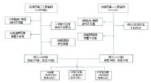
Fig.1
Data inclusion process"
Tab.1
Pathological classification of all ovarian adnexal masses"
| 分类 | 病理类型 | 训练集 | 内部验证集 | 外部测试集 |
|---|---|---|---|---|
| 良性 | 上皮性肿瘤 | 80 | 34 | 15 |
| 生殖细胞肿瘤 | 55 | 22 | 18 | |
| 性索-间质肿瘤 | 16 | 6 | 4 | |
| 混合性肿瘤 | 3 | 1 | 1 | |
| 非肿瘤性囊肿 | 75 | 37 | 21 | |
| 小计 | 229 | 99 | 59 | |
| 恶性 | 上皮性肿瘤 | 100 | 43 | 27 |
| 生殖细胞肿瘤 | 4 | 1 | 2 | |
| 性锁-间质肿瘤 | 17 | 7 | 5 | |
| 转移性肿瘤 | 13 | 5 | 4 | |
| 混合性肿瘤 | 2 | 1 | 0 | |
| 交界性 | 53 | 22 | 13 | |
| 小计 | 189 | 79 | 51 |
Tab.2
Clinical baseline characteristics after dataset division"
| 指标 | 训练集(n = 418) | 内部验证集(n = 178) | t/χ2值 | P值* | 外部测试集(n = 110) | t/χ2值 | P值# |
|---|---|---|---|---|---|---|---|
| 年龄/岁 | 45.48 ± 13.75 | 44.67 ± 14.21 | 0.63 | 0.527 | 40.80 ± 13.64 | 2.42 | 0.015 |
| AFP/(μg/L) | 12.63 ± 30.04 | 11.54 ± 29.45 | 0.40 | 0.691 | 9.96 ± 29.53 | 0.84 | 0.402 |
| CA125/(U/mL) | 88.98 ± 54.69 | 87.41 ± 49.44 | 0.32 | 0.752 | 94.78 ± 71.09 | 0.92 | 0.358 |
| CA199/(U/mL) | 45.51 ± 33.45 | 47.70 ± 35.02 | 0.71 | 0.482 | 40.35 ± 59.63 | 1.07 | 0.287 |
| CEA/(μg/L) | 23.41 ± 5.71 | 22.47 ± 5.30 | 0.44 | 0.661 | 19.81 ± 5.45 | 0.96 | 0.341 |
| HE4/(pmol/L) | 106.22 ± 38.52 | 111.32 ± 35.48 | 1.06 | 0.349 | 117.31 ± 51.36 | 2.32 | 0.021 |
| 肿瘤最大径/mm | 81.15 ± 36.71 | 79.83 ± 36.66 | 0.37 | 0.292 | 69.72 ± 27.52 | 2.17 | 0.032 |
| 绝经/[例(%)] | 258(61.72) | 120(67.47) | 1.16 | 0.283 | 77(70.00) | 1.13 | 0.285 |
| 阴道流血/[例(%)] | 39(9.33) | 19(10.61) | 0.39 | 0.351 | 14(12.58) | 1.41 | 0.227 |
Tab.3
Logistic regression analysis results of associations between clinical features and disease status"
| 特征变量 | 单因素 | 多因素 | ||
|---|---|---|---|---|
| OR(95%CI) | P值 | OR(95%CI) | P值 | |
| 年龄 | 1.01(1.00 ~ 1.01) | 0.039 | 1.01(1.00 ~ 1.01) | 0.028 |
| 绝经情况 | 4.21(2.58 ~ 6.87) | 0.002 | 2.79(1.45 ~ 5.49) | < 0.001 |
| 阴道流血情况 | 1.03(0.67 ~ 1.58) | 0.820 | ||
| AFP | 1.00(1.00 ~ 1.01) | 0.275 | ||
| CA125 | 1.06(1.03 ~ 1.08) | 0.032 | 1.04(1.02 ~ 1.07) | 0.034 |
| CA199 | 1.00(1.00 ~ 1.00) | 0.431 | ||
| CEA | 1.00(1.00 ~ 1.01) | 0.276 | ||
| HE4 | 1.06(1.03 ~ 1.09) | 0.014 | 1.06(1.04 ~ 1.08) | < 0.001 |
| 肿瘤最大径 | 1.01(1.00 ~ 1.01) | 0.015 | 1.01(1.00 ~ 1.02) | 0.038 |
Tab.4
O-RADS classification results by three physicians for all ultrasound images"
| 类别 | 医生A | 医生B | 医生C | 最终共识 |
|---|---|---|---|---|
| O-RADS 2 | 142(20.17) | 149(21.02) | 141(32.30) | 147(20.90) |
| O-RADS 3 | 217(30.82) | 214(30.25) | 220(31.10) | 215(30.40) |
| O-RADS 4 | 230(32.53) | 224(31.68) | 220(31.10) | 227(32.10) |
| O-RADS 5 | 117(16.48) | 119(16.75) | 125(17.75) | 117(16.60) |
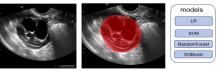
Fig.2
Using ITK-SNAP to delineate the mask region for feature extraction"
Tab.5
Diagnostic performance of different models"
| 数据集 | 模型 | 准确率 | 灵敏度 | 特异度 | AUC(95%CI) | P值 |
|---|---|---|---|---|---|---|
| 训练集 | O-RADS模型 | 0.77 | 0.82 | 0.76 | 0.86(0.81 ~ 0.92) | < 0.001 |
| O-RADS临床模型 | 0.83 | 0.85 | 0.81 | 0.90(0.86 ~ 0.95) | 0.008 | |
| 联合模型 | 0.89 | 0.90 | 0.89 | 0.95(0.92 ~ 0.98) | ||
| 内部验证集 | O-RADS模型 | 0.79 | 0.82 | 0.74 | 0.85(0.80 ~ 0.91) | 0.003 |
| O-RADS临床模型 | 0.84 | 0.86 | 0.82 | 0.88(0.82 ~ 0.93) | 0.023 | |
| 联合模型 | 0.86 | 0.88 | 0.87 | 0.92(0.88 ~ 0.96) | ||
| 外部测试集 | O-RADS模型 | 0.78 | 0.81 | 0.71 | 0.83(0.77 ~ 0.89) | < 0.001 |
| O-RADS临床模型 | 0.83 | 0.84 | 0.82 | 0.85(0.79 ~ 0.90) | 0.015 | |
| 联合模型 | 0.85 | 0.85 | 0.84 | 0.89(0.83 ~ 0.94) |
| [1] |
WEI Y F, NING L, XU Y L, et al.Worldwide patterns and trends in ovarian cancer incidence by histological subtype: A population-based analysis from 1988 to 2017[J]. E Clin Med, 2024, 79: 102983. doi: 10.1016/j.eclinm.2024.102983 .
doi: 10.1016/j.eclinm.2024.102983 |
| [2] |
WANG Y, WANG Z, ZHANG Z H, et al. Burden of ovarian cancer in China from 1990 to 2030: A systematic analysis and comparison with the global level[J]. Front Public Health, 2023, 11: 1136596. doi: 10.3389/fpubh.2023.1136596 .
doi: 10.3389/fpubh.2023.1136596 |
| [3] |
秦绪颖, 王瑞国, 王稳, 等. 卵巢肿瘤良恶性风险评估方法学中国专家共识(2024年版)[J].中国实用妇科与产科杂志, 2024, 40(3): 312-320. doi: 10.19538/j.fk2024030113 .
doi: 10.19538/j.fk2024030113 |
| [4] |
KOUTRAS A, PERROS P, PROKOPAKIS I, et al. Advantages and limitations of ultrasound as a screening test for ovarian cancer[J]. Diagnostics (Basel), 2023, 13(12): 2078. doi: 10.3390/diagnostics13122078 .
doi: 10.3390/diagnostics13122078 |
| [5] |
GUO Y, PHILLIPS C H, SUAREZ-WEISS K, et al. Interreader agreement and intermodality concordance of O-RADS US and MRI for assessing large, complex ovarian-adnexal cysts[J]. Radiol Imaging Cancer, 2022, 4(5): e220064. doi: 10.1148/rycan.220064 .
doi: 10.1148/rycan.220064 |
| [6] |
AKKAYA H, DEMIREL E, DILEK O, et al. Ovarian-adnexal reporting and data system MRI scoring: Diagnostic accuracy, interobserver agreement, and applicability to machine learning[J]. Br J Radiol, 2025, 98(1166): 254-261. doi: 10.1093/bjr/tqae221 .
doi: 10.1093/bjr/tqae221 |
| [7] |
MATSAS A, STEFANOUDAKIS D, TROUPIS T, et al. Tumor markers and their diagnostic significance in ovarian cancer[J]. Life (Basel), 2023, 13(8): 1689. doi: 10.3390/life13081689 .
doi: 10.3390/life13081689 |
| [8] |
SILVERWOOD S M, BACKER G, GALLOWAY A, et al. Assessing the rates of false-positive ovarian cancer screenings and surgical interventions associated with screening tools: A systematic review[J]. BMJ Oncol, 2024, 3(1): e000404. doi: 10.1136/bmjonc-2024-000404 .
doi: 10.1136/bmjonc-2024-000404 |
| [9] |
SALIMA S, RACHMAWATI A, HARSONO A, et al. Ovarian cancer-self assessment: An innovation for early detection and risk assessment of ovarian cancer[J]. Asian Pac J Cancer Prev, 2022, 23(8): 2643-2647. doi: 10.31557/APJCP.2022.23.8.264 .
doi: 10.31557/APJCP.2022.23.8.264 |
| [10] |
LI B, SUN M, YAO P, et al. Radiogenomics: A valuable tool for the clinical assessment and research of ovarian cancer[J]. J Comput Assist Tomogr, 2022, 46(3): 371-378. doi: 10.1097/RCT.0000000000001279 .
doi: 10.1097/RCT.0000000000001279 |
| [11] |
ZHOU J, CAO W, WANG L, et al. Application of artificial intelligence in the diagnosis and prognostic prediction of ovarian cancer[J]. Comput Biol Med, 2022, 146: 105608. doi: 10.1016/j.compbiomed.2022.105608 .
doi: 10.1016/j.compbiomed.2022.105608 |
| [12] |
何尚鹏, 黄炜贤, 江燕辉, 等. 超声特征与超声影像组学对BethesdaⅢ类甲状腺结节良恶性再评价的价值[J]. 实用医学杂志, 2025, 41(12): 1892-1898. doi: 10.3969/j.issn.1006-5725.2025.12.018 .
doi: 10.3969/j.issn.1006-5725.2025.12.018 |
| [13] |
HIETT A K, SONEK J D, GUY M, et al. Performance of IOTA Simple Rules, Simple Rules risk assessment, ADNEX model and O-RADS in differentiating between benign and malignant adnexal lesions in North American women[J]. Ultrasound Obstet Gynecol, 2022, 59(5): 668-676. doi: 10.1002/uog.24777 .
doi: 10.1002/uog.24777 |
| [14] |
JHA P, GUPTA A, BARAN T M, et al. Diagnostic performance of the ovarian-adnexal reporting and data system (O-RADS) ultrasound risk score in women in the United States[J]. JAMA Netw Open, 2022, 5(6): e2216370. doi: 10.1001/jamanetworkopen.2022.16370 .
doi: 10.1001/jamanetworkopen.2022.16370 |
| [15] |
HAN J, WEN J, HU W. Comparison of O-RADS with the ADNEX model and IOTA SR for risk stratification of adnexal lesions: A systematic review and meta-analysis[J]. Front Oncol, 2024, 14: 1354837. doi: 10.3389/fonc.2024.1354837 .
doi: 10.3389/fonc.2024.1354837 |
| [16] |
苏漫婷, 吴曼丽, 张曼, 等. 超声造影提高O-RADS 4~5类附件肿块诊断准确性的多中心回顾性研究[J]. 新医学, 2025, 56(9): 872-881. doi: 10.12464/j.issn.0253-9802.2025-0091 .
doi: 10.12464/j.issn.0253-9802.2025-0091 |
| [17] |
GRANT E G. Adding contrast-enhanced US to O-RADS: A route to improved specificity?[J]. Radiology, 2023, 308(2): e231483. doi: 10.1148/radiol.231483 .
doi: 10.1148/radiol.231483 |
| [18] |
刘芳欣, 王洲, 李健, 等. 超声O-RADS分类联合超声造影及血清CA125和HE4检测诊断绝经后卵巢肿物的应用价值[J]. 实用肿瘤杂志, 2023, 38(4): 392-397. doi: 10.13267/j.cnki.syzlzz.2023.062 .
doi: 10.13267/j.cnki.syzlzz.2023.062 |
| [19] |
何丽英, 昌禹豪, 马强, 等. 基于超声影像组学和临床特征构建的联合模型诊断早期卵巢癌的临床价值[J]. 临床超声医学杂志, 2025, 27(1): 39-47. doi: 10.16245/j.cnki.issn1008-6978.2025.01.016 .
doi: 10.16245/j.cnki.issn1008-6978.2025.01.016 |
| [20] |
STRACHOWSKI L M, JHA P, PHILLIPS C H, et al. O-RADS US v2022: An update from the American College of Radiology's ovarian-adnexal reporting and data system US committee[J]. Radiology, 2023, 308(3): e230685. doi: 10.1148/radiol.230685 .
doi: 10.1148/radiol.230685 |
| [21] |
杜阳春, 郑红雨, 陈海宁, 等. 深度学习超声影像组学列线图模型鉴别Ⅰ和Ⅱ型上皮性卵巢癌[J]. 实用医学杂志, 2025, 41(18): 2920-2927. doi: 10.3969/j.issn.1006-5725.2025.18.020 .
doi: 10.3969/j.issn.1006-5725.2025.18.020 |
| [22] |
王稳, 王兴国, 刘淑娟, 等. 交界性卵巢肿瘤诊治中国专家共识(2022年版)[J]. 中国实用妇科与产科杂志, 2022, 38(12): 1185-1194. doi: 10.19538/j.fk2022120110 .
doi: 10.19538/j.fk2022120110 |
| [23] |
POONYAKANOK V, TANMAHASAMUT P, JAISHUEN A. Prospective comparative trial comparing O-RADS, IOTA ADNEX model, and RMI score for preoperative evaluation of adnexal masses for prediction of ovarian cancer[J]. J Obstet Gynaecol Res, 2023, 49(5): 1412-1417. doi: 10.1111/jog.15624 .
doi: 10.1111/jog.15624 |
| [24] |
LU B, LIU C, QI J, et al. Comparison of contrast-enhanced ultrasound, IOTA simple rules and O-RADS for assessing the malignant risk of sonographically appearing solid ovarian masses[J]. J Gynecol Obstet Hum Reprod, 2023, 52(4): 102564. doi: 10.1016/j.jogoh.2023.102564 .
doi: 10.1016/j.jogoh.2023.102564 |
| [25] |
SHANG J, LEI T, WU L, et al. Comparison of performance between O-RADS, IOTA simple rules risk assessment and ADNEX model in the discrimination of ovarian Brenner tumors[J]. Arch Gynecol Obstet, 2023, 308(3): 961-970. doi: 10.1007/s00404-022-06903-8 .
doi: 10.1007/s00404-022-06903-8 |
| [26] |
HENRY T, SUN R, LEROUSSEAU M, et al. Investigation of radiomics based intra-patient inter-tumor heterogeneity and the impact of tumor subsampling strategies[J]. Sci Rep, 2022, 12(1): 17244. doi: 10.1038/s41598-022-20931-z .
doi: 10.1038/s41598-022-20931-z |
| [27] |
LAN W, HONG J, HUAYUN T. Advances in ovarian cancer radiomics: A bibliometric analysis from 2010 to 2024[J]. Front Oncol, 2024, 14: 1456932. doi: 10.3389/fonc.2024.1456932 .
doi: 10.3389/fonc.2024.1456932 |
| [28] |
WANG D, SU N, WANG R, et al. Serous surface papillary borderline ovarian tumors: Correlation of sonographic features with clinic pathological findings[J]. Ultrasound Obstet Gynecol, 2024, 63(5): 691-698. doi: 10.1002/uog.27454 .
doi: 10.1002/uog.27454 |
| [29] |
MASCILLINI F, MORO F, PASCIUTO T, et al. Imaging in gynecological disease (28): Clinical and ultrasound characteristics of serous and mucinous cystadenomas in the adnexa[J]. Ultrasound Obstet Gynecol, 2025, 66(2): 233-241. doi: 10.1002/uog.29248 .
doi: 10.1002/uog.29248 |
| [30] |
XIE X, MA S X, LUO X D, et al. Automatic recognition of adrenal incidentalomas using a two-stage cascade network: A multicenter study[J]. Ann Med, 2025, 57(1). doi: 0.1080/07853890.2025.2540596 .
doi: 0.1080/07853890.2025.2540596 |
| [1] | Yao ZHOU,Qianbin DAI,Wei LONG,Kaixiang HU,Rui WU,Biqi FU. Expression and significance of circular RNA hsa_circ_0001707 in ankylosing spondylitis [J]. The Journal of Practical Medicine, 2026, 42(2): 295-302. |
| [2] | Danhui LAI,Yanhui JIANG,Siting YE,Shulian ZHUANG,Shuang YANG,Wen XUE,Jianxing ZHANG. Analysis of prediction of carotid in-stent restenosis based on ultrasonographic carotid plaque radiomics [J]. The Journal of Practical Medicine, 2025, 41(5): 742-750. |
| [3] | Yutang HUANG,Weiqin DU,Dong YUAN,Tiantian LEI,Chunjie WEN,Lanxiang. WU. The mechanism of JUP promoting the malignant progression of high⁃grade serous ovarian cancer [J]. The Journal of Practical Medicine, 2025, 41(24): 3848-3859. |
| [4] | Li XIN,Weibin WANG,Xinrong WEI,Qingqing PEI,Hua. WEI. The impact of the number of negative lymph node resections on the overall survival and recurrence rate of patients with ovarian cancer [J]. The Journal of Practical Medicine, 2025, 41(21): 3412-3421. |
| [5] | Guoliang WEN,Hang FANG,Wei. ZHANG. Value of conventional radiological features and ct radiomics features in differentiating parotid adenolymphoma from malignant tumors [J]. The Journal of Practical Medicine, 2025, 41(21): 3442-3448. |
| [6] | Yangchun DU,Hongyu ZHENG,Haining CHEN,Wenwen GUO,Jinxiu YAO,Tongliu LAN,Yanju XIAO. Ultrasound⁃based deep learning radiomics nomogram to differentiate type Ⅰ and type Ⅱ epithelial ovarian cancer [J]. The Journal of Practical Medicine, 2025, 41(18): 2920-2927. |
| [7] | Zhaoyang WANG,Nan. ZHANG. Constructing a Nomogram prediction model for early recurrence of hepatocellular carcinoma radical hepatectomy based on CT imaging omics and traditional Chinese medicine tongue image features [J]. The Journal of Practical Medicine, 2025, 41(16): 2590-2596. |
| [8] | Huiliang CAI,Qianying ZHANG,Ying HUANG,Weisheng PENG,Chengli WANG,Cuiting YANG,Na DENG,Sizhu ZHANG,Nina XU,Xiaobing HAN. Assessments of ki⁃67 expression in hepatocellular carcinoma using enhanced MRI intratumoral and peritumoral radiomics and clinical imaging features [J]. The Journal of Practical Medicine, 2025, 41(15): 2311-2319. |
| [9] | Shangpeng HE,Weixian HUANG,Yanhui JIANG,Xiongqiang PENG,Lingcui MENG,Jianxing ZHANG. Value of ultrasound radiomics in re⁃evaluating the benign or malignant of Bethesda Ⅲ nodules [J]. The Journal of Practical Medicine, 2025, 41(12): 1892-1898. |
| [10] | Yekun HE,Ying LONG,Lizhang CEN,Desheng. YAO. Research progress of perineural invasion in gynecological malignant tumors [J]. The Journal of Practical Medicine, 2025, 41(12): 1929-1935. |
| [11] | Xiaoqian WU,Xuexin LIU,Yulan GAO,Zhihua HAO,Leilei GUO,Qian. NIE. Application of immune inflammatory markers combined with magnetic controlled capsule internal examination in the diagnosis of gastric adenocarcinoma and precancerous lesions [J]. The Journal of Practical Medicine, 2024, 40(16): 2333-2339. |
| [12] | Shan′gao HUANG,Yueling WU,Ying. ZHANG. Aiming for the future: The latest advances in targeted therapy for ovarian cancer [J]. The Journal of Practical Medicine, 2024, 40(14): 1901-1907. |
| [13] |
HE Wanshan , HONG Xiaoshan, CHEN Guanqiao, CHEN Bin, WEN Bin, LIN Yu, WEI Zhifu, LUO Xiping.
Screening of gene differential expression of NSUN2 in ovarian cancer cells based on transcriptome sequenc⁃ ing technology [J]. The Journal of Practical Medicine, 2023, 39(9): 1079-1085. |
| [14] | Ping YIN,Hanzi XU,Chenjing. ZHU. Application of ctDNA in gynecological malignant cancer [J]. The Journal of Practical Medicine, 2023, 39(17): 2153-2158. |
| [15] | Lin LIU,Runrong LI,Shipeng. GONG. Efficacy and safety of PEG⁃rhG⁃CSF for preventing neutropenia after chemotherapy with doxorubicin plus carboplatin in epithelial ovarian cancer patients: A retrospective clinical study [J]. The Journal of Practical Medicine, 2023, 39(17): 2236-2240. |
| Viewed | ||||||
|
Full text |
|
|||||
|
Abstract |
|
|||||