

The Journal of Practical Medicine ›› 2026, Vol. 42 ›› Issue (6): 999-1007.doi: 10.3969/j.issn.1006-5725.2026.06.012
• Chronic Disease Control • Previous Articles Next Articles
Mengxue ZANG1,Tianqi HAN1,Ying DAI1,Haiyan CHEN2,Lizhi PENG2,Yongming JIANG2,Guixin YANG2,Hui LIANG1,Xuebin LI1,2,Jianmin HUANG2( )
)
Received:2025-12-09
Revised:2026-01-14
Accepted:2026-01-15
Online:2026-03-25
Published:2026-03-26
Contact:
Jianmin HUANG
E-mail:bshuangjianmin@126.com
CLC Number:
Mengxue ZANG,Tianqi HAN,Ying DAI,Haiyan CHEN,Lizhi PENG,Yongming JIANG,Guixin YANG,Hui LIANG,Xuebin LI,Jianmin HUANG. Study on the role and mechanism of inhibiting miR-203a-3p expression in collateral circulation reconstruction in rats with cerebral ischemia-reperfusion injury model[J]. The Journal of Practical Medicine, 2026, 42(6): 999-1007.
Tab.1
Primer sequence"
| 靶基因 | 序列 |
|---|---|
miR203a?3p PIK3CA PI3K AKT Survivin | Forward:5′-CGCGGTGAAATGTTTAGGAC-3′ Reverse:5′-ATCCAGTGCAGGGTCCGAGG-3′ Forward:5'-AAATGAAAGCTCACTCTGG-3' Reverse:5'-TGTGCAATTCCTATGCAATC-3' Forward:5′-AACGTGCCGATCCTACAGTC-3′ Reverse:5′-AGGTCCAGAGATTCAGCCTC-3′ Forward:5′-GGAGACGATGGACTTCCGGT-3′ Reverse:5′-ACTCGTTCATGGTCACACGG-3′ Forward:5'-TAAGCCACTTGTCCCAGCTT-3' Reverse:5'-CTCATCCACTCCCTTCCTCA-3' |
U6 GAPDH | Forward:5′-CCTGCTTCGGCAGCACA-3′ Reverse:5′-AACGCTTCACGAATTTGCGT-3′ Forward:5′-AGACCTCTATGCCAACACAGTGC-3′ Reverse:5′-GAGCCACCAATCCACACAGAGT-3′ |
Fig.1
The binding diagram of miR-203a-3p and PIK3CA-3'UTR target site"

Tab.2
Comparison of neurological deficit scores among groups"
| 组别 | n | Longa评分 |
|---|---|---|
| 假手术组 | 10 | 0.00 ± 0.00 |
| 模型组 | 10 | 2.10 ± 0.57? |
| antagomiR-NC组 | 10 | 2.10 ± 0.86? |
| antagomiR-203a-3p组 | 10 | 1.10 ± 0.57? # & |
Tab.3
Comparison of cerebral infarct volume and MBPU in the cerebral cortex among different groups"
| 组别 | n | 脑梗死体积/% | 大脑皮层MBPU/PU |
|---|---|---|---|
| 假手术组 | 5 | 0.00 ± 0.00 | 1 265.63 ± 250.54 |
| 模型组 | 5 | 26.19 ± 2.68* | 771.13 ± 186.25* |
| antagomiR-NC组 | 5 | 28.03 ± 2.63* | 710.71 ± 256.16* |
| antagomiR-203a-3p组 | 5 | 21.49 ± 2.59* # & | 1 095.37 ± 185.88# & |
Fig.2
TTC staining of brain tissue from rats in each group"
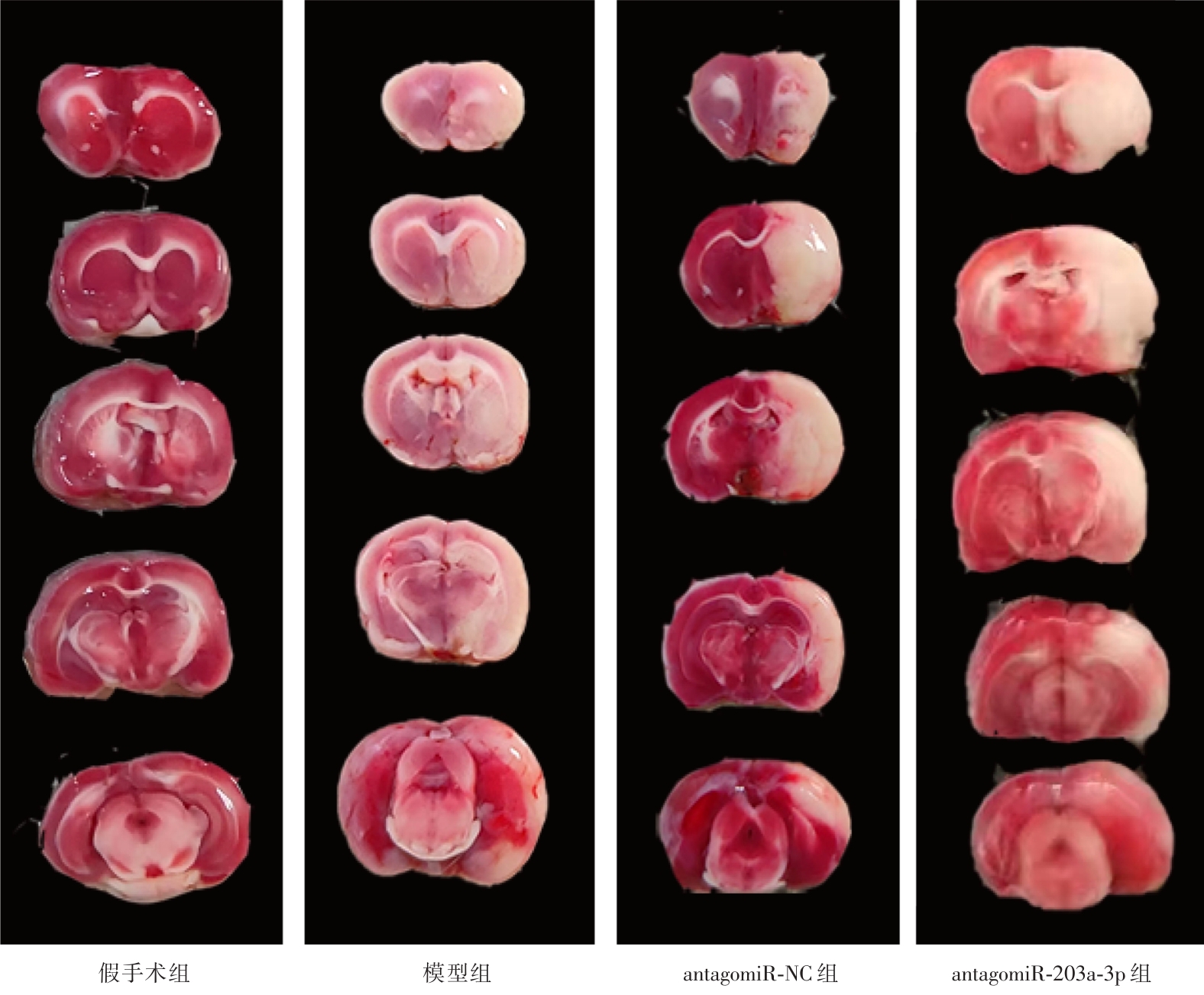

Fig.3
Changes in collateral circulation in rats of each group"
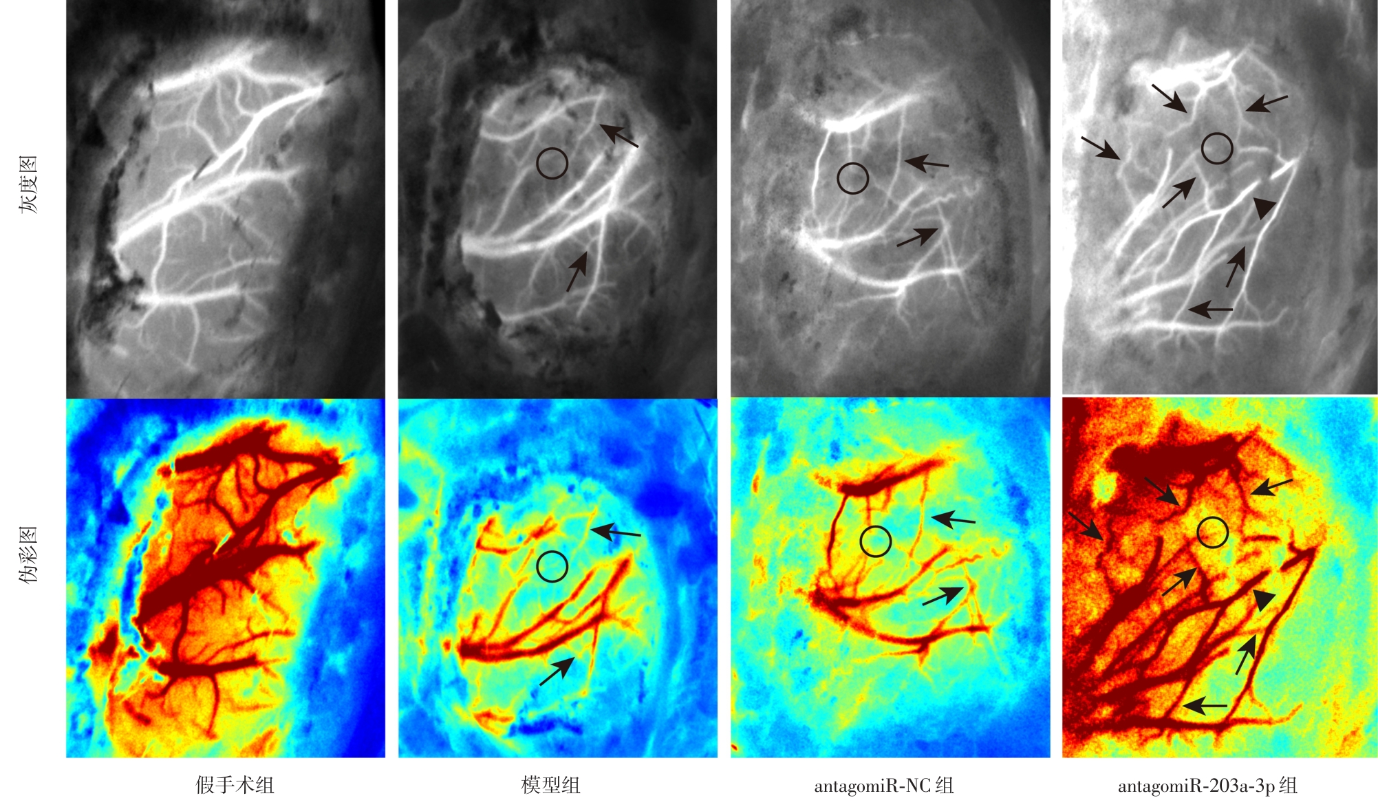
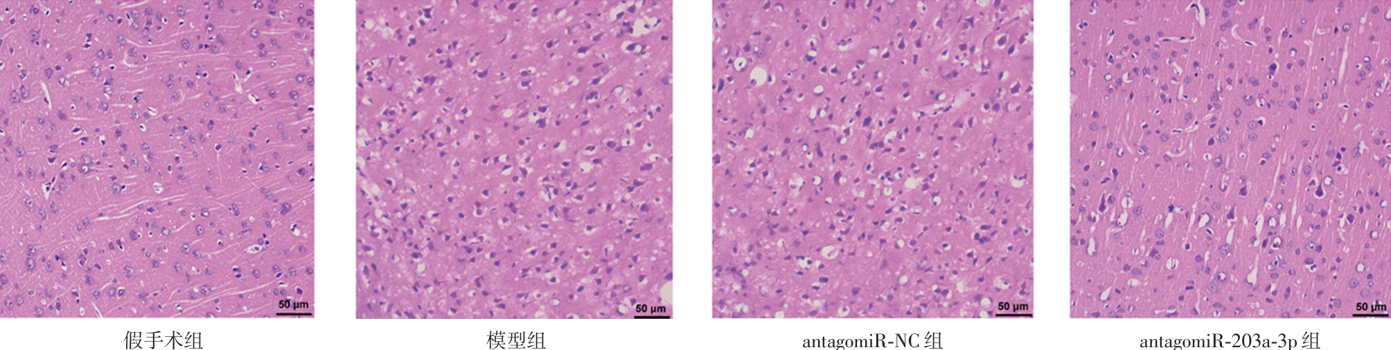
Fig.4
Pathological morphology of brain tissue in each group of rats(HE,× 400)"
Tab.4
Comparison of miR-203a-3p, PIK3CA, PI3K, AKT, and Survivin mRNA expression in the brain tissue of rats among the groups"
| 组别 | miR-203a-3p | PIK3CA | PI3K | AKT | Survivin |
|---|---|---|---|---|---|
| 假手术组 | 3.03 ± 0.35 | 0.49 ± 0.10 | 0.64 ± 0.09 | 0.57 ± 0.11 | 0.59 ± 0.06 |
| 模型组 | 1.20 ± 0.28? | 1.09 ± 0.17? | 1.21 ± 0.19? | 1.20 ± 0.16? | 1.25 ± 0.12? |
| antagomiR-NC组 | 1.19 ± 0.07? | 1.13 ± 0.10? | 1.11 ± 0.08? | 1.12 ± 0.09? | 1.26 ± 0.11? |
| antagomiR-203a-3p组 | 0.24 ± 0.10? # & | 2.15 ± 0.43? # & | 3.68 ± 0.38? # & | 5.12 ± 0.44? # & | 3.27 ± 0.41? # & |
Tab.5
Comparison of PIK3CA, p-PI3K/PI3K, p-AKT/AKT, and Survivin protein expression in the brain tissue of rats among the groups"
| 组别 | PIK3CA | p-PI3K/PI3K | p-AKT/AKT | Survivin |
|---|---|---|---|---|
| 假手术组 | 0.50 ± 0.05 | 0.33 ± 0.05 | 0.41 ± 0.08 | 0.32 ± 0.04 |
| 模型组 | 1.00 ± 0.14? | 0.74 ± 0.10? | 0.89 ± 0.10? | 0.67 ± 0.13? |
| antagomiR-NC组 | 1.04 ± 0.09? | 0.74 ± 0.11? | 0.92 ± 0.13? | 0.68 ± 0.14? |
| antagomiR-203a-3p组 | 1.80 ± 0.32? # & | 1.34 ± 0.07? # & | 1.69 ± 0.29? # & | 1.33 ± 0.12? # & |

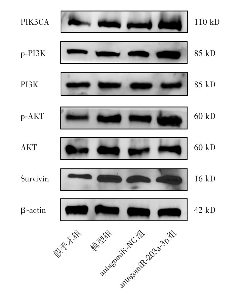
Fig.5
Western blot analysis of PIK3CA, p-PI3K, PI3K, p-AKT, AKT, and Survivin protein expression in brain tissues across groups"
| [1] |
MAIDA C D, NORRITO R L, RIZZICA S, et al. Molecular Pathogenesis of Ischemic and Hemorrhagic Strokes: Background and Therapeutic Approaches [J]. Int J Mol Sci, 2024,25(12):6297. doi:10.3390/ijms25126297 .
doi: 10.3390/ijms25126297 |
| [2] |
OH M, LEE M. Clinical Implications of Prominent Cortical Vessels on Susceptibility-Weighted Imaging in Acute Ischemic Stroke Patients Treated with Recanalization Therapy [J]. Brain Sci, 2022,12(2):184. doi:10.3390/brainsci12020184 .
doi: 10.3390/brainsci12020184 |
| [3] |
LI X, WANG F, ZHAO Z, et al. A SCANO Nomogram for Individualized Prediction of the Probability of 1-Year Unfavorable Outcomes in Chinese Acute Ischemic Stroke Patients [J].Front Neurol, 2020,11:531. doi:10.3389/fneur.2020.00531 .
doi: 10.3389/fneur.2020.00531 |
| [4] |
LI X, LI S, MA C, et al. Preparation of baicalin-loaded ligand-modified nanoparticles for nose-to-brain delivery for neuroprotection in cerebral ischemia [J]. Drug Deliv, 2022,29(1):1282-1298. doi:10.1080/10717544.2022.2064564 .
doi: 10.1080/10717544.2022.2064564 |
| [5] |
HE Q, WANG Y, FANG C, et al. Advancing stroke therapy: A deep dive into early phase of ischemic stroke and recanalization [J]. CNS Neurosci Ther, 2024,30(2):e14634. doi:10.1111/cns.14634 .
doi: 10.1111/cns.14634 |
| [6] |
AL-AJLAN F S, ALKHIRI A, ALAMRI A F, et al. Golden Hour Intravenous Thrombolysis for Acute Ischemic Stroke: A Systematic Review and Meta‐Analysis [J]. Ann Neurol, 2024,96(3):582-590. doi:10.1002/ana.27007 .
doi: 10.1002/ana.27007 |
| [7] |
姜浩, 程凯理, 陈柳. 血清血小板反应蛋白-1联合阿尔伯塔中风项目早期CT评分对大动脉闭塞性急性脑梗死患者预后的评估价值 [J]. 实用医学杂志, 2025,41(16):2561-2567. doi:10.3969/j.issn.1006-5725.2025.16.018 .
doi: 10.3969/j.issn.1006-5725.2025.16.018 |
| [8] |
ZHANG X, DUAN G, ZHANG H, et al. Safety and effectiveness assessment of endovascular recanalization fornon‐acute middle cerebral artery occlusion [J].CNS Neurosci Ther, 2024,30(3):e14426. doi:10.1111/cns.14426 .
doi: 10.1111/cns.14426 |
| [9] |
MAGUIDA G, SHUAIB A. Collateral Circulation in Ischemic Stroke: An Updated Review [J].J Stroke, 2023,25(2):179-198. doi:10.5853/jos.2022.02936 .
doi: 10.5853/jos.2022.02936 |
| [10] |
HUANG J, LI X, ZHAO J, et al. Association of BIRC5 Gene Polymorphism with the Collateral Circulation and Severity of Large Artery Atherosclerotic Stroke [J]. Int J Clin Pract, 2022,2022(1):9177545. doi:10.1155/2022/9177545 .
doi: 10.1155/2022/9177545 |
| [11] |
李雪斌, 黄建敏, 云艳芳, 等.生存素抑制剂YM155对脑梗死大鼠微血管密度及血管新生相关基因表达的影响 [J]. 实用医学杂志, 2022, 38 (8):946-951. doi:10.3969/j.issn.1006-5725.2022.08.007 .
doi: 10.3969/j.issn.1006-5725.2022.08.007 |
| [12] |
李雪斌, 黄建敏, 陈海燕, 等. 过表达BIRC5基因对氧糖剥夺/复氧诱导的脑微血管内皮细胞活性及VEGF表达的影响[J]. 右江民族医学院学报, 2024,46(1):1-7. doi:10.3969/j.issn. 1001-5817.2024.01.001 .
doi: 10.3969/j.issn. 1001-5817.2024.01.001 |
| [13] |
朱腾腾. miR-203a-3p通过PIK3CA/PI3K/Akt通路调控PASMCs的增殖和迁移在PAH中的机制研究 [D]. 长沙:中南大学, 2023. doi:10.27661/d.cnki.gzhnu.2023.000221 .
doi: 10.27661/d.cnki.gzhnu.2023.000221 |
| [14] |
HU Z, ZHUO L, LI Y, et al. MicroRNA‑203a‑3p suppresses endothelial cell proliferation and invasion, and promotes apoptosis in hemangioma by inactivating the VEGF‑mediated PI3K/AKT pathway [J]. Exp Ther Med, 2022,24(5):644. doi:10.3892/etm. 2022.11581 .
doi: 10.3892/etm. 2022.11581 |
| [15] |
胡琳, 杨艳娟, 李洁滢, 等. 红景天苷通过激活PI3K/Akt和抑制NF ⁃ κ B信号通路改善川崎病冠状动脉损伤 [J]. 实用医学杂志, 2022, 38 (21): 2657-2662. doi:10.3969/j.issn.1006-5725.2022.21.005 .
doi: 10.3969/j.issn.1006-5725.2022.21.005 |
| [16] |
GREITHER T, KOSER F, HOLZHAUSEN H, et al. MiR-155-5p and MiR-203a-3p Are Prognostic Factors in Soft Tissue Sarcoma [J]. Cancers (Basel), 2020,12(8):2254. doi:10.3390/cancers12082254 .
doi: 10.3390/cancers12082254 |
| [17] |
ENTEZARI M, SOLTANI B M, SADEGHIZADEH M. MicroRNA-203a inhibits breast cancer progression through the PI3K/Akt and Wnt pathways [J]. Sci Rep, 2024,14(1):4715. doi:10.1038/s41598-024-52940-5 .
doi: 10.1038/s41598-024-52940-5 |
| [18] |
ZHUO L, HU Z, CHANG J, et al. MicroRNA‑203a‑3p improves bleomycin and pingyangmycin sensitivity by inactivating the PI3K/AKT pathway in hemangioma [J]. Exp Ther Med, 2024,27(2). doi:10.3892/etm.2024.12369 .
doi: 10.3892/etm.2024.12369 |
| [19] |
袁柳媚, 卢小叶, 云夏, 等. 线栓法制备大鼠大脑中动脉闭塞脑缺血模型的体会 [J]. 中国医药科学, 2022,12(2): 7-10. doi:10.3969/j.issn.2095-0616.2022.02.003 .
doi: 10.3969/j.issn.2095-0616.2022.02.003 |
| [20] |
LI Q, ZHANG H, LIU X. Didymin Alleviates Cerebral Ischemia-Reperfusion Injury by Activating the PPAR Signaling Pathway [J]. Yonsei Med J, 2022,63(10):956. doi:10.3349/ymj. 2022.0040 .
doi: 10.3349/ymj. 2022.0040 |
| [21] |
WANG L, SHI Q, XUE Y, et al. Albumin levels and cerebral collateral circulation in patients with acute ischemic stroke due to intracranial arteriosclerotic: A propensity score-matched analysis [J].Medicine (Baltimore), 2024,103(21):e38254. doi:10.1097/MD.0000000000038254 .
doi: 10.1097/MD.0000000000038254 |
| [22] |
YUN S W, KIM W Y, LEE J B. Correlation between Serum Erythropoietin and Cerebral Collateral Flow in Acute Ischemic Stroke Patient [J].Korean J Fam Med, 2023,44(1):53-57. doi:10.4082/kjfm.22.0117 .
doi: 10.4082/kjfm.22.0117 |
| [23] |
陈英道, 李海宁, 张岐平, 等. 脑侧支循环对急性脑梗死患者机械取栓术后疗效及预后的影响 [J]. 实用医学杂志, 2021, 37 (12): 1563-1568. doi:10.3969/j.issn.1006-5725.2021. 12.011 .
doi: 10.3969/j.issn.1006-5725.2021. 12.011 |
| [24] |
CAI W, LIU S, LIU Z, et al. Downregulation of lung miR-203a-3p expression by high-altitude hypoxia enhances VEGF/Notch signaling [J]. Aging (Albany NY), 2020, 12(5): 4247-4267. doi:10.18632/aging.102878 .
doi: 10.18632/aging.102878 |
| [25] |
JEON Y S, KIM H J, ROH H G, et al. Impact of Collateral Circulation on Futile Endovascular Thrombectomy in Acute Anterior Circulation Ischemic Stroke [J]. J Korean Neurosurg Soc, 2024,67(1):31-41. doi:10.3340/jkns.2023.0139 .
doi: 10.3340/jkns.2023.0139 |
| [26] |
LEE J M, SHIN Y J, BYOUNG-SOO S, et al. A case series of acute ischemic strokes with contralateral perfusion time delay on brain computed tomography [J].Medicine (Baltimore), 2023,102(19):e33790. doi:10.1097/MD.0000000000033790 .
doi: 10.1097/MD.0000000000033790 |
| [27] |
HAN G, LI D, WANG J, et al. Adaptive window space direction laser speckle contrast imaging to improve vascular visualization [J]. Biomed Opt Express, 2023, 14(6): 3086-3099. doi:10.1364/BOE.488054 .
doi: 10.1364/BOE.488054 |
| [28] |
KIM Y, CHOI W J, OH J, et al. Compact Smartphone-Based Laser Speckle Contrast Imaging Endoscope Device for Point-of-Care Blood Flow Monitoring [J]. Biosensors (Basel), 2022,12(6):398. doi:10.3390/bios12060398 .
doi: 10.3390/bios12060398 |
| [29] |
LI K, GAO Z, GUO Y, et al. Preconditioning exercise reduces brain damage of ischemic stroke in rats via PI3K-AKT pathway by bioinformatic analysis [J]. Exp Brain Res, 2024, 242(4): 869-878. doi:10.1007/s00221-024-06778-y .
doi: 10.1007/s00221-024-06778-y |
| [30] |
YANG X, LI X, LIN Q, et al. Up-regulation of microRNA-203 inhibits myocardial fibrosis and oxidative stress in mice with diabetic cardiomyopathy through the inhibition of PI3K/Akt signaling pathway via PIK3CA [J].Gene, 2019,715:143995. doi:10.1016/j.gene.2019.143995 .
doi: 10.1016/j.gene.2019.143995 |
| [31] |
YU L, FU J, YU N, et al. Long noncoding RNA MALAT1 participates in the pathological angiogenesis of diabetic retinopathy in an oxygen-induced retinopathy mouse model by sponging miR-203a-3p [J]. Can J Physiol Pharmacol, 2020,98(4):219-227. doi:10.1139/cjpp-2019-0489 .
doi: 10.1139/cjpp-2019-0489 |
| [32] |
XU Y, TAN X, WANG D, et al. Elevated survivin expression in peripheral blood mononuclear cells is central to collateral formation in coronary chronic total occlusion [J]. Int J Mol Med, 2015,35(6):1501-1510. doi:10.3892/ijmm.2015.2154 .
doi: 10.3892/ijmm.2015.2154 |
| [33] |
MA J, MA Y, SHUAIB A, et al. Improved collateral flow and reduced damage after remote ischemic perconditioning during distal middle cerebral artery occlusion in aged rats [J]. Sci Rep, 2020,10(1):12392. doi:10.1038/s41598-020-69122-8 .
doi: 10.1038/s41598-020-69122-8 |
| [34] |
LV H, LIU X, CHEN W, et al. Yangxue Jiedu Fang Ameliorates Psoriasis by Regulating Vascular Regression via Survivin/PI3K/Akt Pathway [J]. J Immunol Res, 2021,2021:1-18. doi:10.1155/2021/4678087 .
doi: 10.1155/2021/4678087 |
| [35] |
CANDIDO S, SALEMI R, PICCININ S, et al. The PIK3CA H1047R Mutation Confers Resistance to BRAF and MEK Inhibitors in A375 Melanoma Cells through the Cross-Activation of MAPK and PI3K-Akt Pathways [J].Pharmaceutics, 2022,14(3):590. doi:10.3390/pharmaceutics14030590 .
doi: 10.3390/pharmaceutics14030590 |
| [36] |
LI Q, TANG Y, ZUO J, et al. CENP-H as a new prognostic biomarker for tumors: A real-world literature review [J]. Front Oncol, 2025,15:1521988. doi:10.3389/fonc.2025.1521988 .
doi: 10.3389/fonc.2025.1521988 |
| [1] | Yue SUN,Na GE,Xue ZHAO,Tengfei GUO,Jiawei LIU,Wenlong ZHANG,Peng. ZHOU. Effect of ursolic acid extracted from Hippophae Rhamnoides L. on repair of sciatic nerve injury in rats [J]. The Journal of Practical Medicine, 2023, 39(24): 3158-3162. |
| [2] | Yi AN,Yanfang YUN,Guixin YANG,Haiyan CHEN,Yongming JIANG,Dongxu HUANG,Xiaorong MO,Xiaolan LI,Baoyin WEI,Yingjie ZHOU,Xuebin LI,Jianmin. HUANG. Relationship between non⁃high density lipoprotein cholesterol and leptomeningeal collaterals in patients with acute middle cerebral artery occlusion [J]. The Journal of Practical Medicine, 2023, 39(24): 3200-3204. |
| [3] | Jiashan LI,Debin YANG,Zhifeng. PENG. Effect of chronic high fat diet on brain injury in ischemia/reperfusion rats and its mechanisms [J]. The Journal of Practical Medicine, 2023, 39(20): 2579-2583. |
| [4] |
HUANG Jianmin, YUN Yanfang, YANG Guixin, CHEN Haiyan, JIANG Yongming, HUANG Dongxu, LIU Jie, MO Xiaorong, LI Xuebin..
Effects of survivin inhibitor YM155 on microvessel density and angiogenesis⁃related gene expression in rats with cerebral infarction [J]. The Journal of Practical Medicine, 2022, 38(8): 946-951. |
| [5] |
QIU Xianxian, AN Nini, JIANG Kehua, PENG Jinpu, ZHAO Yinyin, SUN Fa..
Reproductive dysfunction induced by PM2.5 exposure through NF ⁃ κB signaling pathway in male rats [J]. The Journal of Practical Medicine, 2022, 38(7): 836-847. |
| [6] |
ZHOU Yongxin, ZHAI Wenjing, JIA Zhiqiang, ZHAO Xiaoguang, WANG Lei, FANG Liping, ZHAI Shafei, HUANG Tao. .
Exosomes derived from miR⁃210⁃5p⁃modified mesenchymal stem cells promote recovery after spinal cord injury in rats [J]. The Journal of Practical Medicine, 2022, 38(6): 711-714. |
| [7] |
ZHENG Qiang, TANG Yong, ZHOU Chengji, GU Zi, LIU Shiping..
The effect and mechanism of lactulose on acute lung injury in hemorrhagic shock rats [J]. The Journal of Practical Medicine, 2022, 38(5): 577-582. |
| [8] |
PAN Qi, ZHANG Wangming, LUO Fei, XU Ruxiang. .
Alterations of firing activity of the ventromedial thalamic nucleus in rats with unilateral 6⁃OHDA lesions [J]. The Journal of Practical Medicine, 2022, 38(3): 324-329. |
| [9] |
NONG Changliang, HUANG Chuhong, FENG Jinming, XU Qianhui, HE Xiao, MO Jinli, SHI Xuekai, HUANG Yuwei..
Improvement of hyperoxia ⁃induced lung injury via ginsenoside Rb1 in newborn rats and its mechanism [J]. The Journal of Practical Medicine, 2022, 38(2): 145-149. |
| [10] |
WEN Hailin, YANG Jingzhe, MENG Xiangxi, ZHANG Xiangyun..
miR⁃320 reduces intestinal mucosal injury in burn injury rats by inhibiting TLR4/NF⁃κB signaling path⁃ way [J]. The Journal of Practical Medicine, 2022, 38(16): 2031-2036. |
| [11] |
ZHANG Kai, LI Wenjun.
Expression of HO⁃1 mRNA and HO⁃1 in sepsis rats [J]. The Journal of Practical Medicine, 2021, 37(6): 713-717. |
| [12] |
LI Wanmei, YANG Xubin, HUANG Yiwen, CHEN Xueyan..
Effect of blood glucose fluctuation on cardiovascular autonomic neuropathy in diabetic rats [J]. The Journal of Practical Medicine, 2021, 37(16): 2065-2068. |
| [13] |
CHEN Yingdao, LI Haining, ZHANG Qiping, LI Yuying, LIANG Bingsong, RAO Yuan, CHEN Xiaoling. .
Influence of collateral circulation on curative effect and prognosis of patients with acute cerebral infarction after mechanical thrombectomy [J]. The Journal of Practical Medicine, 2021, 37(12): 1563-1568. |
| Viewed | ||||||
|
Full text |
|
|||||
|
Abstract |
|
|||||