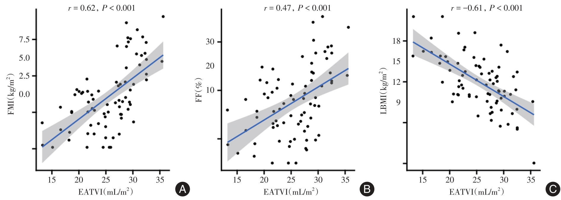
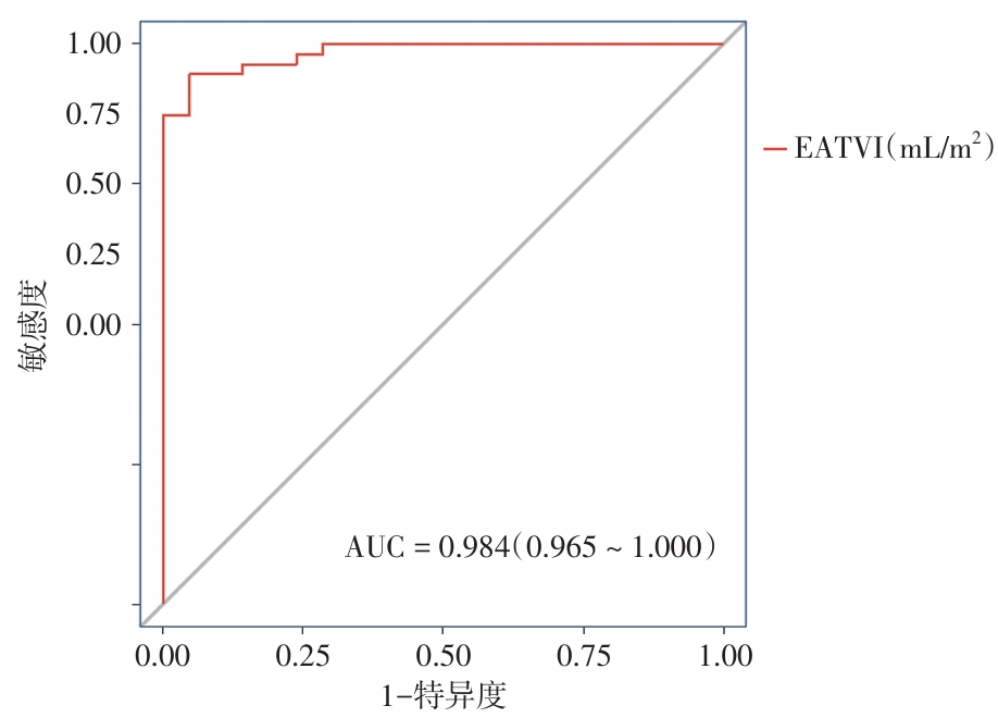
| [1] |
CHANG M, CAI Y, GAO Z, et al. Duchenne muscular dystrophy: Pathogenesis and promising therapies[J]. J Neurol, 2023, 270(8): 3733-3749. doi: 10.1007/s00415-023-11796-x .
doi: 10.1007/s00415-023-11796-x
|
| [2] |
OWENS A T, JESSUP M. Cardioprotection in Duchenne muscular dystrophy[J]. Eur Heart J, 2021, 42(20): 1985-1987. doi: 10.1093/eurheartj/ehab152 .
doi: 10.1093/eurheartj/ehab152
|
| [3] |
中华医学会罕见病分会,北京医学会罕见病分会. Duchenne型肌营养不良多学科管理指南[J]. 中华内科杂志, 2025, 64(9): 812-824. doi: 10.3760/cma.j.cn112138-20250412-00210 .
doi: 10.3760/cma.j.cn112138-20250412-00210
|
| [4] |
罗新林, 樊朝美. Becker型肌营养不良心肌病的诊治进展[J]. 中华心力衰竭和心肌病杂志, 2021, 5(2): 123-127. doi: 10.3760/cma.j.cn101460-20210506-00032 .
doi: 10.3760/cma.j.cn101460-20210506-00032
|
| [5] |
IACOBELLIS G. Epicardial adipose tissue in contemporary cardiology[J]. Nat Rev Cardiol, 2022, 19(9): 593-606. doi: 10.1038/s41569-022-00679-9 .
doi: 10.1038/s41569-022-00679-9
|
| [6] |
PACKER M. Epicardial Adipose Tissue May Mediate Deleterious Effects of Obesity and Inflammation on the Myocardium[J]. J Am Coll Cardiol, 2018, 71(20): 2360-2372. doi: 10.1016/j.jacc.2018.03.509 .
doi: 10.1016/j.jacc.2018.03.509
|
| [7] |
VAN BEIJSTERVELDT IALP, VAN DER STEEN M, DE FLUITER K S, et al. Body composition and bone mineral density by Dual Energy X-ray Absorptiometry: Reference values for young children[J]. Clin Nutr, 2022, 41(1): 71-79. doi: 10.1016/j.clnu.2021.11.010 .
doi: 10.1016/j.clnu.2021.11.010
|
| [8] |
SILVA M C, MAGALHÃES T A, MEIRA Z M, et al. Myocardial Fibrosis Progression in Duchenne and Becker Muscular Dystrophy: A Randomized Clinical Trial[J]. JAMA Cardiol, 2017, 2(2): 190-199. doi: 10.1001/jamacardio.2016.4801 .
doi: 10.1001/jamacardio.2016.4801
|
| [9] |
刘慧, 杨志刚, 郭应坤, 等. 心脏MRI诊断肌营养不良心肌病研究进展[J]. 中国医学影像技术, 2019, 35(10): 1578-1581. doi: 10.13929/j.1003-3289.201904179 .
doi: 10.13929/j.1003-3289.201904179
|
| [10] |
GOELLER M, ACHENBACH S, MARWAN M, et al. Epicardial adipose tissue density and volume are related to subclinical atherosclerosis, inflammation and major adverse cardiac events in asymptomatic subjects[J]. J Cardiovasc Comput Tomogr, 2018, 12(1): 67-73. doi: 10.1016/j.jcct.2017.11.007 .
doi: 10.1016/j.jcct.2017.11.007
|
| [11] |
IACOBELLIS G. Local and systemic effects of the multifaceted epicardial adipose tissue depot[J]. Nat Rev Endocrinol, 2015, 11(6): 363-371. doi: 10.1038/nrendo.2015.58 .
doi: 10.1038/nrendo.2015.58
|
| [12] |
CALCATERRA G, VARGHESE R T, BARONI M, et al. Earlier Prediction of Cardiovascular Risk with Epicardial Fat Assessment[J]. Eur J Intern Med, 2026, 143: 106481. doi:10.1016/j.ejim.2025.106481 .
doi: 10.1016/j.ejim.2025.106481
|
| [13] |
YUAN W, XU H, YU L, et al. Association of increased epicardial adipose tissue derived from cardiac magnetic resonance imaging with myocardial fibrosis in Duchenne muscular dystrophy: A clinical prediction model development and validation study in 283 participants[J]. Quant Imaging Med Surg, 2024, 14(1): 736-748. doi: 10.21037/qims-23-790 .
doi: 10.21037/qims-23-790
|
| [14] |
RABKIN S W. Epicardial fat: Properties, function and relationship to obesity[J]. Obes Rev, 2007, 8(3): 253-261. doi: 10.1111/j.1467-789X.2006.00293.x .
doi: 10.1111/j.1467-789X.2006.00293.x
|
| [15] |
GONZÁLEZ N, MORENO-VILLEGAS Z, GONZÁLEZ-BRIS A, et al. Regulation of visceral and epicardial adipose tissue for preventing cardiovascular injuries associated to obesity and diabetes[J]. Cardiovasc Diabetol, 2017, 16(1): 44. doi: 10.1186/s12933-017-0528-4 .
doi: 10.1186/s12933-017-0528-4
|
| [16] |
FU K, DONG Y, WANG Z, et al. The role of body composition in left ventricular remodeling, reverse remodeling, and clinical outcomes for heart failure with mildly reduced ejection fraction: More knowledge to the "obesity paradox"[J]. Cardiovasc Diabetol, 2024, 23(1): 334. doi: 10.1186/s12933-024-02430-9 .
doi: 10.1186/s12933-024-02430-9
|
| [17] |
王叶宝, 林永萍, 刘玲, 等. 血清miR-210、TSG-6、CTRP3水平与扩张型心肌病患者心肌纤维化及预后的关系[J]. 实用医学杂志, 2025, 41(23): 3690-3696. doi: 10.3969/j.issn.1006-5725.2025.23.009 .
doi: 10.3969/j.issn.1006-5725.2025.23.009
|
| [18] |
VERHAERT D, RICHARDS K, RAFAEL-FORTNEY J A, et al. Cardiac involvement in patients with muscular dystrophies: Magnetic resonance imaging phenotype and genotypic considerations[J]. Circ Cardiovasc Imaging, 2011, 4(1): 67-76. doi: 10.1161/CIRCIMAGING.110.960740 .
doi: 10.1161/CIRCIMAGING.110.960740
|
| [19] |
WEBER F J, LATSHANG T D, BLUM M R, et al. Prognostic factors, disease course, and treatment efficacy in Duchenne muscular dystrophy: A systematic review and meta-analysis[J]. Muscle Nerve, 2022, 66(4): 462-470. doi: 10.1002/mus.27682 .
doi: 10.1002/mus.27682
|
| [20] |
杨晓, 张欢, 张朝晖, 等. 地夫可特改善杜氏肌营养不良症小鼠运动能力及减轻肌肉炎症损伤[J]. 实用医学杂志, 2023, 39(13): 1614-1619, 1626. doi: 10.3969/j.issn.1006-5725.2023.13.004 .
doi: 10.3969/j.issn.1006-5725.2023.13.004
|
| [21] |
MENDONÇA D D, SILVA W V R DA, SOUZA G C, et al. Body Composition and Survival in Patients With Heart Failure: A Systematic Review and Meta-Analysis[J]. JACC Heart Fail, 2025, 13(6): 943-954. doi: 10.1016/j.jchf.2025.01.016 .
doi: 10.1016/j.jchf.2025.01.016
|
| [22] |
冯钰玲, 许华燕, 李春平, 等. 肌营养不良心肌病的影像学研究进展[J]. 实用放射学杂志, 2021, 37(11): 1898-1902. doi: 10.3969/j.issn.1002-1671.2021.11.036 .
doi: 10.3969/j.issn.1002-1671.2021.11.036
|


 )
)