

The Journal of Practical Medicine ›› 2025, Vol. 41 ›› Issue (3): 379-384.doi: 10.3969/j.issn.1006-5725.2025.03.011
• Clinical Research • Previous Articles
Yiyang ZHAI,Yunyi MA,Junying ZHAI,Hongli NIU,Ying. WANG
Received:2024-10-18
Online:2025-02-10
Published:2025-02-19
CLC Number:
Yiyang ZHAI,Yunyi MA,Junying ZHAI,Hongli NIU,Ying. WANG. The relationship between tumor necrosis factor alpha inducible protein 8 family members 2, cell proliferation nuclear antigen expression levels, and clinical pathological parameters and prognosis in endometrial cancer tissue[J]. The Journal of Practical Medicine, 2025, 41(3): 379-384.
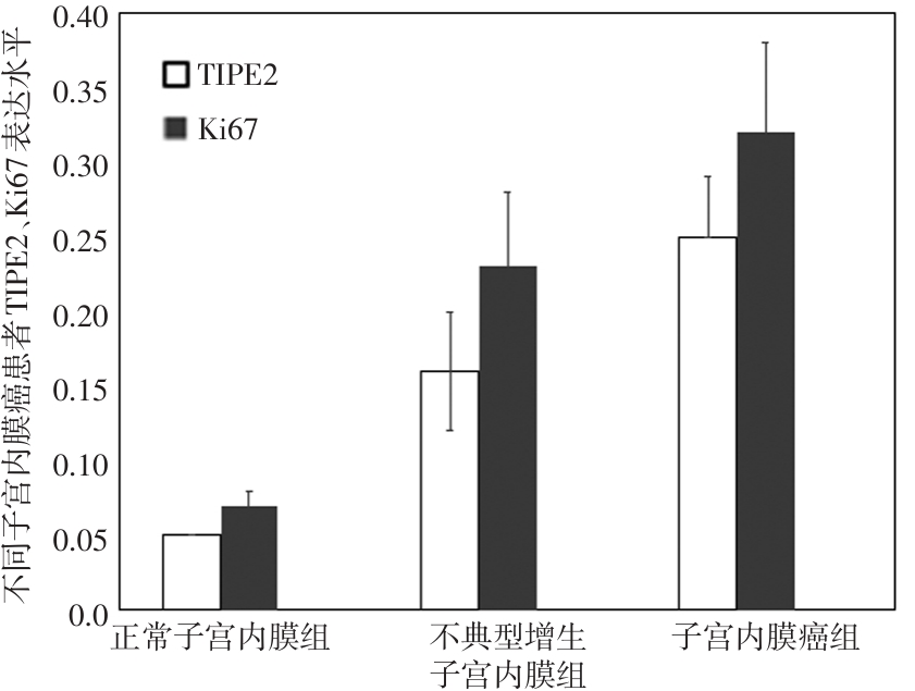
Tab.1
Expression levels of TIPE2 and Ki67 in different patients with EC"
| 组别 | TIPE2 | Ki67 |
|---|---|---|
| 正常子宫内膜组(n = 120) | 0.05 ± 0.00 | 0.07 ± 0.01 |
| 不典型增生子宫内膜组(n = 120) | 0.16 ± 0.04? | 0.23 ± 0.05? |
| EC组(n = 96) | 0.25 ± 0.04?# | 0.32 ± 0.06?# |
| F值 | 1 054.102 | 899.139 |
| P值 | < 0.001 | < 0.001 |

Fig.1
Expression levels of TIPE2 and Ki67 in different patients with EC"
Tab.2
Relationship between expression levels of TIPE2 and Ki67 in EC tissues and clinical pathological parameters"
| 临床病理参数 | 例数 | TIPE表达水平 | F/t值 | P值 | Ki67表达水平 | F/t值 | P值 |
|---|---|---|---|---|---|---|---|
| 组织病理类型 | 1.600 | 0.195 | 0.834 | 0.479 | |||
| 腺癌 | 30 | 0.23 ± 0.04 | 0.28 ± 0.06 | ||||
| 鳞状上皮癌 | 28 | 0.25 ± 0.05 | 0.30 ± 0.07 | ||||
| 浆液性癌 | 12 | 0.26 ± 0.06 | 0.31 ± 0.08 | ||||
| 黏液性癌 | 26 | 0.24 ± 0.04 | 0.30 ± 0.06 | ||||
| FIGO手术病理分期 | 18.803 | <0.001 | 5.629 | 0.005 | |||
| Ⅰ期 | 34 | 0.20 ± 0.03 | 0.27 ± 0.05 | ||||
| Ⅱ期 | 42 | 0.22 ± 0.04* | 0.29 ± 0.06 | ||||
| Ⅲ期 | 20 | 0.26 ± 0.03*# | 0.32 ± 0.04*# | ||||
| 肌层浸润程度 | 1.226 | 0.223 | 1.406 | 0.163 | |||
| <1/2的肌层 | 62 | 0.24 ± 0.03 | 0.30 ± 0.07 | ||||
| ≥1/2的肌层 | 34 | 0.25 ± 0.05 | 0.32 ± 0.06 | ||||
| 淋巴结转移 | 5.601 | <0.001 | 2.715 | 0.008 | |||
| 无 | 55 | 0.21 ± 0.03 | 0.29 ± 0.07 | ||||
| 有 | 41 | 0.25 ± 0.04 | 0.34 ± 0.11 | ||||
| 合并糖尿病 | 1.861 | 0.066 | 1.957 | 0.053 | |||
| 无 | 42 | 0.22 ± 0.04 | 0.32 ± 0.08 | ||||
| 有 | 54 | 0.24 ± 0.06 | 0.29 ± 0.07 | ||||
| 合并高血压 | 0.000 | 1.000 | 0.420 | 0.676 | |||
| 无 | 36 | 0.23 ± 0.05 | 0.31 ± 0.10 | ||||
| 有 | 60 | 0.23 ± 0.04 | 0.32 ± 0.12 | ||||
| 合并不孕 | 0.495 | 0.622 | 0.288 | 0.774 | |||
| 无 | 83 | 0.25 ± 0.07 | 0.30 ± 0.12 | ||||
| 有 | 13 | 0.24 ± 0.05 | 0.29 ± 0.09 | ||||
| 绝经 | 2.181 | 0.032 | 0.890 | 0.376 | |||
| 否 | 46 | 0.24 ± 0.05 | 0.28 ± 0.12 | ||||
| 是 | 50 | 0.27 ± 0.08 | 0.30 ± 0.10 | ||||
| ER | 2.600 | 0.009 | 1.086 | 0.280 | |||
| 阳性 | 57 | 0.20 ± 0.05 | 0.29 ± 0.08 | ||||
| 阴性 | 39 | 0.23 ± 0.06 | 0.31 ± 0.10 | ||||
| PR | 3.160 | 0.002 | 0.490 | 0.626 | |||
| 阳性 | 55 | 0.20 ± 0.05 | 0.30 ± 0.09 | ||||
| 阴性 | 41 | 0.23 ± 0.04 | 0.31 ± 0.11 | ||||
| HER | 0.754 | 0.453 | 0.466 | 0.642 | |||
| 阳性 | 39 | 0.24 ± 0.08 | 0.33 ± 0.12 | ||||
| 阴性 | 57 | 0.23 ± 0.05 | 0.32 ± 0.09 | ||||
| p53 | 1.757 | 0.082 | 2.207 | 0.030 | |||
| 阳性 | 62 | 0.25 ± 0.08 | 0.32 ± 0.07 | ||||
| 阴性 | 34 | 0.22 ± 0.08 | 0.29 ± 0.05 |
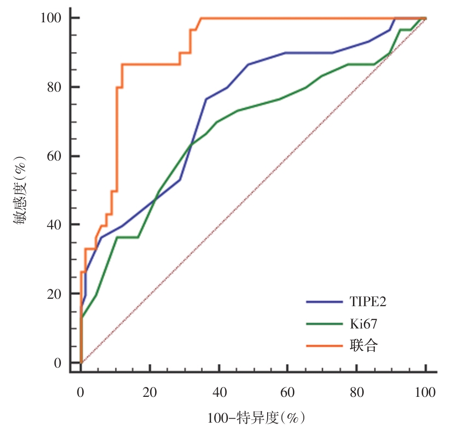
Tab.3
Relationship between the expression levels of TIPE2 and Ki67 in EC tissues and patient prognosis"
| 组别 | TIPE2 | Ki67 |
|---|---|---|
| 良好预后组(n = 66) | 0.23 ± 0.04 | 0.30 ± 0.05 |
| 不良预后组(n = 30) | 0.28 ± 0.05 | 0.35 ± 0.06 |
| t值 | 5.240 | 2.985 |
| P值 | < 0.001 | 0.004 |
Fig.2
ROC curves of the predictive value of individual and combined detection of TIPE2 and Ki67 expression levels in EC tissue for adverse prognosis of EC"
| 1 |
VAN DEN HEERIK A S V M, HOREWEG N, DE BOER S M, et al. Adjuvant therapy for endometrial cancer in the era of molecular classification: Radiotherapy, chemoradiation and novel targets for therapy[J]. Int J Gynecol Cancer, 2021, 31(4): 594-604. doi:10.1136/ijgc-2020-001822
doi: 10.1136/ijgc-2020-001822 |
| 2 |
OAKNIN A, BOSSE T J, CREUTZBERG C L, et al. Endometrial cancer: Esmo clinical practice guideline for diagnosis, treatment and follow-up[J]. Ann Oncol, 2022, 33(9): 860-877. doi:10.1016/j.annonc.2022.05.009
doi: 10.1016/j.annonc.2022.05.009 |
| 3 |
CAI X, ZHANG H, WANG Y, et al. Digital pathology-based artificial intelligence models for differential diagnosis and prognosis of sporadic odontogenic keratocysts[J]. Int J Oral Sci, 2024, 16(1): 16. doi:10.1038/s41368-024-00287-y
doi: 10.1038/s41368-024-00287-y |
| 4 |
CAI Y, WANG B, XU W, et al. Endometrial cancer: Genetic, metabolic characteristics, therapeutic strategies and nanomedicine[J]. Curr Med Chem, 2021, 28(42): 8755-8781. doi:10.2174/0929867328666210705144456
doi: 10.2174/0929867328666210705144456 |
| 5 |
BAI K H, ZHANG Y Y, LI X P, et al. Comprehensive analysis of tumor necrosis factor-α-inducible protein 8-like 2 (tipe2): A potential novel pan-cancer immune checkpoint[J]. CSB J, 2022, 20(3): 5226-5234. doi:10.1016/j.csbj.2022.09.021
doi: 10.1016/j.csbj.2022.09.021 |
| 6 | HAN Z P, YANG Y, CHEN H Y, et al. Tipe2 and pcnp expression abnormalities in peripheral blood mononuclear cells associated with disease activity in rheumatoid arthritis: A meta-analysis[J]. Eur Rev Med Pharmacol Sci, 2021, 25(3): 1242-1249. |
| 7 | 曹蕾, 杨立赟, 田浩, 等. 磁共振弥散成像的ADC直方图对子宫内膜癌分级分期及Ki-67表达的评估价值 [J]. 中国医学计算机成像杂志, 2024, 30(1): 80-85. |
| 8 | 周琦, 吴小华, 刘继红, 等.子宫内膜癌诊断与治疗指南(第四版)[J]. 中国实用妇科与产科杂志, 2018, 34(8): 52-58. |
| 9 | 张学森, 秦凯悦, 李晓菡, 等. 白藜芦醇减轻同型半胱氨酸引起的巨噬细胞免疫应答和增殖的作用[J]. 中国临床药理学杂志, 2024, 40(14): 2038-2042. |
| 10 | 吴兴旗, 党啟华, 徐晶晶, 等. 全自动免疫组化染色仪比较不同抗原修复液对p53的染色结果[J]. 临床与实验病理学杂志, 2024, 40(6): 663-664. |
| 11 |
KARPEL H C, SLOMOVITZ B, COLEMAN R L, et al. Treatment options for molecular subtypes of endometrial cancer in 2023[J]. Curr Opin Obstet Gynecol, 2023, 35(3): 270-278. doi:10.1097/gco.0000000000000855
doi: 10.1097/gco.0000000000000855 |
| 12 |
RIOS-DORIA E, MOMENI-BOROUJENI A, FRIEDMAN C F, et al. Integration of clinical sequencing and immunohistochemistry for the molecular classification of endometrial carcinoma[J]. Gynecol Oncol, 2023, 174(4): 262-272. doi:10.1016/j.ygyno.2023.05.059
doi: 10.1016/j.ygyno.2023.05.059 |
| 13 | XU S, GAO X, QIU J, et al. TIPE2 acts as a tumor suppressor and correlates with tumor microenvironment immunity in epithelial ovarian cancer[J]. Aging (Albany NY), 2023, 15(4): 1052-1073. |
| 14 |
ZHANG H, HAN W J, ZHANG Z L. The Importance of tumor necrosis factor-α-induced protein-8 like-2 in the pathogenesis of cervical cancer and preeclampsia via regulation of cell invasion[J]. Tohoku J Exp Med, 2022, 257(3): 181-191. doi:10.1620/tjem.2022.j026
doi: 10.1620/tjem.2022.j026 |
| 15 |
LASHEN A G, TOSS M S, GHANNAM S F, et al. Expression, assessment and significance of Ki67 expression in breast cancer: an update[J]. J Clin Pathol, 2023, 76(6): 357-364. doi:10.1136/jcp-2022-208731
doi: 10.1136/jcp-2022-208731 |
| 16 | 中国医师协会微无创医学专业委员会, 中国抗癌协会中西整合卵巢癌专业委员会, 张颐, 等. 卵巢肿瘤良恶性风险评估方法学中国专家共识(2024年版)[J]. 中国实用妇科与产科杂志, 2024, 40(3): 312-320. |
| 17 |
ETWEBI Z, GOLDSMITH J R, BOU-DARGHAM M, et al. Tipe2 promotes tumor initiation but inhibits tumor progression in murine colitis-associated colon cancer[J]. Inflamm Bowel Dis, 2022, 28(5): 764-774. doi:10.1093/ibd/izab306
doi: 10.1093/ibd/izab306 |
| 18 |
YANG X, ZHANG M, SU T, et al. Tipe2 inhibits migration and promotes apoptosis as a tumor suppressor in hypopharyngeal carcinoma[J]. Curr Protein Pept Sci, 2022, 23(6): 424-436. doi:10.2174/1389203723666220727090317
doi: 10.2174/1389203723666220727090317 |
| 19 |
REN S, FENG L, LIU H, et al. Gut microbiome affects the response to immunotherapy in non-small cell lung cancer[J]. Thorac Cancer, 2024, 15(14): 1149-1163. doi:10.1111/1759-7714.15303
doi: 10.1111/1759-7714.15303 |
| 20 |
CHEN C, SHEN J, DU Y, et al. Characteristics of gut microbiota in patients with gastric cancer by surgery, chemotherapy and lymph node metastasis[J]. Clin Transl Oncol, 2022, 24(11): 2181-2190. doi:10.1007/s12094-022-02875-y
doi: 10.1007/s12094-022-02875-y |
| 21 | 周丽媛, 施喆, 刘悦君, 等. TIPE2 Ki67在子宫内膜癌组织中的表达及对预后的评估价值[J]. 安徽医学, 2023, 44(11): 1334-1337. |
| 22 | WU R, YU I, TOKUMARU Y, et al. Elevated bile acid metabolism and microbiome are associated with suppressed cell proliferation and better survival in breast cancer[J]. Am J Cancer Res, 2022, 12(11): 5271-5285. |
| [1] | Shenglei ZHANG,Ruicong TIAN,Jingjing JIN,Fan LU,Meijuan CHENG,Yaling BAI,Jinsheng. XU. To study the relationship between lymphocyte subsets and renal clinicopathological features and prognosis in patients with IgA nephropathy [J]. The Journal of Practical Medicine, 2025, 41(3): 352-357. |
| [2] | Yujian CUI,Yuke LI,Sainan ZHU,Shuangling LI,Nan. LI. The association between early peripheral perfusion and acute kidney injury in patients admitted to the intensive care unit following major noncardiac surgery [J]. The Journal of Practical Medicine, 2025, 41(2): 195-201. |
| [3] | Jinlan GUO,Xiaoning ZHANG,Jiahui JIANG,Tao. YUAN. Expression of ACTN1 in cutaneous squamous cell carcinoma and its correlation with clinicopathological features and prognosis [J]. The Journal of Practical Medicine, 2025, 41(2): 232-237. |
| [4] | Ke XU,Mingzhe ZHANG,Mingyang WANG,Song. LIU. Correlation between plasma ADAMTS13 level, disease severity and prognosis in patients with endometriosis [J]. The Journal of Practical Medicine, 2025, 41(2): 238-243. |
| [5] | Shi′an SUN,Yingdong WANG,Xiaofeng CHENG,Yanna DI,Jianhui JIANG,Chaoxu YU. The value of multimodal brain function monitoring based on QEEG and TCD in evaluating hematoma enlargement and prognosis in acute hypertensive intracerebral hemorrhage [J]. The Journal of Practical Medicine, 2025, 41(1): 114-119. |
| [6] | Dandan SHANG,Ping LIU,Lizhen LIU,Yiwei PANG,Chao ZHOU. Diagnosis and advances in individualized management of resistant ovary syndrome and premature ovarian insufficiency [J]. The Journal of Practical Medicine, 2025, 41(1): 146-152. |
| [7] | Xiaomei CHEN,Anqi WANG,Jizhen YANG,Miao YU. Prognosis and immune correlation analysis of m1A/m5C/m6A/m7G regulated genes in gastric cancer [J]. The Journal of Practical Medicine, 2024, 40(9): 1230-1237. |
| [8] | Shihe XIAO,Gang LI,Zhonghai LIU,Zhen. LIU. Expression of ZEB2 and CCL20 in glioma tissue and their correlation with prognosis [J]. The Journal of Practical Medicine, 2024, 40(9): 1262-1267. |
| [9] | Futao DANG,Yingmei TANG,Haiyan FU,Huanlong LI,Jiamin XU,Xu. TAN. The efficacy of artificial (nonbiological) extracorporeal liver support system for primary biliary cholangitis and the effect of this therapy on prognosis [J]. The Journal of Practical Medicine, 2024, 40(7): 955-961. |
| [10] | Xiya MA,Hu JI,Zehua ZHU,Bo PAN,Qiang XIE,Xiaobo. YAO. The predictive value of 18F⁃FDG PET/CT metabolic heterogeneity parameters combined with clinical features for the prognosis of esophageal squamous cell carcinoma before definitive radiochemotherapy [J]. The Journal of Practical Medicine, 2024, 40(7): 966-971. |
| [11] | Min ZHAO,Ping NI,Huiying ZHAI,Xiaoke JIN,Yuqiong. YANG. Analysis of the expression of lymphoid enhancer binding factor 1 in B cell chronic lymphoproliferative disorders [J]. The Journal of Practical Medicine, 2024, 40(7): 984-988. |
| [12] | Yuxin CHENG,Liang LIU,Shiyu DONG,Shengchao LI,Meng ZHANG. Research advances in exosomal proteins, mRNA and non⁃coding RNA regulation of Hepatocellular Carcinoma [J]. The Journal of Practical Medicine, 2024, 40(6): 748-755. |
| [13] | Juan TANG,Yi LI,Liqiong ZHAI,Shaowen LIU,Yong SHEN,Shuo CONG,Yongmei. LIU. The diagnostic value of miR⁃571 levels in blood from the peripherals in liver fibrosis [J]. The Journal of Practical Medicine, 2024, 40(5): 653-657. |
| [14] | Weifeng LIU,Zheng DAI,Yibin ZHOU,Kaiwen FENG,Kai WEI,Gule SUN,Dongrong YANG,Jin. ZHU. The value of urine protein kinase Y⁃linked gene promoter site methylation in early diagnosis of prostate cancer [J]. The Journal of Practical Medicine, 2024, 40(5): 688-694. |
| [15] | Sibo LONG,Yan CHEN,Xintong ZHANG,Yanjun YIN,Limei YANG,Maike ZHENG,Chaohong WANG,Qing SUN,Jun YAN,Yiheng SHI,Guangli SHI,Yan ZHAO,Guirong. WANG. Serum levels of procalcitonin, interleukin⁃6 and interleukin⁃8 in patients with COVID⁃19 infection at admission and their significance in patient prognosis [J]. The Journal of Practical Medicine, 2024, 40(4): 471-475. |
| Viewed | ||||||
|
Full text |
|
|||||
|
Abstract |
|
|||||