

The Journal of Practical Medicine ›› 2025, Vol. 41 ›› Issue (1): 100-107.doi: 10.3969/j.issn.1006-5725.2025.01.017
• Medical Examination and Clinical Diagnosis • Previous Articles
Yaqian DENG1,Wenxiao LI1,Zelin XU1,Jinmei MA1,Tingting DU1,Wen LIU1,Jun LI1,2( )
)
Received:2024-10-16
Online:2025-01-10
Published:2025-01-14
Contact:
Jun LI
E-mail:1287424798@qq.com
CLC Number:
Yaqian DENG,Wenxiao LI,Zelin XU,Jinmei MA,Tingting DU,Wen LIU,Jun LI. Predictive value of growth orientation quantification combined with S⁃Detect technique for axillary lymph node metastasis in breast cancer[J]. The Journal of Practical Medicine, 2025, 41(1): 100-107.
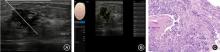
Fig.1
Invasive ductal carcinoma of the breast with axillary lymph node metastasis routine ultrasound, S?Detect examination and pathological findings"
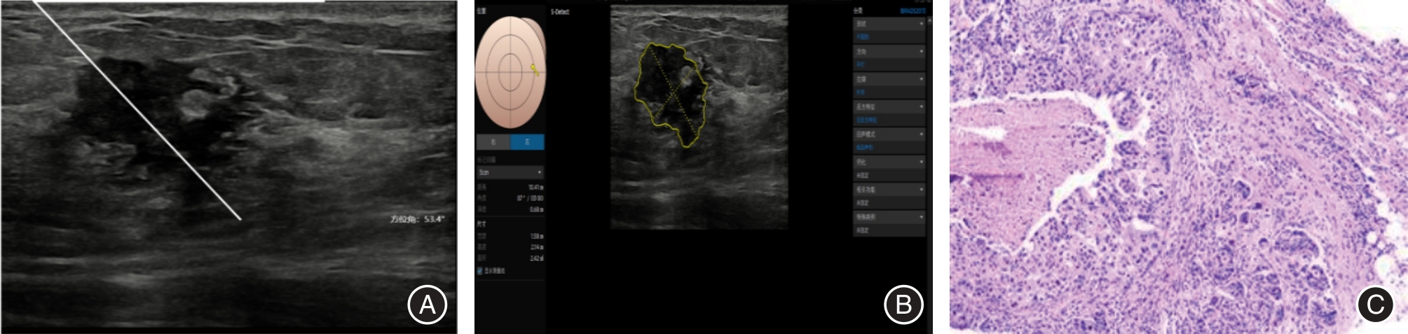

Fig.2
Invasive ductal carcinoma of the breast without axillary lymph node metastasis routine ultrasound, S?Detect examination and pathological findings"
Tab.1
Comparison of pathological types in ALN metastatic and non-metastatic breast cancer patients"
| 项目 | 病理类型 | |||||
|---|---|---|---|---|---|---|
| 浸润性导管癌 | 导管原位癌 | 浸润小叶癌 | 黏液癌 | 乳头状癌 | 髓样癌 | |
| 未转移 | 69 | 25 | 2 | 1 | 3 | 1 |
| 转移 | 55 | 5 | 1 | 1 | 0 | 0 |
| χ2值 | 10.518 | |||||
| P值 | 0.062 | |||||
Tab.2
Comparison of conventional ultrasound and S-Detect characteristics parameters in ALN metastatic and non-metastatic breast cancer patients"
| 指标 | 未转移组 (n = 101) | 转移组 (n = 62) | Z值 | P值 |
|---|---|---|---|---|
| 年龄[M(P25,P75 )]/岁 | 52(42,62) | 55(41,69) | -1.964 | 0.050 |
| 月经状态 | 2.070 | 0.150 | ||
| 绝经 | 85(84.2) | 57(91.9) | ||
| 未绝经 | 16(15.8) | 5(8.1) | ||
| 最大径 | 26.844 | < 0.001 | ||
| ≤ 2 cm | 65(64.4) | 14(37.1) | ||
| > 2 cm | 36(35.6) | 48(62.9) | ||
| 象限 | 2.601 | 0.627 | ||
| 内上 | 19(18.8) | 7(11.3) | ||
| 内下 | 9(8.9) | 7(11.3) | ||
| 外上 | 58(57.4) | 41(66.1) | ||
| 外下 | 12(11.9) | 6(9.7) | ||
| 乳晕 | 3(3.0) | 1(1.6) | ||
| 组织学分级 | 1.598 | 0.450 | ||
| 低 | 4(4.0) | 1(1.6) | ||
| 中 | 65(64.4) | 45(72.6) | ||
| 高 | 32(31.6) | 16(25.8) | ||
| 边界 | 6.628 | 0.010 | ||
| 清晰 | 39(38.6) | 12(19.4) | ||
| 不清晰 | 62(61.4) | 50(80.6) | ||
| 边缘 | 4.843 | 0.028 | ||
| 光整 | 19(16.8) | 4(6.5) | ||
| 不光整 | 82(83.2) | 58(93.5) | ||
| 形状 | 2.280 | 0.131 | ||
| 规则 | 12(11.9) | 3(4.8) | ||
| 不规则 | 89(88.1) | 59(95.2) | ||
| 钙化 | 5.851 | 0.016 | ||
| 无 | 62(61.4) | 26(41.9) | ||
| 有 | 39(38.6) | 36(58.1) | ||
| 血流分级 | 4.392 | 0.036 | ||
| 0 ~Ⅰ级 | 46(45.5) | 18(29.0) | ||
| Ⅱ~Ⅲ级 | 55(54.5) | 44(71.0) | ||
| 内部回声 | 0.013 | 0.909 | ||
| 均匀 | 12(11.9) | 7(11.3) | ||
| 不均匀 | 89(88.1) | 55(88.7) | ||
| 后方回声 | 0.397 | 0.529 | ||
| 未衰减 | 54(53.5) | 30(48.4) | ||
| 衰减 | 47(46.5) | 32(51.6) | ||
| 方位角/[M(P25,P75)] | 27.7(33,35) | 39.2(42,97) | -3.042 | 0.002 |
Tab.3
Assignments for multivariate Logistic regression analysis of influencing factors for ALN metastasis in breast cancer"
| 特征参数 | 变量名 | 赋值 |
|---|---|---|
| 最大径 | X1 | 0=最大径≤ 2 cm,1=最大径> 2 cm |
| 年龄 | X2 | 连续变量 |
| 边界 | X3 | 0=边界清晰,1=边界不清晰 |
| 边缘 | X4 | 0=边缘光整,1=边缘不光整 |
| 钙化 | X5 | 0=无钙化,1=有钙化 |
| 方位角 | X6 | 连续变量 |
| 血流分级 | X7 | 0=0~Ⅰ级,1=Ⅱ~Ⅲ级 |
Tab.4
Multivariate logistic regression analysis of influencing factors for ALN metastasis in breast cancer"
| 观察指标 | B | SE | Wald χ2 | P值 | OR(95%CI) | AUC(95%CI) |
|---|---|---|---|---|---|---|
| 最大径 | 2.299 | 0.445 | 26.662 | < 0.001 | 9.961(4.163 ~ 23.836) | 0.709(0.633 ~ 0.777) |
| 年龄 | 0.022 | 0.021 | 1.124 | 0.289 | 1.022(0.981 ~ 1.065) | - |
| 边界 | 1.171 | 0.476 | 6.060 | 0.014 | 3.227(1.270 ~ 8.199) | 0.596(0.517 ~ 0.672) |
| 边缘 | 2.137 | 0.721 | 8.783 | 0.003 | 8.475(2.062 ~ 34.827) | 0.562(0.482 ~ 0.639) |
| 钙化 | 1.397 | 0.437 | 10.212 | 0.001 | 4.044(1.717 ~ 9.529) | 0.597(0.518 ~ 0.673) |
| 方位角 | 0.034 | 0.009 | 13.239 | < 0.001 | 1.034(1.016 ~ 1.053) | 0.642(0.563 ~ 0.716) |
| 血流分级 | 0.470 | 0.444 | 1.122 | 0.290 | 1.600(0.670 ~ 3.821) | - |
| 常量 | -7.995 | 1.615 | 24.498 | - | - | - |
| 预测模型 | - | - | - | - | - | 0.869(0.807 ~ 0.917) |
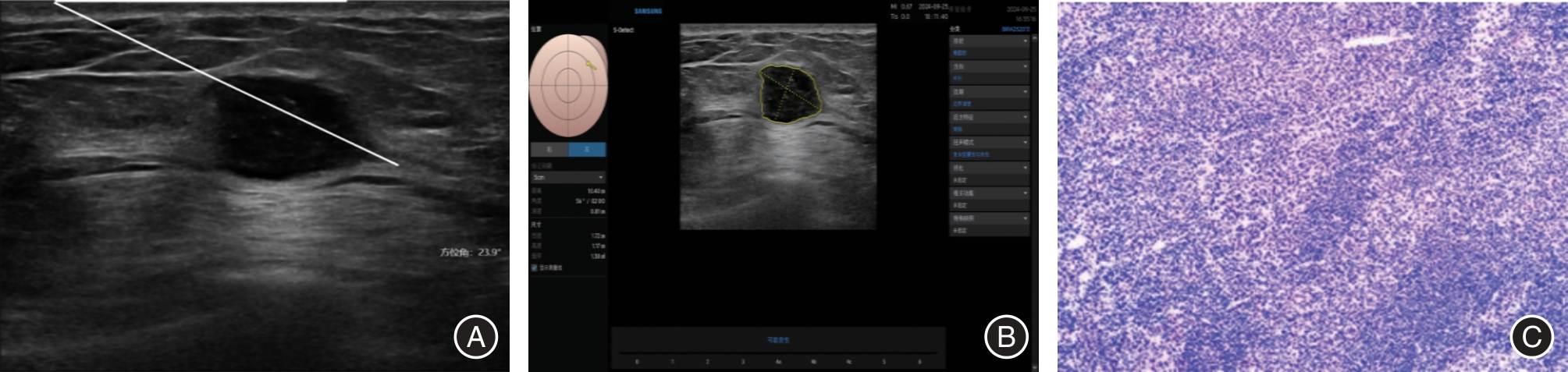
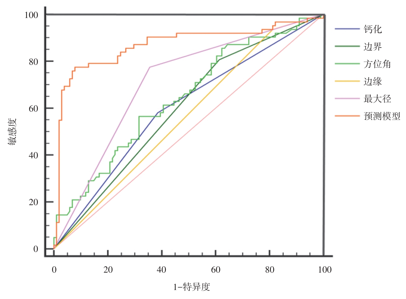
Fig.3
The ROC curves of each parameter alone and in combination for predicting ALN metastasis"
Tab.5
Prediction of ALN metastasis in breast cancer by combined prediction models versus pathological findings"
| 预测模型 | 病理 | |
|---|---|---|
| 转移(n = 30) | 未转移(n = 50) | |
| 转移 | 23 | 4 |
| 未转移 | 7 | 46 |
| Kappa值 | 0.701 | |
| P值 | < 0.005 | |
| 1 |
HYUNA S, JACQUES F, SIEGEL R L, et al.Global cancer statistics2020: Globocan estimates of incidence and mortality worldwide for 36 cancers in 185 countries[J]. Cancer J Clin, 2021, 71(3):209-249. doi:10.3322/caac.21660
doi: 10.3322/caac.21660 |
| 2 |
TO B, ISAAC D, ANDRECHEK E R. Studying Lymphatic Metastasis in Breast cancer: Current Models, Strategies, and Clinical Perspectives[J].J Mammary Gland Biol Neoplasia, 2020, 25(3):191-203. doi:10.1007/s10911-020-09460-5
doi: 10.1007/s10911-020-09460-5 |
| 3 |
MARINO M A, AVENDANO D, ZAPATA P, et al. Lymph node imagingin patients with primary breast cancer: Concurrent diagnostic tools[J].Oncologist, 2020, 25(2):e231-e242. doi:10.1634/theoncologist.2019-0427
doi: 10.1634/theoncologist.2019-0427 |
| 4 |
ZHU Y Y, JIA Y Y, PANG W J, et al. Ultrasound contrast-enhancedpatterns of sentinel lymph nodes: Predictive value for nodal statusand metastatic burden in early breast cancer[J]. Quant Imaging Med Surg, 2023, 13(1):160-170. doi:10.21037/qims-22-234
doi: 10.21037/qims-22-234 |
| 5 |
LI J, LI H, GUAN L, et al. The value of preoperative sentinellymph node contrast-enhanced ultrasound for breast cancer: Alarge, multicenter trial[J]. BMC Cancer, 2022, 22(1):455. doi:10.1186/s12885-022-09551-y
doi: 10.1186/s12885-022-09551-y |
| 6 |
HANKÓ-BAUER O, PODOLEANU C, GEORGESCU R, et al.The accuracy of the preoperative axillary ultrasound examination in predicting the status of the sentinel lymph node involvement in patients with infiltrating breast carcinoma[J]. Chirurgia(Bucur), 2019, 114(3):384-391. doi:10.21614/chirurgia.114.3.384
doi: 10.21614/chirurgia.114.3.384 |
| 7 |
BARTOLOTTA T V, ORLANDO A A M, SPATAFORA L, et al. S-Detect characterization of focal breast lesions according to the US BI RADS lexion:A pictorial essay[J]. J Ultrasound, 2020, 23 (2) :207-215. doi:10.1007/s40477-020-00447-w
doi: 10.1007/s40477-020-00447-w |
| 8 |
GU Y, TIAN J W, RAN H T, et al. The utility of the fifth edition of the BI-RADS ultrasound lexicon in category 4 breast lesions:A prospective multicenter study in China[J]. Acad Radiol, 2022, 29 :S26–S34. doi:10.1016/j.acra.2020.06.027
doi: 10.1016/j.acra.2020.06.027 |
| 9 |
GUO Q, ZHANG L, DI Z, et al. Assessing risk category of breast cancer by ultrasound imaging characteristics[J]. Ultrasound Med Biol, 2018, 44(4):815–824. doi:10.1016/j.ultrasmedbio.2017.12.001
doi: 10.1016/j.ultrasmedbio.2017.12.001 |
| 10 | FENG C, ZHAN Y, SHAO H, et al. Postoperative expressions of TRACP5b and CA125 in patients with breast cancerand their values for monitoring bone metastasis[J]. J BUON, 2020, 25(2):688-695. |
| 11 | 韩文, 李卫民, 吴晓明, 等. 纵横比大于1乳腺肿块的X线再评估及临床、超声特征分析[J]. 实用医学杂志, 2021, 37(24):3179-3183. |
| 12 |
WATANABE T, YAMAGUCHI T, TOHNO E, et al. B-mode ultrasound diagnostic flowchart for solid breast masses: JABTSBC-01 study[J]. J Med Ultrason, 2021, 48(1):71-81. doi:10.1007/s10396-020-01072-0
doi: 10.1007/s10396-020-01072-0 |
| 13 |
WANG B, ZHU L, HE C, et al. Growth pattern can be used as a new characteristic to predict malignancy in breast cancer[J].Breast Cancer, 2020, 27(3):445-455. doi:10.1007/s12282-019-01041-7
doi: 10.1007/s12282-019-01041-7 |
| 14 |
WANG H, ZHAN W, CHEN W, et al. Sonography with vertical orientation feature predicts worse disease outcome in triple negative breast cancer[J]. Breast,2020,49:33-40. doi:10.1016/j.breast.2019.10.006
doi: 10.1016/j.breast.2019.10.006 |
| 15 |
CHEN K, WU S.The utility of quantifying the orientation of breast masses in ultrasound imaging[J]. Sci Rep, 2024, 14(1):4578. doi:10.1038/s41598-024-55298-w
doi: 10.1038/s41598-024-55298-w |
| 16 | 桑田, 张海俊, 曹玉文, 等. Logistic回归分析乳腺癌常规超声征象与腋窝淋巴结转移的关系[J].中国医学影像技术, 2021, 37(8):1158-1162. |
| 17 | 石丽楠, 曹春莉, 桑田, 等. 基于乳腺癌超声特征及临床病理指标的列线图预测腋窝淋巴结转移风险[J]. 中国医学影像学杂志, 2024, 32(4):332-338. |
| 18 |
ZONG Q, DENG J, GE W, et al. Establishment of simple nomograms for predicting axillary lymph node involvement in early breast cancer[J]. Cancer Manag Res, 2020, 12:2025-2035. doi:10.2147/cmar.s241641
doi: 10.2147/cmar.s241641 |
| 19 |
LIU H, XU G, YAO M H, et al. Association of conventional ultrasound, elastography and clinicopathological factors with axillary lymph node status in invasive ductal breast carcinoma with sizes > 10 mm [J]. Oncotarget, 2018, 9(2):2819-2828. doi:10.18632/oncotarget.18969
doi: 10.18632/oncotarget.18969 |
| 20 |
LI X L, XU H X, LI D D, et al. A risk model based on ultra-sound,ultrasound elastography,and histologic parameters for predicting axillary lymph node metastasis in breast in-vasive ductal carcinoma [J]. Sci Rep, 2017, 7(1):3029-3039. doi:10.1038/s41598-017-03582-3
doi: 10.1038/s41598-017-03582-3 |
| 21 |
YI C B, DING Z Y, DENG J, et al.Combining the ultrasound features of primary tumor and axillary lymph nodes can reduce false-negative rate dueing the prediction of high axillary node burden in BI-RADS category 4 or 5 breast cancer lesions[J]. Ultrasound Med Biol, 2020, 46(8):1941-1948. doi:10.1016/j.ultrasmedbio.2020.04.003
doi: 10.1016/j.ultrasmedbio.2020.04.003 |
| 22 |
XUE S, ZHAO Q, TAI M, et al.Correlation between breast ultrasound microcalcification and the prognosis of breast cancer[J]. J Healthc Eng, 2021, 2021:6835963. doi:10.1155/2021/6835963
doi: 10.1155/2021/6835963 |
| 23 |
OGRADY S, MORGAN M P.Microcalcifications in breastcancer: From pathophysiology to diagnosis and prognosis[J]. Biochim Biophys Acta Rev Cancer, 2018, 1869(2):310-320. doi:10.1016/j.bbcan.2018.04.006
doi: 10.1016/j.bbcan.2018.04.006 |
| 24 | 熊朝月, 周敏, 何小芳, 等. 基于超声特征联合临床资料的预测模型对早期乳腺癌患者腋窝淋巴结转移的评估价值[J]. 实用临床医药杂志, 2022, 26(12):14-18, 22. |
| 25 |
YU X Q, HAO X Y, WAN J, et al. Correlation between ultrasound appearance of small breast cancer and axillary lymph node metastasis[J]. Ultrasound Med Biol, 2018, 44(2):342-349. doi:10.1016/j.ultrasmedbio.2017.09.020
doi: 10.1016/j.ultrasmedbio.2017.09.020 |
| 26 |
LI L, YU T, SUN J, et al. Prediction of the number of metastatic axillary lymph nodes in breast cancer by radiomic signature based on dynamic contrast-enhanced MRI[J]. Acta Radiol, 2022, 63(8):1014-1022. doi:10.1177/02841851211025857
doi: 10.1177/02841851211025857 |
| 27 |
YI C B, DING Z Y, DENG J, et al. Combining the ultrasound features of primary tumor and axillary lymph nodes can reduce false-negative rate during the prediction of high axillary node burden in BI-RADS category 4 or 5 breast cancer lesions[J]. Ultrasound Med Biol, 2020, 46(8):1941-1948. doi:10.1016/j.ultrasmedbio.2020.04.003
doi: 10.1016/j.ultrasmedbio.2020.04.003 |
| 28 |
ZHANG H, SUI X, ZHOU S, et al. Correlation of Conventional Ultrasound Characteristics of Breast Tumors With Axillary Lymph Node Metastasis and Ki-67 Expression in Patients With Breast Cancer[J]. J Ultrasound Med, 2019, 38(7):1833-1840. doi:10.1002/jum.14879
doi: 10.1002/jum.14879 |
| 29 |
XIONG J, ZUO W, WU Y, et al. ultrasonography and clinicopathological features of breast cancer in predicting axillary lymph node metastases[J]. BMC Cancer, 2022, 22(1):1155. doi:10.1186/s12885-022-10240-z
doi: 10.1186/s12885-022-10240-z |
| 30 | 郑碧玉, 张晓东, 苏毅明, 等. 年轻乳腺癌69例超声征象与腋窝淋巴结转移的关系[J]. 超声影像学杂志, 2022, 31(6):525-531. |
| 31 |
ZHAO Q, SUN J W, ZHOU H, et al. Pre-operative Conventional Ultrasound and Sonoelastography Evaluation for Predicting Axillary Lymph Node Metastasis in Patients with Malignant Breast Lesions[J]. Ultrasound Med Biol, 2018, 44 (12): 2587-2595. doi:10.1016/j.ultrasmedbio.2018.07.017
doi: 10.1016/j.ultrasmedbio.2018.07.017 |
| 32 |
ZHU A Q, LI X L, AN L W, et al. Predicting axillary lymph node metastasis in patients with breast invasive ductal carcinoma with negative axillary ultrasound results using conventional altrasound and contrast-enhanced ultrasound[J]. J Ultrasound Med, 2020, 39(10):2059-2070. doi:10.1002/jum.15314
doi: 10.1002/jum.15314 |
| Viewed | ||||||
|
Full text |
|
|||||
|
Abstract |
|
|||||