

The Journal of Practical Medicine ›› 2026, Vol. 42 ›› Issue (8): 1332-1340.doi: 10.3969/j.issn.1006-5725.2026.08.004
• Chronic Disease Control • Previous Articles
Jing HUANG1,Shuang HAN2,Xuemei ZHAO1,Yuan CHEN3,Yinfeng QIAN1( )
)
Received:2025-12-03
Online:2026-04-25
Published:2026-04-28
Contact:
Yinfeng QIAN
E-mail:894206876@qq.com
CLC Number:
Jing HUANG,Shuang HAN,Xuemei ZHAO,Yuan CHEN,Yinfeng QIAN. Cone beam CT image measurement of alveolar bone changes in patients with periodontitis and its clinical significance[J]. The Journal of Practical Medicine, 2026, 42(8): 1332-1340.
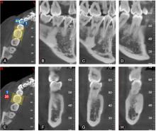
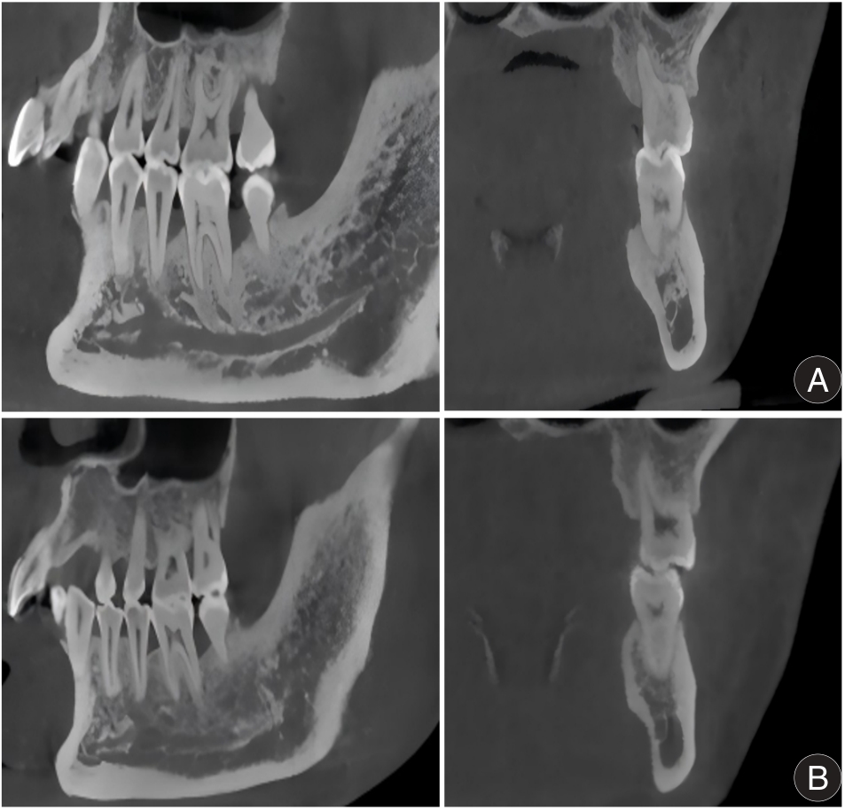
Fig.1
BDH measurement maps"
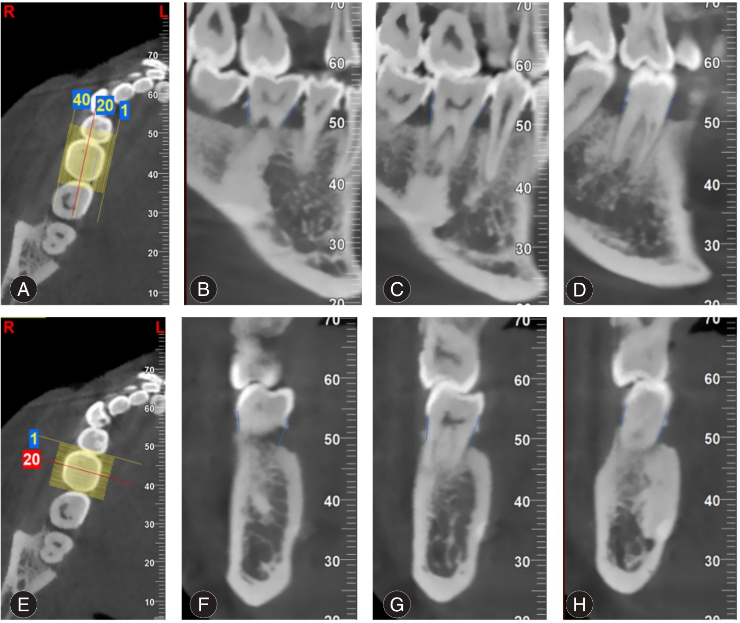
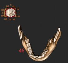
Fig.2
Three-dimensional measurement map"

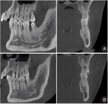
Fig.3
BMD measurement maps"
Tab.1
Comparison of BDH and BMD in patients of different genders before and after treatment"
| 性别 | n | BDH | BMD | ||
|---|---|---|---|---|---|
| 治疗前 | 治疗后 | 治疗前 | 治疗后 | ||
| 男 | 1 744 | 4.67 ± 0.92 | 4.48 ± 0.46* | 923.58 ± 103.25 | 956.28 ± 113.85* |
| 女 | 1 020 | 4.25 ± 0.89 | 3.87 ± 0.43* | 852.22 ± 109.52 | 913.26 ± 115.82* |
| t值 | - | 11.721 | 34.453 | 17.142 | 9.525 |
| P值 | - | < 0.001 | < 0.001 | < 0.001 | < 0.001 |
Tab.2
Comparison of BDH in patients of different ages before and after treatment"
| 年龄 | n | 近中 | 远中 | 颊侧 | 舌侧 | ||||
|---|---|---|---|---|---|---|---|---|---|
| 治疗前 | 治疗后 | 治疗前 | 治疗后 | 治疗前 | 治疗后 | 治疗前 | 治疗后 | ||
| 30岁以下 | 696 | 2.96 ± 0.78 | 2.89 ± 0.81 | 2.73 ± 0.75 | 2.68 ± 0.79 | 2.48 ± 0.78 | 2.42 ± 0.72 | 2.23 ± 0.69 | 2.18 ± 0.61 |
| 30~50岁 | 1 128 | 3.65 ± 1.05& | 3.43 ± 0.99*& | 3.41 ± 1.127 | 3.12 ± 0.86*& | 2.91 ± 0.82& | 2.67 ± 0.76*& | 2.34 ± 0.83& | 2.07 ± 0.81*& |
| 50岁以上 | 940 | 6.38 ± 1.28&# | 6.04 ± 1.29*&# | 6.12 ± 1.14&# | 5.76 ± 1.22*&# | 5.48 ± 1.02&# | 5.13 ± 1.12*&# | 4.82 ± 1.13&# | 4.52 ± 1.02*&# |
| F值 | - | 2 483.167 | 2 229.879 | 2 587.084 | 2 576.744 | 3 017.860 | 2 571.409 | 2 364.783 | 2 517.917 |
| P值 | - | < 0.001 | < 0.001 | < 0.001 | < 0.001 | < 0.001 | < 0.001 | < 0.001 | < 0.001 |
Tab.4
Comparison of BDH in patients with different severities before and after treatment"
| 项目 | n | 近中 | 远中 | 颊侧 | 舌侧 | ||||
|---|---|---|---|---|---|---|---|---|---|
| 治疗前 | 治疗后 | 治疗前 | 治疗后 | 治疗前 | 治疗后 | 治疗前 | 治疗后 | ||
| 轻度 | 778 | 2.90 ± 0.71 | 2.65 ± 0.73* | 2.76 ± 0.70 | 2.55 ± 0.72* | 2.59 ± 0.74 | 2.47 ± 0.65* | 2.32 ± 0.56 | 2.26 ± 0.64* |
| 中度 | 1 003 | 3.73 ± 0.91& | 3.54 ± 0.94*& | 3.62 ± 1.06& | 3.51 ± 0.98*& | 3.49 ± 0.87& | 3.28 ± 0.85*& | 3.33 ± 0.92& | 3.13 ± 0.88*& |
| 重度 | 983 | 6.49 ± 1.39&# | 6.35 ± 1.21*&# | 6.31 ± 1.26&# | 6.09 ± 1.13*&# | 6.07 ± 1.21&# | 5.87 ± 1.06*&# | 5.92 ± 1.15&# | 5.77 ± 1.22*&# |
| F值 | - | 2 867.352 | 3 443.790 | 2 826.656 | 3 216.370 | 3 135.207 | 3 699.494 | 3 619.088 | 3 299.546 |
| P值 | - | < 0.001 | < 0.001 | < 0.001 | < 0.001 | < 0.001 | < 0.001 | < 0.001 | < 0.001 |
Tab.6
Comparison of BDH in different positions before and after treatment"
| 项目 | n | 近中 | 远中 | 颊侧 | 舌侧 | ||||
|---|---|---|---|---|---|---|---|---|---|
| 治疗前 | 治疗后 | 治疗前 | 治疗后 | 治疗前 | 治疗后 | 治疗前 | 治疗后 | ||
| 前牙 | 1 126 | 2.86 ± 0.65 | 2.81 ± 0.73 | 2.73 ± 0.72 | 2.68 ± 0.71 | 2.60 ± 0.67 | 2.55 ± 0.72 | 2.71 ± 0.76 | 2.67 ± 0.65 |
| 前磨牙 | 756 | 3.64 ± 0.95& | 3.41 ± 0.91*& | 3.41 ± 0.94& | 3.12 ± 0.90*& | 3.05 ± 0.81& | 2.71 ± 0.83*& | 3.25 ± 0.73& | 2.76 ± 0.82*& |
| 磨牙 | 882 | 6.02 ± 1.38&# | 5.71 ± 1.29*&# | 5.59 ± 1.21&# | 5.15 ± 1.13*&# | 5.21 ± 0.92&# | 4.81 ± 1.07*&# | 5.45 ± 1.23&# | 5.13 ± 1.19*&# |
| F值 | - | 2 504.250 | 2 261.929 | 2 297.706 | 1 945.174 | 2 874.429 | 1 900.150 | 2 289.235 | 2 185.919 |
| P值 | - | < 0.001 | < 0.001 | < 0.001 | < 0.001 | < 0.001 | < 0.001 | < 0.001 | < 0.001 |
| [1] |
NITYA K, SHREE H K, ARYA A, et al. Detection of clinically relevant resistance genes in subgingival biofilms of chronic periodontitis: A cross-sectional molecular surveillance study[J]. J Oral Biol Craniofac Res,2026,16(1):167-173. doi:10.1016/J.JOBCR.2025.12.002 .
doi: 10.1016/J.JOBCR.2025.12.002 |
| [2] |
YANG M, MA Y, QU X, et al. Inhibition of sclerostin activity promotes bone regeneration in experimental periodontal disease: A systematic review and meta-analysis of animal models[J]. Exp Ther Med,2026,31(2):31. doi:10.3892/ETM.2025.13026 .
doi: 10.3892/ETM.2025.13026 |
| [3] |
SHANTININGSIH R R, WIDYANINGRUM R, DIBA F S, et al. Investigating anterior and posterior alveolar trabecular patterns on periapical radiographs: Insights into bone mineral density in postmenopausal women[J]. J Oral Biol Craniofac Res,2025,15(5):1077-1082. doi:10.1016/J.JOBCR.2025.06.025 .
doi: 10.1016/J.JOBCR.2025.06.025 |
| [4] |
MEHRU M, SHARMA R, KUMAR V, et al. Estimation of age using pulp/tooth area ratio using orthopantomographs and intra-oral periapical radiographs[J]. Bioinformation,2024,20(12):2040-2044. doi:10.6026/9732063002002040 .
doi: 10.6026/9732063002002040 |
| [5] |
TURKYILMAZ I, FELDMAN Z D, SUER T B. Imaging techniques in dental implant planning: Understanding the paradigm shift from periapical radiograph to cone beam computed tomography (CBCT)[J].Prim Dent J,2024,13(4):50-52. doi:10.1177/20501684241272282 .
doi: 10.1177/20501684241272282 |
| [6] |
丁子凌, 刘昕, 杨晓喻, 刘楚峰. 华南地区成人上颌中切牙与牙槽骨相对位置关系CBCT分析[J]. 口腔疾病防治, 2024, 32(2): 116-122.doi:10.12016/J.ISSN.2096-1456.2024.02.005 .
doi: 10.12016/J.ISSN.2096-1456.2024.02.005 |
| [7] |
CHHABRA A, RAMYA K P, PRATHAP B S, et al. Comparative evaluation of gutta-percha adaptation in different obturation techniques using cone-beam computed tomography: A randomized controlled trial[J]. J Conserv Dent Endod,2025,28(12):1264-1271.doi:10.4103/JCDE.JCDE_729_25 .
doi: 10.4103/JCDE.JCDE_729_25 |
| [8] |
SHILADITYA S, SUBHANKAR G, NILANJANA S, et al. Comparison of root canal morphology in intraoral periapical radiographs and cone-beam computed tomography: An in vitro study[J]. Indian J Dent Sci,2024,16(1):17-24 doi:10.4103/IJDS.IJDS_73_23 .
doi: 10.4103/IJDS.IJDS_73_23 |
| [9] |
SHAO J L, YU Y, LYU C X, et al. Introduction and interpretation of the European Federation of Periodontology S3 level clinical practice guideline for treatment of periodontitis[J]. Chin J Stomatol,2022,57(12):1202-1208. doi:10.3760/CMA.J.CN112144-20220719-00394 .
doi: 10.3760/CMA.J.CN112144-20220719-00394 |
| [10] |
ALJOGHAIMAN E, ALZAHRANI A, ALBARQI R, et al. Association between diabetes and vertical bone defects in periodontitis using cone beam computed tomography: A cross-sectional study in the eastern province, saudi arabia[J]. Clinics Pract,2025,15(5):95-95. doi:10.3390/CLINPRACT15050095 .
doi: 10.3390/CLINPRACT15050095 |
| [11] |
JIN H, WU D, CHEN L. 3-Dimensional Radiographic Analysis Of Mandibular Homonym Premolars Using CBCT[J]. Int Dent J,2025, 75(S1): 104201-104201.doi:10.1016/J.IDENTJ.2025.104201 .
doi: 10.1016/J.IDENTJ.2025.104201 |
| [12] |
VELURI S, GOTTUMUKKALA S V N S, PENMETSA G, et al. Retrospective analysis of the relationship between Schneiderian membrane thickness and periodontitis severity using cone beam computed tomography (CBCT)[J]. Dent Med Probl,2025,62(1):65-72. doi:10.17219/DMP/147105 .
doi: 10.17219/DMP/147105 |
| [13] |
杨婷婷,黄一丹,杨蓉蓉,等. 2型糖尿病合并慢性牙周炎患者血清LRG1、LDH与牙周指标和牙周病变程度的关系[J]. 实用医学杂志,2024,40(16):2250-2255. doi:10.3969/j.issn.1006-5725.2024.16.009 .
doi: 10.3969/j.issn.1006-5725.2024.16.009 |
| [14] |
FENG G, LIAO Z, WANG Y, et al. Galactose-modified small molecule modulator targets RORα to enhance circadian rhythm and alleviate periodontitis-associated alveolar bone loss[J]. Bone Res,2025,13(1):91.doi:10.1038/S41413-025-00445-W .
doi: 10.1038/S41413-025-00445-W |
| [15] |
HONG G H, NGUYEN H, ALKHAMEES A, et al. Non-surgical treatment of adult skeletal class III patient with severe alveolar bone loss by sequential segmental displacement and third molar extraction: A case report[J]. Int Orthod,2025,24(2):101087.doi:10.1016/J.ORTHO.2025.101087 .
doi: 10.1016/J.ORTHO.2025.101087 |
| [16] |
DYLAN D, MENIK P, BRAMMA K, et al. Digital Subtraction radiography in the evaluation of early alveolar bone density changes using dried mandible: An original radiological in vitro study[J]. J Indian Acad Oral Med Radiol,2025,37(3):303-307. doi:10.4103/JIAOMR.JIAOMR_50_25 .
doi: 10.4103/JIAOMR.JIAOMR_50_25 |
| [17] |
WU L, YUAN Y, YU Y. Effects of Nd∶YAP laser-assisted periodontal therapy on masticatory function and gingival circulation indices in patients with chronic periodontitis[J]. Lasers Med Sci,2025,40(1):494. doi:10.1007/S10103-025-04752-W .
doi: 10.1007/S10103-025-04752-W |
| [18] |
RAFIEIZADEH S, LARI S, MALEKI M M, et al. Investigation of the correlation between radiomorphometric indices in cone-beam computed tomography images and dual X-ray absorptiometry bone density test results in postmenopausal women[J]. BMC Med Imaging,2025,25(1):203.doi: 10.1186/s12880-025-01739-5 .
doi: 10.1186/s12880-025-01739-5 |
| [19] |
刘南南,徐隽,古力克孜·台外库力,等. 维吾尔族与汉族上颌前牙区牙槽骨密度的CBCT测量分析[J]. 广东医学,2021,42(2):184-187. doi:10.13820/j.cnki.gdyx.20203064 .
doi: 10.13820/j.cnki.gdyx.20203064 |
| [20] |
ERKERT A J, BENITEZ K B, LILL Y, et al. Cone-beam computed tomography-guided volumetric assessment of secondary alveolar bone grafting[J]. Sci Rep,2025,15(1):29766-29766. doi:10.1038/S41598-025-15083-9 .
doi: 10.1038/S41598-025-15083-9 |
| [21] |
OKUBO S, MIYABE S, KISE Y, et al. A logistic regression model for predicting osteoporosis using alveolar bone mineral density measured on intraoral radiographs combined with panoramic mandibular cortical index[J]. J Clin Med,2025,14(20):7198-7198.doi:10.3390/JCM14207198 .
doi: 10.3390/JCM14207198 |
| [22] |
MISTRETTA F, MAGNINI A, CINCI L, et al. A systematic review and meta-analysis on the concept of bone quality in dento-maxillofacial Cone Beam Computed Tomography[J]. La Radiol Med,2025,130(8):1-14.doi:10.1007/S11547-025-02052-5 .
doi: 10.1007/S11547-025-02052-5 |
| [23] |
BEGUM R, MALIK R, GUPTA K, et al. Quantitative assessment of bone density at the borders of radiolucent mandibular lesions using cone-beam computed tomography: Correlations with lesion aggressiveness[J]. Cureus,2025,17(6):e86111-e86111.doi:10.7759/CUREUS.86111 .
doi: 10.7759/CUREUS.86111 |
| [24] |
王晖,方伟,周平,等. 应用CBCT测量慢性牙周炎患者口服维生素D3牙槽骨密度改变的临床研究[J]. 口腔医学, 2019, 39(10): 912-915. doi:10.13591/j.cnki.kqyx.2019.10.008 .
doi: 10.13591/j.cnki.kqyx.2019.10.008 |
| [25] |
ZHANG Y, XU B, PENG C, et al. Expression of autophagy and apoptosis during orthodontic tooth movement alveolar bone remodeling in rats with varied periodontal conditions[J]. Int Orthod, 2025, 24(1): 101076. doi: 10.1016/J.ORTHO.2025.101076 .
doi: 10.1016/J.ORTHO.2025.101076 |
| [26] |
MIADILI M, LI X, ZHANG Y, et al. The impact of jawbone regions (molar area, premolar area, anterior area) and bone density on the accuracy of robot-assisted dental implantation: A preliminary study[J]. Front Bioeng Biotechnol,2025,25(13):1536957-1536957. doi: 10.3389/FBIOE.2025.1536957 .
doi: 10.3389/FBIOE.2025.1536957 |
| [27] |
MARTINS C A L, SZALEWSKI L, PAŁKA K, et al. Repeatability of gray value-based bone density measurements in cone beam computed tomography (CBCT) images under different acquisition protocols[J]. BMC Oral Health,2025,25(1): 1549-1549. doi:10.1186/S12903-025-06760-2 .
doi: 10.1186/S12903-025-06760-2 |
| [1] | Maoqing WANG,Wenhui LI,Meihuang. CAI. Effect of compound Xueshuantong combined with viaminate of oral lichen planus complicated with chronic periodontitis [J]. The Journal of Practical Medicine, 2024, 40(21): 3076-3081. |
| [2] | Bin ZHANG,Wei HU,Rongzhen TAN,Panpan YANG,Jun HU,Zhong YUAN,Gongtao. JIANG. Effects of Yishen Huayu Xugu prescription combined with Dixumab on IL-6, β-CTX and bone mineral density in elderly patients operated for osteoporotic lumbar vertebral compression fractures [J]. The Journal of Practical Medicine, 2024, 40(19): 2766-2771. |
| [3] | Tingting YANG,Yidan HUANG,Rongrong YANG,Ying YANG,Jing. ZHANG. Study on the relationship between serum LRG1, LDH with periodontal indexes and periodontal lesions in patients with type 2 diabetes and chronic periodontitis [J]. The Journal of Practical Medicine, 2024, 40(16): 2250-2255. |
| [4] | Junlu ZHAO,Zhai LIU,Deyuan ZHAO,Guanwei NIE,Qingyun. REN. Quantitative CT evaluation for thoracic vertebral bone density and age⁃related bone loss [J]. The Journal of Practical Medicine, 2024, 40(10): 1429-1433. |
| [5] |
DENG Jiajie, YANG Yan, CAI Yulan, WU Min, WAN Ling, YANG Jia..
Study on the relationship between bone mineral density and serum 25(OH)D and Irisin in elderly men with different degrees of sarcopenia [J]. The Journal of Practical Medicine, 2023, 39(3): 321-325. |
| [6] |
LIU Yingtao# , WANG Dejun, WANG Wei, ZHANG Liyong, TAN Kai, YAN Jia, HUANG Yumei..
Point ⁃ through ⁃ point acupotomy on shoulder joint motion during frozen periodontitis of shoulder and its long ⁃ term efficacy observation [J]. The Journal of Practical Medicine, 2023, 39(2): 230-235. |
| [7] | WANG Peipei, HUA Fei, HUANG Xia, GE Junyi, ZHAO Lu, GU Min. . Research progress of oral microenvironment in patients with type 2 diabetes mellitus and chronic periodon⁃ titis [J]. The Journal of Practical Medicine, 2023, 39(10): 1320-1324. |
| [8] |
LI Xiaohai, XIE Guangyou, LIANG Lisong, LIU Jiangyong, ZENG Xianchun, WANG Rongpin..
Bone mineral density of lumbar spine QCT and spinal fragility fractures in middle⁃aged and elderly people in Guizhou province [J]. The Journal of Practical Medicine, 2023, 39(1): 60-65. |
| [9] |
YOU Yuehua, ZHOU Shanyu, YUAN Bo, WANG Wei, HUANG Lin, YANG Xingmin, YIN Chengfang, SHI Ling, HUANG Yongqi..
Clinical effects of iRoot BP plus and mineral trioxide aggregate for apical barrier of permanent teeth in adults [J]. The Journal of Practical Medicine, 2021, 37(7): 869-873. |
| Viewed | ||||||
|
Full text |
|
|||||
|
Abstract |
|
|||||