

The Journal of Practical Medicine ›› 2026, Vol. 42 ›› Issue (1): 94-100.doi: 10.3969/j.issn.1006-5725.2026.01.012
• Chronic Disease Control • Previous Articles Next Articles
Abudimijiti XIEYIDAI,Xiangxin SONG,Aizezi DILIREBA,Yibulaiyin HASIYETI( )
)
Received:2025-09-05
Online:2026-01-10
Published:2026-01-14
Contact:
Yibulaiyin HASIYETI
E-mail:hsyt930927@126.com
CLC Number:
Abudimijiti XIEYIDAI,Xiangxin SONG,Aizezi DILIREBA,Yibulaiyin HASIYETI. Research on the correlation between specific indicators of serum ferroptosis and Alzheimer′s disease[J]. The Journal of Practical Medicine, 2026, 42(1): 94-100.
Tab.1
Comparison of general data between the two groups"
| 项目 | AD组(n = 37) | 对照组(n = 35) | χ2 /t值 | P值 |
|---|---|---|---|---|
| 性别 | 1.345 | 0.246 | ||
| 男 | 14(37.8) | 18(51.4) | ||
| 女 | 23(62.2) | 17(48.6) | ||
| 年龄(x ± s)/岁 | 70.35 ± 9.917 | 71.29 ± 8.659 | 0.425 | 0.672 |
| BMI(x ± s)/(kg/m2) | 23.606 ± 2.832 | 25.494 ± 3.029 | 2.879 | 0.005 |
| 教育水平 | 4.890 | 0.087 | ||
| 文盲 | 1(2.7) | 5(14.3) | ||
| 小学 | 10(27.0) | 13(37.1) | ||
| 初中级以上 | 26(70.3) | 17(48.6) | ||
| 吸烟史 | 8(21.6) | 10(28.6) | 0.463 | 0.496 |
| 饮酒史 | 6(16.2) | 6(17.1) | 0.011 | 0.916 |
| 高血压 | 25(67.6) | 22(62.9) | 0.176 | 0.675 |
| 冠心病 | 6(16.2) | 10(28.6) | 1.589 | 0.208 |
| 糖尿病 | 12(32.4) | 7(20.0) | 1.431 | 0.232 |
| MMSE(x ± s)/分 | 16.30 ± 6.311 | 24.91 ± 3.302 | 7.198 | < 0.001 |
| MoCA(x ± s)/分 | 12.38 ± 6.188 | 23.66 ± 4.399 | 8.868 | < 0.001 |
Tab.2
Comparison of two groups of serum ferroptosis-related indicators"
| 项目 | AD组(n = 37) | 对照组(n = 35) | t值 | P值 |
|---|---|---|---|---|
| GPX4/(μU/mL) | 21.767 ± 9.609 | 31.345 ± 10.553 | 4.031 | < 0.001 |
| GSH/(μmol/L) | 23.573 ± 8.862 | 34.380 ± 19.673 | 2.977 | 0.005 |
| 血清铁/(μmol/L) | 13.727 ± 4.975 | 14.528 ± 4.767 | 0.697 | 0.488 |
| Hcy/(μmol/L) | 13.554 ± 5.011 | 12.980 ± 6.822 | 0.408 | 0.684 |
Tab.3
The correlation between serum ferroptosis-related indicators and MMSE and MoCA scores"
| 项目 | GSH | GPX4 | Hcy | 血清铁 |
|---|---|---|---|---|
| MMSE评分 | 0.157 | 0.233* | -0.144 | 0.231 |
| MoCA评分 | 0.163 | 0.296* | -0.078 | 0.252* |
Tab.4
Univariate and multivariate binary logistic regression analysis of single and multiple factors influencing the occurrence of AD"
| 项目 | 单因素二元logistic回归分析 | 多因素二元logistic回归分析 | |||||||||||
|---|---|---|---|---|---|---|---|---|---|---|---|---|---|
| β | SE | Wald | P值 | OR值 | OR 95%CI | β | SE | Wald | P值 | OR值 | OR 95%CI | ||
| BMI | -0.233 | 0.088 | 6.985 | 0.008 | 0.792 | 0.667~0.942 | -0.230 | 0.098 | 5.498 | 0.019 | 0.794 | 0.655~0.963 | |
| GSH | -0.068 | 0.026 | 6.596 | 0.010 | 0.935 | 0.888~0.984 | -0.047 | 0.029 | 2.635 | 0.105 | 0.954 | 0.902~1.010 | |
| GPX4 | -0.097 | 0.029 | 11.095 | 0.001 | 0.907 | 0.857~0.961 | -0.087 | 0.032 | 7.577 | 0.006 | 0.917 | 0.862~0.975 | |
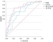
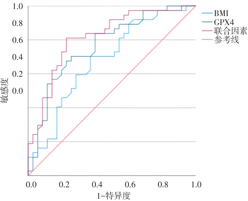
Fig.1
ROC curves for diagnosing AD by BMI, GPX4 and combined factors"
| [1] |
JI Q, CHEN J, LI Y, et al. Incidence and prevalence of Alzheimer's disease in China: A systematic review and meta-analysis [J]. Eur J Epidemiol, 2024, 39(7): 701-714. doi: 10.1007/s10654-024-001144-2 .
doi: 10.1007/s10654-024-001144-2 |
| [2] |
SHARMA A, RUDRAWAR S, BHARATE S B, et al. Recent advancements in the therapeutic approaches for Alzheimer's disease treatment: Current and future perspective [J]. RSC Med Chem, 2025, 16(2): 652-693. doi: 10.1039/d4md00630e .
doi: 10.1039/d4md00630e |
| [3] |
YAN N, ZHANG J. Iron Metabolism, Ferroptosis, and the Links With Alzheimer's Disease [J]. Front Neurosci, 2019, 13: 1443. doi: 10.3389/fnins.2019.01443 .
doi: 10.3389/fnins.2019.01443 |
| [4] |
KIM A C, LIM S, KIM Y K. Metal Ion Effects on Aβ and Tau Aggregation [J]. Int J Mol Sci, 2018, 19(1):128. doi: 10.3390/ijms19010128 .
doi: 10.3390/ijms19010128 |
| [5] |
WANG F, WANG J, SHEN Y, et al. Iron Dyshomeostasis and Ferroptosis: A New Alzheimer's Disease Hypothesis? [J]. Front Aging Neurosci, 2022, 14: 830569. doi: 10.3389/fnagi. 2022. 830569 .
doi: 10.3389/fnagi. 2022. 830569 |
| [6] |
ZHANG Y, WANG M, CHANG W. Iron dyshomeostasis and ferroptosis in Alzheimer's disease: Molecular mechanisms of cell death and novel therapeutic drugs and targets for AD [J]. Front Pharmacol, 2022, 13: 983623. doi: 10.3389/fphar.2022.983623 .
doi: 10.3389/fphar.2022.983623 |
| [7] |
YOO M H, GU X, XU X M, et al. Delineating the role of glutathione peroxidase 4 in protecting cells against lipid hydroperoxide damage and in Alzheimer's disease [J]. Antioxid Redox Signal, 2010, 12(7): 819-827. doi: 10.1089/ars.2009.2891 .
doi: 10.1089/ars.2009.2891 |
| [8] |
HAMBRIGHT W S, FONSECA R S, CHEN L, et al. Ablation of ferroptosis regulator glutathione peroxidase 4 in forebrain neurons promotes cognitive impairment and neurodegeneration [J]. Redox Biol, 2017, 12: 8-17. doi: 10.1016/j.redox.2017.01.021 .
doi: 10.1016/j.redox.2017.01.021 |
| [9] |
丁赛能, 马小茜, 赵倩华. 阿尔茨海默病诊断标准的变迁:2021年版国际工作组标准解读 [J]. 神经病学与神经康复学杂志, 2021, 17(4): 135-139. doi: 10.12022/jnnr.2021-0096 .
doi: 10.12022/jnnr.2021-0096 |
| [10] |
Association Alzheimer's. 2023 Alzheimer's disease facts and figures [J]. Alzheimers Dement, 2023, 19(4): 1598-1695. doi: 10.1002/alz.13016 .
doi: 10.1002/alz.13016 |
| [11] |
张洪铭, 冯丽娜, 孙保亮. 阿尔茨海默病与铁死亡相关性的研究进展 [J]. 神经损伤与功能重建, 2024, 19(2): 105-108. doi: 10.16780/j.cnki.sjssgncj.20230797 .
doi: 10.16780/j.cnki.sjssgncj.20230797 |
| [12] |
HASSANNIA B, VAN COILLIE S, VANDEN BERGHE T. Ferroptosis: Biological Rust of Lipid Membranes [J]. Antioxid Redox Signal, 2021, 35(6): 487-509. doi: 10.1089/ars.2020.8175 .
doi: 10.1089/ars.2020.8175 |
| [13] |
卢刚刚, 李生龙, 赵永强, 等. 氧化应激与铁死亡在糖尿病型阳痿中的相关性研究进展 [J]. 实用医学杂志, 2024, 40(16): 2229-2235. doi:10.3969/j.issn.1006-5725.2024.16.006 .
doi: 10.3969/j.issn.1006-5725.2024.16.006 |
| [14] |
GAMMELLA E, RECALCATI S, RYBINSKA I, et al. Iron-induced damage in cardiomyopathy: Oxidative-dependent and independent mechanisms [J]. Oxid Med Cell Longev, 2015, 2015: 230182. doi: 10.1155/2015/230182 .
doi: 10.1155/2015/230182 |
| [15] |
王准, 孙谕莹, 黄汉昌. 氧化应激与阿尔茨海默病的病理关系及干预措施 [J]. 生命科学, 2023, 35(4): 519-528. doi: 10.13376/j.cbls/2023061 .
doi: 10.13376/j.cbls/2023061 |
| [16] |
刘汝雄, 杨万镇, 涂杰, 等. 铁死亡调控蛋白GPX4的小分子抑制剂研究进展 [J]. 药学实践与服务, 2024, 42(9): 375-378. doi: 10.12206/j.issn.2097-2024.202312075 .
doi: 10.12206/j.issn.2097-2024.202312075 |
| [17] |
王灿雨, 刘晓龙, 孙玉, 等. 铁死亡及其在神经退行性疾病中的研究进展 [J]. 安徽医科大学学报, 2023, 58(10): 1801-1806. doi: 10.19405/j.cnki.issn1000-1492.2023.10.034 .
doi: 10.19405/j.cnki.issn1000-1492.2023.10.034 |
| [18] |
YAO S, QUAN Y. Research progress of ferroptosis pathway and its related molecular ubiquitination modification in liver cancer [J]. Front Oncol, 2025, 15: 1502673. doi: 10.3389/fonc. 2025. 1502673 .
doi: 10.3389/fonc. 2025. 1502673 |
| [19] |
LI J, CAO F, YIN H L, et al. Ferroptosis: Past, present and future [J]. Cell Death Dis, 2020, 11(2): 88. doi: 10.1038/s41419-020-2298-2 .
doi: 10.1038/s41419-020-2298-2 |
| [20] |
SHI J, CHEN D, WANG Z, et al. Homocysteine induces ferroptosis in endothelial cells through the systemXc(-)/GPX4 signaling pathway [J]. BMC Cardiovasc Disord, 2023, 23(1): 316. doi: 10.1186/s12872-023-03342-4 .
doi: 10.1186/s12872-023-03342-4 |
| [21] |
李涛, 于志超, 赵静雯, 等. 铁死亡在阿尔茨海默病中的研究进展 [J]. 重庆医科大学学报, 2023, 48(5): 508-511. doi: 10.13406/j.cnki.cyxb.003222 .
doi: 10.13406/j.cnki.cyxb.003222 |
| [22] |
樊赟, 窦润鹏, 胡久略, 等. 茯苓酸通过Nrf2/SLC7A11/GPX4信号通路调控铁死亡改善阿尔茨海默病大鼠认知障碍的研究 [J]. 中国全科医学, 2024, 27(2): 177-183. doi: 10.12114/j.issn.1007-9572.2023.0326 .
doi: 10.12114/j.issn.1007-9572.2023.0326 |
| [23] |
MANDAL P K, GOEL A, BUSH A I, et al. Hippocampal glutathione depletion with enhanced iron level in patients with mild cognitive impairment and Alzheimer's disease compared with healthy elderly participants [J]. Brain Commun, 2022, 4(5): fcac215. doi: 10.1093/braincomms/fcac215 .
doi: 10.1093/braincomms/fcac215 |
| [24] |
邓翕仁, 曾道君, 张官鹏, 等. 黄芩苷通过PGE2在脑缺血再灌注损害小鼠认知功能中的作用研究 [J]. 实用医学杂志, 2023, 39(15): 1881-1887. doi:10.3969/j.issn.1006-5725. 2023. 15.005 .
doi: 10.3969/j.issn.1006-5725. 2023. 15.005 |
| [25] |
LIU Y, CHEN Z, LI B, et al. Supplementation with γ-glutamylcysteine (γ-GC) lessens oxidative stress, brain inflammation and amyloid pathology and improves spatial memory in a murine model of AD [J]. Neurochem Int, 2021, 144: 104931. doi: 10.1016/j.neuint.2020.104931 .
doi: 10.1016/j.neuint.2020.104931 |
| [26] |
LI D D, ZHANG W, WANG Z Y, et al. Serum Copper, Zinc, and Iron Levels in Patients with Alzheimer's Disease: A Meta-Analysis of Case-Control Studies [J]. Front Aging Neurosci, 2017, 9: 300. doi: 10.3389/fnagi.2017.00300 .
doi: 10.3389/fnagi.2017.00300 |
| [27] |
李华妮, 刘长河, 郭晓燕, 等. 心脑舒通胶囊抑制铁死亡减轻大鼠脑缺血再灌注损伤的机制 [J]. 中国药房, 2025, 36(3): 306-311. doi: 10.6039/j.issn.1001-0408.2025.03.08 .
doi: 10.6039/j.issn.1001-0408.2025.03.08 |
| [28] |
MCCADDON A, HUDSON P, HILL D, et al. Alzheimer's disease and total plasma aminothiols [J]. Biol Psychiatry, 2003, 53(3): 254-260. doi: 10.1016/s0006-3223(02)01451-8 .
doi: 10.1016/s0006-3223(02)01451-8 |
| [29] |
GAO L, JIANG Z, CAI Z, et al. Brain iron deposition analysis using susceptibility weighted imaging and its association with body iron level in patients with mild cognitive impairment [J]. Mol Med Rep, 2017, 16(6): 8209-8215. doi: 10.3892/mmr. 2017.7668 .
doi: 10.3892/mmr. 2017.7668 |
| [30] |
WANG Z X, TAN L, WANG H F, et al. Serum Iron, Zinc, and Copper Levels in Patients with Alzheimer's Disease: A Replication Study and Meta-Analyses [J]. J Alzheimers Dis, 2015, 47(3): 565-581. doi: 10.3233/JAD-143108 .
doi: 10.3233/JAD-143108 |
| [31] |
桂琴, 刘峰, 董春霞. 血清Hcy水平与阿尔茨海默病患者认知功能障碍的相关性 [J]. 中国医学创新, 2023, 20(5): 162-166. doi:10.3969/j.issn.1674-4985.2023.05.037 .
doi: 10.3969/j.issn.1674-4985.2023.05.037 |
| [32] |
MCCADDON A, MILLER J W. Homocysteine-a retrospective and prospective appraisal [J]. Front Nutr, 2023, 10: 1179807. doi: 10.3389/fnut.2023.1179807 .
doi: 10.3389/fnut.2023.1179807 |
| [33] |
黄勉, 李琳, 李芬, 等. 血浆同型半胱氨酸、脂联素水平与阿尔茨海默病患者认知功能障碍程度的相关性 [J]. 神经损伤与功能重建, 2020, 15(8): 483-485. doi: 10.16780/j.cnki.sjssgncj.20181400 .
doi: 10.16780/j.cnki.sjssgncj.20181400 |
| [34] |
LIN C H, LANE H Y. Plasma Glutathione Levels Decreased with Cognitive Decline among People with Mild Cognitive Impairment (MCI): A Two-Year Prospective Study [J]. Antioxidants (Basel), 2021, 10(11):1839.doi: 10.3390/antiox10111839 .
doi: 10.3390/antiox10111839 |
| [35] |
KANG S Y, KIM Y J, JANG W, et al. Body mass index trajectories and the risk for Alzheimer's disease among older adults [J]. Sci Rep, 2021, 11(1): 3087. doi: 10.1038/s41598-021-82593-7 .
doi: 10.1038/s41598-021-82593-7 |
| [36] |
BELL S P, LIU D, SAMUELS L R, et al. Late-Life Body Mass Index, Rapid Weight Loss, Apolipoprotein E ε4 and the Risk of Cognitive Decline and Incident Dementia [J]. J Nutr Health Aging, 2017, 21(10): 1259-1267. doi: 10.1007/s12603-017-0906-3 .
doi: 10.1007/s12603-017-0906-3 |
| [37] |
张悦. 1.老年时期体重波动与认知功能下降的关系 2.肥胖代谢表型与多发性骨髓瘤再入院风险的关系 [D].济南:山东大学,2023. doi: 10.27272/d.cnki.gshdu.2023.007006 .
doi: 10.27272/d.cnki.gshdu.2023.007006 |
| [38] |
PANDEY N, YANG Z, CIEZA B, et al. Plasma phospho-tau217 as a predictive biomarker for Alzheimer's disease in a large south American cohort [J]. Alzheimers Res Ther, 2025, 17(1): 1. doi: 10.1186/s13195-024-01655-w .
doi: 10.1186/s13195-024-01655-w |
| [39] |
THIJSSEN E H, LA JOIE R, STROM A, et al. Plasma phosphorylated tau 217 and phosphorylated tau 181 as biomarkers in Alzheimer's disease and frontotemporal lobar degeneration: A retrospective diagnostic performance study [J]. Lancet Neurol, 2021, 20(9): 739-752. doi: 10.1016/S1474-4422(21)00214-3 .
doi: 10.1016/S1474-4422(21)00214-3 |
| [1] | Yuying SONG,Saimaiti KHALIBINUR,Xu ZHOU,Xiangxin SONG,Yidai Abdigiti XIE,Aizezi DILRABA,Yibulayin. HASYATI. Quantitative susceptibility mapping analysis of brain iron deposition characteristics and their correlation with cognitive function in Alzheimer's disease patients [J]. The Journal of Practical Medicine, 2025, 41(20): 3267-3275. |
| [2] | Cong LIU,Fei ZHAI,Min LI,Xiaoli ZHANG,Han SHI,Ningning GUO,Changhong WANG. Association between smoking status, cognitive function, and personality traits in first⁃episode male patients with schizophrenia [J]. The Journal of Practical Medicine, 2025, 41(12): 1922-1928. |
| [3] | Song LI,Xingyou HE,Dian HE,Bo WANG,Yu ZHAN,Jingjing. SUN. The joint efficacy of NBP and LIPost C in treatment of elderly patients with large atherosclerotic cerebral infarction [J]. The Journal of Practical Medicine, 2024, 40(9): 1286-1292. |
| [4] | Na LI,Yihua BAI,Hongying JIANG,Feng ZHANG,Meng LI,Jiao YANG. Frailty of patients with long⁃term maintenance dialysis and its influencing factors [J]. The Journal of Practical Medicine, 2024, 40(3): 330-335. |
| [5] | Jinhui XU,Mailong YUAN,Tao ZHOU,Mingsheng ZHANG,Yaqi. LI. Effects of sevoflurane or propofol anesthesia maintenance on renal function and postoperative cognition in patients undergoing liver transplantation [J]. The Journal of Practical Medicine, 2024, 40(24): 3509-3514. |
| [6] |
FANG Xue, SHAO Wei, XU Huifang, LI Daoxin, WANG Jing..
A study on improvement in cognitive function after cerebral infarction based on 2⁃Cl⁃MGV⁃1/BDNF⁃TrkB pathway [J]. The Journal of Practical Medicine, 2023, 39(7): 819-826. |
| [7] | Yaxin YANG,Mao HUANG,Xin WANG,Yaru ZHI,Xuemeng SHI,Hongjin REN,Wanying. PENG. Effect of head acupuncturing therapy combined with massaging key head acupoints on language and cognitive function in children with autism [J]. The Journal of Practical Medicine, 2023, 39(23): 3132-3136. |
| [8] | Suzhen YE,Xiaoyang WANG,Hongzhao BAI,Xuezhen ZHOU,Haiyan LI. Clinical efficacy and influencing factor of Remote Ischemic Postconditioning in Patients with Post Stroke Fatigue [J]. The Journal of Practical Medicine, 2023, 39(21): 2812-2816. |
| [9] | DENG Xiren , ZENG Daojun, ZHANG Guanpeng, DUAN Xiaoxia. . The effect of baicalin on cognitive function of cerebral ischemia-reperfusion injury in mice through PGE2 [J]. The Journal of Practical Medicine, 2023, 39(15): 1881-1887. |
| [10] |
GUO Fei, HUANG Yunhui, HUANG Yujing, NIU Huifang, DU Ailing. .
Systematic review of efficacy and safety of xingnaojing injection combined with alteplase for ischemic stroke [J]. The Journal of Practical Medicine, 2022, 38(2): 206-211. |
| [11] |
YUAN Bingkun, LIAO Shengwu, WU Chengkai, PENG Cheng, CHEN Lei, LI Wenyuan..
Study on the relationship between daily sleep duration and the prevalence of hyperuricemia among urban workers in Guangzhou [J]. The Journal of Practical Medicine, 2022, 38(15): 1948-1953. |
| [12] |
YANG Bo, ZHANG Maorui, ZHOU Jun.
Effects of propofol preconditioning on cognitive function in septic rats [J]. The Journal of Practical Medicine, 2021, 37(7): 845-850. |
| [13] |
LONG Qijia, LV Zongxia, SHI Ke, LI Chunyan, ZHENG Jinou..
Analysis of hippocampus in patients with anti⁃NMDAR encephalitis by voxel⁃based morphology and func⁃ tional connectivity [J]. The Journal of Practical Medicine, 2021, 37(18): 2366-2370. |
| [14] |
LI Jia, LIN Yin, JI Xunqi, KANG Yanhai, DONG Jie..
Association of APOE and MTHFR C677T gene polymorphisms with schizophrenia and cognitive function in patients [J]. The Journal of Practical Medicine, 2021, 37(18): 2391-2394. |
| [15] |
LI Jia, LIN Yin, JI Xunqi, KANG Yanhai, DONG Jie..
Association of APOE and MTHFR C677T gene polymorphisms with schizophrenia and cognitive function in patients [J]. The Journal of Practical Medicine, 2021, 37(18): 2391-2394. |
| Viewed | ||||||
|
Full text |
|
|||||
|
Abstract |
|
|||||